CD3 - The Marker the Antibodies
- On This Page
- The T cell activation mediator
- CD3 antibody images by application
- CD3 Antibodies and Multicolor Kits
- References
s

s


The T cell activation mediator
CD3 is a member of the immunoglobulin superfamily and acts as a mediator of signal transduction. It is a multimeric protein composed of four distinct polypeptide chains (ε, γ, δ, ζ). Each of these chains contain immunoreceptor tyrosine activation motifs (ITAMS) which are required for initiation of signaling cascades, as they recruit protein tyrosine kinases, signaling intermediates and adapter molecules. CD3 is expressed by a high-percentage of circulating peripheral T cells forming a complex with the T cell receptor (TCR). As CD3 is present at all stages of T cell development, it is a highly effective T cell marker. Read our CD3 mini-review for detailed information on CD3 including its structure alongside that of the TCR, the genes involved in its expression, its function and the signal transduction pathways mediated by the CD3 complex.
For more information about T cells refer to the following resources available:
- T cell overview; including antibodies available to this important immune cell
- T cell receptor mini-review for detailed information on TCR activation, signaling, development and diversity
- Human T cell lineage posters
- Human T cell marker selection and expression guide
- Mouse T cell lineage posters
- Mouse T cell marker selection and expression guide
- Our CD3 antibody range
Here we look at anti-CD3 clones and their recommended applications.
Anti-human CD3 clones
- Clone CD3-12, specifically recognizes a conserved epitope within the cytoplasmic sequence of the human CD3ε chain. The epitope on the epsilon polypeptide is conserved among many species resulting in antibody clone, CD3-12, having a very broad species cross reactivity for the CD3 marker. Applications for this antibody include flow cytometry, immunofluorescence/immunocytochemistry, immunohistochemistry – frozen, immunohistochemistry – paraffin sections and western blotting
- Clone MEM-57 recognizes an extracellular epitope of the human CD3 complex containing either δ-ε or γ-ε subunit complexes (Dave et al. 1998). Applications for this antibody include flow cytometry and immunoprecipitation
- CloneUCHT1 binds to a region in the ectodomain of human CD3ε and has been shown to bind to a discontinuous epitope near an acidic region of CD3ε opposite the dimer interface; as shown by crystallographic studies of the CD3ε/δ dimer complexed with a single chain UCHT1 antibody fragment (Arnett et al. 2004). Applications for this antibody include flow cytometry and immunohistochemistry – frozen
- Clone OKT3 recognizes T-cell surface glycoprotein CD3 epsilon chain (CD3ε). CD3ε is a subunit within the multiprotein cell surface receptor TCR-CD3 complex, which is expressed on the surface of mature T cells, NKT cells and thymocytes (Mariuzza et al. 2020). Commonly used in flow cytometry in the phenotyping human T cells, and immunohistochemistry - frozen.
- Clone SK7 recognizes the T-cell surface glycoprotein CD3 epsilon chain (CD3ɛ), also known as T-cell surface antigen T3/Leu-4 epsilon. For use in flow cytometry.
Anti-mouse CD3 clones
- Clone KT3 may be used to trigger proliferation and cytotoxicity of CD3 positive cells. Applications for this antibody include flow cytometry and immunohistochemistry – frozen
- Clone 145-2C11 is useful for in vitro blocking and activation assays, as well as apoptosis induction and in vitro T cell depletions. Contrary to KT3, clone 145-2C11 induces cytokine release and morbidity in vivo (Vossen et al. 1995). Applications for this antibody include flow cytometry, immunofluorescence/immunocytochemistry, immunohistochemistry – frozen, immunoprecipitation and western blotting
Anti-rat CD3 clones
- Clone 1F4 has been demonstrated to induce proliferation of T cells in cell culture (Nicolls et al. 1993). Mouse anti-rat CD3, clone 1F4 does not react with rat B cells. Applications for this antibody include flow cytometry, immunohistochemistry – frozen, immunohistochemistry – paraffin sections and immunoprecipitation
CD3 antibodies are also available for dog, pig, chicken and rhesus monkey.
CD3 antibody images by application
Flow cytometry
Anti-mouse CD3 V CD4
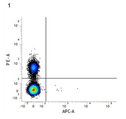
Fig. 1. RPE conjugated rat anti-mouse CD4 (MCA4635PE) and APC conjugated rat IgG2a isotype control.
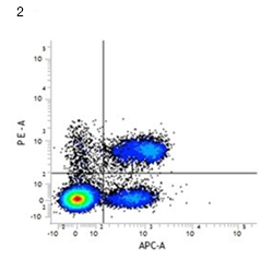
Fig. 2. RPE conjugated rat anti-mouse CD4 (MCA4635PE) and APC conjugated rat anti-mouse CD3 (MCA500APC). All experiments performed on murine splenocytes in the presence of murine SeroBlock (BUF041A).
Immunohistochemistry
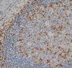
Anti-human CD3
Fig 3. Staining of a formalin fixed paraffin embedded human tonsil with rat anti-human CD3 antibody, clone CD3-12 (MCA1477) following heat mediated antigen retrieval.
CD3 Antibodies and Multicolor Kits
| Description | Target | Format | Clone | Applications | Citations | Code |
|---|
References
- Arnett KL et al. (2004). Crystal structure of a human CD3-epsilon/delta dimer in complex with a UCHT1 single-chain antibody fragment. Proc Natl Acad Sci U S A 101, 16268-16273.
- Dave VP et al. (1998). Altered functional responsiveness of thymocyte subsets from CD3delta-deficient mice to TCR-CD3 engagement. Int Immunol 10, 1481-1490.
- Nicolls MR et al. (1993). Induction of long-term specific tolerance to allografts in rats by therapy with an anti-CD3-like monoclonal antibody. Transplantation 55, 459-468.
- Vossen AC et al. (1995). Fc receptor binding of anti-CD3 monoclonal antibodies is not essential for immunosuppression, but triggers cytokine-related side effects. Eur J Immunol 25, 1492-1496.
- Mariuzza et al. (2020). The structural basis of T-cell receptor (TCR) activation: An enduring enigma. J Biol Chem.