Phospho-Specific PrecisionAb Antibodies

- On This Page
- Highly specific antibodies
- Anti-protein antibodies
- Protein phosphorylation in disease research
- Phospho-Specific PrecisionAb Antibodies range
s
Western Blot University
s
Cancer Pathway Posters

Compiling key proteins involved in key signaling pathways: cGAS-STING, EGF R, mTOR, NF-kB, and PI3/AKT pathways.
s
What are R-loops?
Highly Specific Antibodies to Detect Phosphorylation Events
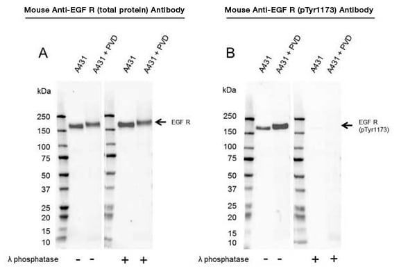
Bio-Rad’s new Phospho-Specific Validated PrecisionAb Antibodies have been validated by comparing phosphorylation levels in both treated versus untreated cell lysates and by dephosphorylating proteins on western blot membranes (Figure 1).
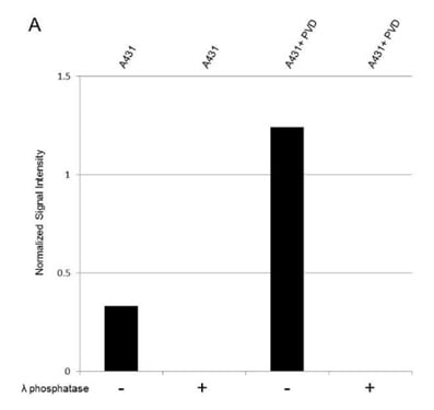
The phosphorylation level quantification we undertake enables you to quickly determine the impact of cellular treatments and provides you with an easy way to judge the antibody specificity (Figure 2).

Fig. 1. Western blot analysis of whole cell lysates probed with A, Mouse Anti-EGF Receptor Antibody (VMA00061) or B, Mouse Anti-EGF Receptor (pTyr1173) Antibody (VMA00752). It is followed by detection with HRP conjugated Goat Anti-Mouse IgG (1/10,000, STAR207P). Membranes were treated with (+) and without (-) lambda protein phosphatase as indicated and visualized on the ChemiDoc MP Imaging System.

Fig. 2. Total Protein Normalization.
Normalized
signal intensity of EGF R (pTyr1173) from PVD treated and untreated lysates, treated with (+) or without (-) lambda protein phosphatase. Lambda protein phosphatase dephosphorylates serine, threonine and tyrosine residues. Normalized signal intensity was calculated by dividing the TPN-normalized signal intensity for each phosphoprotein by the TPN-normalized intensity of the corresponding total protein. Learn more about TPN Total Protein Normalization.
 Western Blot Detection of Phosphorylation Events
?
 Detection of Phosphorylated Proteins by Western Blotting
?
Unsure What Total Anti-Protein Antibody to Use as a Control in Your Phospho Western Blots?
To showcase the different binding characteristics of phospho-specific and total protein antibodies, we have compared Phospho-Specific PrecisionAb Antibodies against total PrecisionAb Antibodies; this enables you to assess the impact of the different treatments and provides a control recommendation. The total and phospho-specific antibody can be purchased individually.
Protein Phosphorylation in Disease Research
Phosphorylation is involved in the regulation of multiple biological processes and appropriate phosphorylation is crucial for cell homeostasis. Kinases are enzymes that catalyze the transfer of a phosphate group to a specific protein, while phosphatases remove a phosphate group. Various diseases are caused by mutations in specific kinases (Table 1) and phosphatases. Mutation and activation state, defined by phosphorylation, can have a significant impact on a clinical course (Magkou et al. 2008). Cyclosporin and rapamycin were the first drugs to act by inhibiting protein phosphatases or kinases (Cohen 2001). As we learn more about protein phosphorylation and how it functions, we can develop more drugs that target protein kinases and phosphatases.
Table 1. Examples of diseases caused by mutations in specific protein kinases (Cohen 2001).
Disease |
Kinase |
|---|---|
|
Autosomal recessive SCID |
|
|
X-Linked SCID |
|
|
Non-Hodgkins lymphoma |
Alk1 kinase |
|
Peutz-Jeghers syndrome |
Lkb1 kinase |
|
Li-Fraumeni syndrome |
Chk2 kinase |
Much attention has been placed on the development of drugs targeting kinases as cancer treatments, resulting in small-molecule kinase inhibitors. In addition, a therapeutic monoclonal antibody called cetuximab, which blocks the interaction of epidermal growth factor (EGF) with EGF R, is used to treat colorectal, head, and neck cancers. Part of the challenge in developing effective treatments is understanding complex signaling pathways and providing robust tools to study the proteins involved. The EGF R pathway, for example, involves 211 biochemical reactions and 322 signaling molecules (Oda et al. 2005).
Phosphorylation can also influence protein folding and aggregation, and therefore plays a key role in neurogenerative disorders. As an example, cerebrospinal fluid (CSF) phosphorylated tau is a core biomarker for Alzheimer’s disease (Suarez-Calvet et al. 2020).
Using a specific antibody directed towards the phosphorylation site of your protein of interest is the easiest way to study protein phosphorylation. Find below a selection of our highly validated Phospho-Specific PrecisionAb Antibodies to enable generation of robust data.
Phospho-Specific PrecisionAb Antibodies Range
| Description | Target | Format | Clone | Applications | Citations | Code |
|---|
References
- Cohen P (2001). The role of protein phosphorylation in human health and disease. Eur J Biochem 268, 5001-5010.
- Magkou C et al. (2008). Expression of the epidermal growth factor receptor (EGFR) and the phosphorylated EGFR in invasive breast carcinomas. Breast Cancer Res 10, R49.
- Oda K et al. (2005). A comprehensive pathway map of epidermal growth factor receptor signaling. Mol Syst Biol 1, 2005.0010.
- Suarez-Calvert M (2020). Novel tau biomarkers phosphorylated at T181, T217 or T231 rise in the initial stages of the preclinical Alzheimer’s continuum when only subtle changes in Aβ pathology are detected. EMBO Mol Med. 12, e12921.


