CD19 - The Marker, the Antibodies
- On This Page
- Overview
- Anti-human CD19 clones
- Anti-mouse CD19 clones
- Images by application
- CD19 antibodies
- References
s

s


Overview
CD19 is a 95 kDa type I transmembrane glycoprotein containing two C2 type Ig-like domains in the N-terminal extracellular region and several potential phosphorylation sites for tyrosine together with a single serine in the cytoplasmic region. CD19 is also known as B-lymphocyte surface antigen B4. CD19 is expressed on virtually all B cells with the exception of plasma cells and is also found on follicular dendritic cells (de Rie et al. 1989).
CD19 has an essential regulatory role in B cell differentiation, proliferation and subsequent antibody production. Where CD19 forms a multimolecular complex with CD21, CD81 and MHC class II that binds to the B cell receptor (Horváth et al. 1998). When activated CD19 becomes phosphorylated, this then enables binding to Src family tyrosine kinases, to PI3 kinase and Vav, which then leads to the activation of signaling pathways involved in B cell stimulation (Buhl and Cambier 1999. Fujimoto et al. 2000. O’Rourke et al. 1998).
Anti-human CD19 clones
- Clone LE-CD19 antibody (MCA2454) recognizes an epitope within the C-terminal tail sequence of the CD19 molecule. Applications for this antibody include; ELISA, flow cytometry, immunohistochemistry – paraffin section, immunoprecipitation and western blotting
- Clone HIB19 antibody recommended applications include; flow cytometry, immunofluorescence and immunohistochemistry – frozen
- Clone SJ25-C1 antibody (MCA1235C) is recommended for use in flow cytometry. Clones SJ25-C1 and HIB19 recognize overlapping epitopes
- Clone LT19 antibody (MCA1940) recommended applications include; flow cytometry, immunohistochemistry – frozen and immunoprecipitation
- Clone Bu12 antibody (MCA2495) recommended applications include; flow cytometry and immunoprecipitation
Anti-mouse CD19 clones
- Clone 6D5 antibody (MCA1439) recognizes the same epitope as clone ID3. Applications for this antibody include; flow cytometry, immunohistochemistry – frozen and immunoprecipitation
- Clone ID3 antibody recognizes the same epitope as clone 6D5. Applications for this antibody include; flow cytometry, functional assay, immunohistochemistry – frozen and immunoprecipitation
- Clone MB19-1 antibody staining is not as bright as that observed using clone 6D5. Recommended applications include: flow cytometry, functional assay and Immunoprecipitation
CD19 antibody images by application
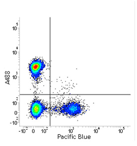
Flow cytometry
Anti-human CD19 V CD4
Red cell lysed human blood stained with CD19PB (MCA1940) and CD4A488 (MCA1267) gated on lymphocytes in the presence of human seroblock (BUF070A).
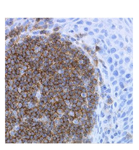
Immunohistochemistry
Anti-human CD19
Immunoperoxidase staining of formalin-fixed, paraffin-embedded human tonsil with mouse anti-human CD19, clone LE-CD19 (MCA2454), followed by visualisation using the Histar Detection System (STAR3000).
CD19 Antibodies
| Description | Target | Format | Clone | Applications | Citations | Code |
|---|
Searching for your antibody of choice is made easy by the use of the sophisticated filter table, which allows you to select for specific antibody parameters. If you are unable to find your desired antibody simply contact us as we are adding new product all the time and we will be happy to help advise you.
References
- Buhl AM and Cambier JC (1999). Phosphorylation of CD19 Y484 and Y515, and linked activation of phosphatidylinositol 3-kinase, are required for B cell antigen receptor-mediated activation of Bruton's tyrosine kinase. J Immunol. 162(8), 4438-4446.
- de Rie MA et al. (1989). Regulatory role of CD19 molecules in B-cell activation and differentiation. Cell Immunol. 118(2), 368-381.
- Fujimoto M et al. (2000). CD19 regulates Src family protein tyrosine kinase activation in B lymphocytes through processive amplification. Immunity. 13(1), 47-57.
- Horváth G et al. (1998). CD19 is linked to the integrin-associated tetraspans CD9, CD81, and CD82. J Biol Chem. 273(46), 30537-30543.
- O'Rourke LM et al. (1998). CD19 as a membrane-anchored adaptor protein of B lymphocytes: costimulation of lipid and protein kinases by recruitment of Vav. Immunity. 8(5), 635-645.