CD3 antibody | 1F4
Mouse anti Rat CD3
- Product Type
- Monoclonal Antibody
- Clone
- 1F4
- Isotype
- IgM
- Specificity
- CD3


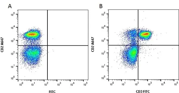
Figure B. Alexa Fluor® 647 conjugated Mouse anti Rat CD2 antibody, clone OX-34 (MCA154A647) and FITC conjugated Mouse anti Rat CD3 antibody, clone 1F4 (MCA772F). All experiments performed on red cell lysed rat blood gated on lymphoid cells in the presence of 10% rat serum.
Data acquired on the ZE5 Cell Analyzer.
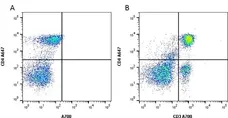
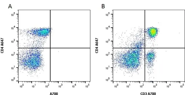
Figure B. Alexa Fluor® 647 conjugated Mouse anti Rat CD4 antibody, clone clone W3/25 (MCA55A647) and Alexa Fluor® 700 conjugated Mouse anti Rat CD3 antibody, clone 1F4 (MCA772A700). All experiments performed on red cell lysed rat blood gated on lymphoid cells in the presence of 10% rat serum.
Data acquired on the ZE5 Cell Analyzer.
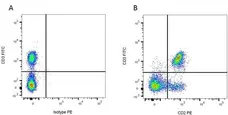
Figure B. FITC conjugated Mouse anti Rat CD3 antibody, clone 1F4 (MCA772F) and RPE conjugated Mouse anti Rat CD2 antibody, clone OX-34 (MCA154PE). All experiments performed on red cell lysed rat blood gated on mononuclear cells. All experiments performed on red cell lysed rat blood gated on lymphoid cells in the presence of 10% rat serum.
Data acquired on the ZE5 Cell Analyzer.
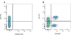
Figure B. FITC conjugated Mouse anti Rat CD3 antibody, clone 1F4 (MCA772F) and RPE conjugated Mouse anti Rat CD2 antibody, clone OX-34 (MCA154PE). All experiments performed on red cell lysed rat blood gated on mononuclear cells. All experiments performed on red cell lysed rat blood gated on lymphoid cells in the presence of 10% rat serum.
Data acquired on the ZE5 Cell Analyzer.
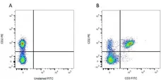
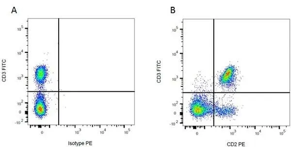
Figure B. RPE conjugated Mouse anti Rat CD2 antibody, clone OX-34 (MCA154PE) and FITC conjugated Mouse anti Rat CD3 antibody, clone 1F4 (MCA772F). All experiments performed on red cell lysed rat blood gated on mononuclear cells.
Data acquired on the ZE5 Cell Analyzer.

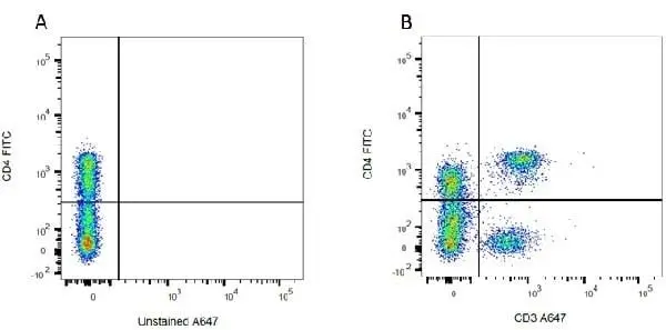
Figure B. Alexa Fluor® 647 conjugated Mouse anti Rat CD3 antibody, clone 1F4 (MCA772A647) and FITC conjugated Mouse anti Rat CD4 (MCA55F). All experiments performed on red cell lysed rat blood gated on mononuclear cells.
Data acquired on the ZE5 Cell Analyzer.
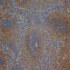
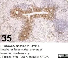
Mouse anti Rat CD3 antibody, clone 1F4 (MCA772) used for the identification of CD3 expressing lymphocytes in rat spleen by immunohistochemistry on formalin fixed, paraffin embedded tissue sections following EDTA mediated antigen retrieval.
Image caption:
CD3/ 1F4/ AbD serotec/ MCA772, Spleen/ Rat.
From: Furukawa S, Nagaike M, Ozaki K.
Databases for technical aspects of immunohistochemistry.
J Toxicol Pathol. 2017 Jan;30(1):79-107.
This image is from an open access article distributed under the terms of the Creative Commons Attribution License.
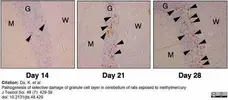
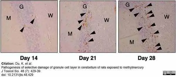
Mouse anti Rat CD3 antibody, clone 1F4 (MCA772) used to label lymphocytes in rat brain by immunohistochemistry on formalin fixed, paraffin embedded tissue sections.
Image caption:
Infiltration of granule cell layer of cerebellar cortex of rats treated with methylmercury by cytotoxic T-lymphocytes. [A] Immunostaining with a CD 3 antibody to detect T-lymphocyte infiltration into the granule cell layer of rats treated with methylmercury (×100). Arrowheads indicate CD 3-positive cells (T lymphocytes). T lymphocytes were detected at days 14, 21, and 28 after treatment with methylmercury. M, molecular layer; G, granular layer; W, white matter.
From: Du K, Hirooka T, Sasaki Y, Yasutake A, Hara T, Yamamoto C, Fujiwara Y, Shinoda Y, Fujie T, Katsuda S, Eto K, Kaji T.
Pathogenesis of selective damage of granule cell layer in cerebellum of rats exposed to methylmercury.
J Toxicol Sci. 2023;48(7):429-439.
doi: 10.2131/jts.48.429.
This image is from an open access article distributed under terms of a Creative Commons Attribution License.
Filter by Application:
F P Reset| Mouse anti Rat CD3 antibody, clone 1F4 recognizes rat CD3, a ~25 kDa antigen which is found on rat T-cells. Mouse anti Rat CD3, clone 1F4 does not react with rat B cells. In immunohistology it stains rat thymus tissues strongly in the medulla and weakly in the cortex. Functionally the addition of the antibody to a culture of rat T cells induces the proliferation of T-cells in the presence of PMA. |
- Target Species
- Rat
- Product Form
- Purified IgM - liquid
- Preparation
- MCA772: Purified IgM prepared by ammonium sulphate precipitation from tissue culture supernatant
- MCA772GA: Purified IgM prepared by ammonium sulphate precipitation from tissue culture supernatant.
- Buffer Solution
- Phosphate buffered saline
- Preservative Stabilisers
0.09% Sodium Azide - Immunogen
- F344 rat T cells stimulated with PMA (TPA) and calcium ionophore
- Approx. Protein Concentrations
- MCA772: IgM concentration 1.0 mg/ml
- MCA772GA: IgM concentration 1 mg/ml
- Fusion Partners
- Spleen cells from immunized BALB/c mice were fused with cells of the P3-X63-Ag8.653 mouse myeloma cell line.
- Regulatory
- For research purposes only
- Guarantee
- 12 months from date of despatch
This product is shipped at ambient temperature. It is recommended to aliquot and store at -20°C on receipt. When thawed, aliquot the sample as needed. Keep aliquots at 2-8°C for short term use (up to 4 weeks) and store the remaining aliquots at -20°C.
Avoid repeated freezing and thawing as this may denature the antibody. Storage in frost-free freezers is not recommended.
Avoid repeated freezing and thawing as this may denature the antibody. Storage in frost-free freezers is not recommended.
This product has been reported to work in the following applications. This information is derived from testing within our laboratories, peer-reviewed publications or personal communications from the originators. Please refer to references indicated for further information. For general protocol recommendations, please visit the antibody protocols page.
| Application Name | Verified | Min Dilution | Max Dilution |
|---|---|---|---|
| Flow Cytometry |  |
1/10 | 1/25 |
| Immunohistology - Frozen | |
1/10 | 1/25 |
| Immunohistology - Paraffin 1 | |
1/10 | |
| Immunoprecipitation | |
- 1This clone is suitable for use on paraffin embedded material following antigen retrieval (McKechnie N.M. et al. 1997).
Where this antibody has not been tested for use in a particular technique this does not necessarily exclude its use in such procedures. Suggested working dilutions are given as a guide only. It is recommended that the user titrates the antibody for use in their own system using appropriate negative/positive controls.
- Flow Cytometry
- Use 10ul of the suggested working dilution to label 106 cells in 100ul.
| Description | Product Code | Applications | Pack Size | List Price | Your Price | Quantity | |
|---|---|---|---|---|---|---|---|
| Goat anti Mouse IgM:HRP | 102005 | C E IB | 1 ml |

|
Log in | ||
| List Price | Your Price | ||||||
|
|
Log in | ||||||
| Description | Goat anti Mouse IgM:HRP | ||||||
| Goat anti Mouse IgM:Alk. Phos.(Human Adsorbed) | STAR138A | C E P WB | 1 ml |
|
Log in | ||
| List Price | Your Price | ||||||
|
|
Log in | ||||||
| Description | Goat anti Mouse IgM:Alk. Phos.(Human Adsorbed) | ||||||
Source Reference
-
Tanaka, T. et al. (1989) Characterization of a CD3-like rat T cell surface antigen recognized by a monoclonal antibody.
J Immunol. 142 (8): 2791-5.
References for CD3 antibody
-
Nicolls, M.G. et al. (1992) Induction of long-term specific tolerance to allografts in rats by therapy with an anti-CD3-like monoclonal antibody.
Transplantation 55: 459-68. -
McKechnie NM et al. (1997) Immunization with the cross-reactive antigens Ov39 from Onchocerca volvulus and hr44 from human retinal tissue induces ocular pathology and activates retinal microglia.
J Infect Dis. 176 (5): 1334-43. -
Candolfi, M. et al. (2007) Intracranial glioblastoma models in preclinical neuro-oncology: neuropathological characterization and tumor progression.
J Neurooncol. 85: 133-48. -
Lohwasser, C. et al. (2009) Role of the receptor for advanced glycation end products in hepatic fibrosis.
World J Gastroenterol. 15: 5789-98. -
Sanchez-Guajardo, V. et al. (2010) Microglia acquire distinct activation profiles depending on the degree of alpha-synuclein neuropathology in a rAAV based model of Parkinson's disease.
PLoS One. 5: e8784. -
Beck, K.D. et al (2010) Quantitative analysis of cellular inflammation after traumatic spinal cord injury: evidence for a multiphasic inflammatory response in the acute to chronic environment.
Brain. 133: 433-47. -
Echeverry, S. et al. (2011) Peripheral Nerve Injury Alters Blood-Spinal Cord Barrier Functional and Molecular Integrity through a Selective Inflammatory Pathway.
J Neurosci. 31: 10819-28. -
Takahashi, Y. et al. (2017) Rituximab protects podocytes and exerts anti-proteinuric effects in rat adriamycin-induced nephropathy independent of B-lymphocytes.
Nephrology (Carlton). 22 (1): 49-57.
View The Latest Product References
-
Sun, J. et al. (2017) Pentapeptide PLNPK ameliorates adjuvant arthritis and inhibits T cell activation by suppressing Lck and PI3K activities
Int J Clin Exp Pathol 10(5): 5252-62. -
Du, K. et al. (2023) Pathogenesis of selective damage of granule cell layer in cerebellum of rats exposed to methylmercury
J Toxicolog Sci. 48 (7): 429-39.
- RRID
- AB_321258
- UniProt
- P19377
- Q64159
- Entrez Gene
- Cd3d
- Cd3g
- GO Terms
- GO:0016021 integral to membrane
- GO:0004888 transmembrane receptor activity
- GO:0007166 cell surface receptor linked signaling pathway
- GO:0045059 positive thymic T cell selection
- GO:0007163 establishment or maintenance of cell polarity
- GO:0015031 protein transport
- GO:0042981 regulation of apoptosis
- GO:0046982 protein heterodimerization activity
View more products with CD3 specificity
Please Note: All Products are "FOR RESEARCH PURPOSES ONLY"
View all Anti-Rat ProductsAlways be the first to know.
When we launch new products and resources to help you achieve more in the lab.
Yes, sign me up