-
US | en
- Products
- Applications
- Flow Cytometry
- Flow Cytometry Explained
- Fluorophores
- Antibody Conjugation Kits

Antibody Conjugation Kits
Fluorophores, Enzymes, and Biotin/Streptavidin Conjugations
Conjugation of proteins or antibodies of interest with various molecular labels is often necessary in biological research to facilitate detection or quantification of the labeled protein, its binding partners, or antigens in the case of labeled antibodies. Molecular labels such as fluorophores or biotin are covalently conjugated to proteins or antibodies through reactions with thiol or amine groups.
With the LYNX Rapid and Rapid Plus Conjugation Kits and ReadiLink Antibody Conjugation Kits, Bio-Rad offers two complementary, fast, and easy-to-use antibody conjugations systems that cover a wide variety of labels including the common fluorophores as well as enzymes and biotin conjugations. Save valuable time by quickly conjugating your label of choice using these kits with minimal hands-on time.

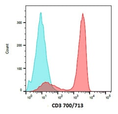
Fig.1. Peripheral blood lymphocytes were stained with CD3 (MCA463) conjugated to ReadiLink 700/713 Antibody Labeling Kit (1351008) to detect T cells. Data acquired on the ZE5 Cell Analyzer.
LYNX Kits
LYNX Rapid and Rapid Plus Conjugation Kits are available in a wide range of options including common fluorophores, enzymes, and biotin/streptavidin conjugations, suitable for use in flow cytometry, ELISA, western blotting, immunohistochemistry, and immunofluorescence. Identical protocols for small and large scale (up to milligram quantities of antibody) conjugations allow for fast one-step conjugation of proteins and antibodies using a method that is superior to traditional kits and techniques. In contrast to conventional conjugation, LYNX Conjugation Kits offer consistent results with no post-conjugation clean-up process required; therefore, the antibodies are ready to use immediately.
Conjugate your antibodies in 3 easy steps with only 30 seconds hands-on time and no purification required. The total conjugation time is fifteen minutes with the LYNX Rapid Plus Conjugation Kits or 3 hours with the LYNX Rapid Conjugation Kits, however the reaction may run overnight without any adverse effects.
Learn more about LYNX KitsReadiLink Kits
ReadiLink Antibody Conjugation Kits offer ten unique fluorescence conjugation options for small volumes. Quickly label your antibody of interest in two easy steps in just over an hour with the unique chemistry of ReadiLink Dyes. The excitation wavelengths and narrow emissions spectra of the fluorophores ranging from violet to far red are ideal for multicolor flow cytometry. The infrared excitable conjugations are suitable for westerns, ELISA, and in-vivo imaging.
Learn more about ReadiLink KitsOur Full Conjugation Kit Range
| Description | Specificity | Target | Format | Host | Isotype | Clone | Applications | Citations | Product Type | Code | Validation Types |
|---|
Degree of Labeling (DOL) Calculation
 Want to know how much fluorophore is conjugated to your antibody?
Easily calculate the fluorophore:protein (F:P) ratio using these 4 steps.
You may need to know the amount of fluorophore conjugated to your antibody for control purposes. To determine the fluorophore : protein ratio you will need to calculate the molar concentrations of both the fluorophore and the protein based on absorbance at known wavelengths in a spectrophotometer and then express this as a ratio. This ratio will be an indication of the average number of dye molecules conjugated to your antibody in your solution.
- Remove any excess fluorophore. Excess fluorophore will interfere with the absorbance measurement.
-
Determine the concentration of protein. Measure the absorbance of the protein at A280 nm. Divide this value by the molar extinction coefficient for your protein.
(e.g. mouse IgG has an extinction coefficient of 203,000 M-1 cm-1). -
Determine the fluorophore concentration. Measure the absorbance of the fluorophore at the wavelength of maximal excitation (e.g. for FITC use 495 nm wavelength). Divide this value by the molar extinction coefficient for the dye.
(e.g. FITC has an extinction coefficient of 75,000 M-1 cm-1). - Determine the labeling. Divide the fluorophore concentration by the protein concentration to calculate the F/P ratio.
