Multiplex Fluorescent Western Blotting
Fluorescent Immunoblot Imaging
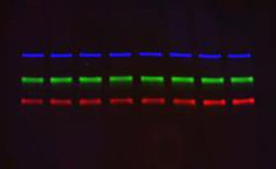
The increasing need to maximize data from a single sample has led to the development of fluorescent based multiplexing assays. Multiplex fluorescent western blotting enables the simultaneous analysis of multiple proteins in a single sample on the same blot, obtaining as much information as possible in one experiment.
The benefits of fluorescent western blotting multiplexing are:
- Multiple samples are analyzed under the same experimental conditions
- Results in context by simultaneous analysis of several proteins and parameters
- Greater consistency of readout using fluorescent dyes rather than the variability of an enzymatic reaction
- Amount of information collected is maximized, using as little of your rare/precious sample as possible
- Cost and time savings
Bio-Rad offers a new generation of multiplex western blotting reagents and equipment, that includes:
- Fluorescent StarBright Blue 700 and 520 Secondary Antibodies
- hFAB Rhodamine Anti-Housekeeping Protein Primary Antibodies
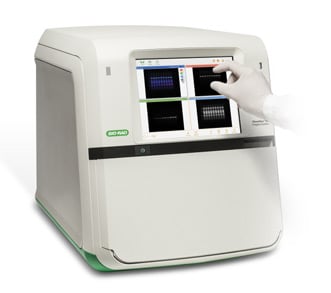
- Imaging with ChemiDoc MP Imaging System
Read the introductory guide for detailed information on multiplex fluorescent western blotting.
StarBright Blue Secondary Antibodies
Labeled with exceptionally bright and stable semiconducting nanoparticles. StarBright Dyes’ fluorescence emission is restricted to a narrow spectral band, making them ideal for multiplex fluorescence imaging for western blotting. They are excited with blue light (440–470 nm) and are available in two colors: StarBright Blue 520 emits in the green range of the spectrum (emission maximum: 520 nm) and StarBright Blue 700 maximally emits in the far red/near infrared region of the spectrum (700 nm).
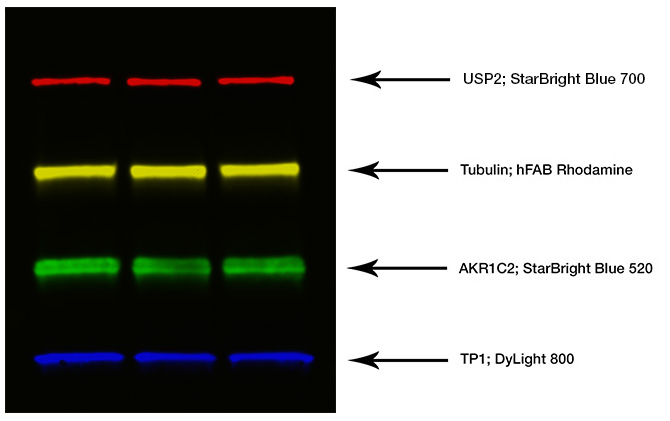
Fig. 1. Composite blot image for quadruple western blotting.
Color |
Target Protein |
Primary Antibody |
Secondary Antibody |
|---|---|---|---|
|
Red |
USP2 |
Rabbit Anti-Human USP2 |
Goat Anti-Rabbit IgG StarBright Blue 700 |
|
Yellow |
Tubulin |
Anti-Tubulin hFAB-Rhodamine |
Not required |
|
Green |
AKR1C2 |
Mouse Anti-Human AKR1C2 |
Goat Anti-Mouse IgG StarBright Blue 520 |
|
Blue |
TP1 |
Goat Anti-Human TP1 (biotinylated) |
DyLight 800 Streptavidin |
Figure 1 shows a composite blot image with four distinct proteins visualized using StarBright Blue 520 and StarBright Blue 700 Secondary Antibodies, in combination with Dylight 800 and a rhodamine labeled recombinant human hFAB antibody, for the detection of popular housekeeping proteins on the ChemiDoc MP Imaging System.
PrecisionAb Antibodies were chosen for this multiplex western blot because of their superior performance and reproducibility, having been rigorously tested and selected for high specificity, high sensitivity, and consistency.
Learn more about the validation process and customers’ experience using PrecisionAb Antibodies and the full range available.
Advantages of StarBright Blue Secondary Antibodies compared to other traditional fluorophores:
- Highly sensitive fluorescent detection — 2- to 4-fold lower limit of detection than traditional fluorophores
- Short exposure times — 50- to 100-fold shorter (seconds vs. minutes)
- Easy multiplexing options — compatible with stain-free gel and blot imaging detection and hFAB Rhodamine Antibodies as well as antibodies labeled with traditional fluorophores
Instructions for use, tips, troubleshooting and further information are available in the StarBright Blue Antibodies instruction manual and Comprehensive Guide to Fluorescence Western Blotting instruction manual.
StarBright Blue 700 Secondary Antibody Range
# Catalog |
Description |
|---|---|
|
Goat Anti-Mouse IgG StarBright Blue 700, 400 µl |
|
|
Goat Anti-Mouse IgG StarBright Blue 700, 80 µl |
|
|
Goat Anti-Rabbit IgG StarBright Blue 700, 400 µl |
|
|
Goat Anti-Rabbit IgG StarBright Blue 700, 80 µl |
StarBright Blue 520 Secondary Antibody Range
# Catalog |
Description |
|---|---|
|
Goat Anti-Mouse IgG StarBright Blue 520, 400 µl |
|
|
Goat Anti-Mouse IgG StarBright Blue 520, 80 µl |
|
|
Goat Anti-Rabbit IgG StarBright Blue 520 400 µl |
|
|
Goat Anti-Rabbit IgG StarBright Blue 520, 80 µl |
hFAB Rhodamine Housekeeping Protein Fluorescent Primary Antibodies
hFAB antibodies are primary antibodies, directly labeled with green-excitable rhodamine, which recognize the major housekeeping proteins (HKPs) β-Tubulin, β-Actin, and GAPDH. Although these antibodies have been raised against human immunogens, they are compatible for the design of multiplexing experiments with mouse and rabbit antibodies, as these housekeeping proteins are evolutionarily conserved.
These Fab fragments have engineered advantages:
- Ensure product consistency and data reproduciblity
- Free-up your multiplexing species choices for your primary targets: being preconjugated, they do not require secondary detection, and are not recognized by conventional secondary detection reagents so they can be used alongside any secondary antibodies without interference
- Facilitate load normalization without the need for a secondary antibody: hFAB Rhodamine Antibodies give a linear response in the common loading range of 10–50 μg of cell lysate, enabling different quantities (low and high) to be easily distinguished
-
Are optimized for multiplexing (fluorescent properties: excitation 530 nm/emission 580 nm):
- 3-color multiplexing with no crosstalk, when using StarBright Dyes for western blotting; the green fluorescence is intermediate between the blue fluorescence of StarBright 700 and StarBright 520
- 5-color multiplexing in combination with DyLight 800 and Alexa 647 conjugated antibodies (Figure 2)
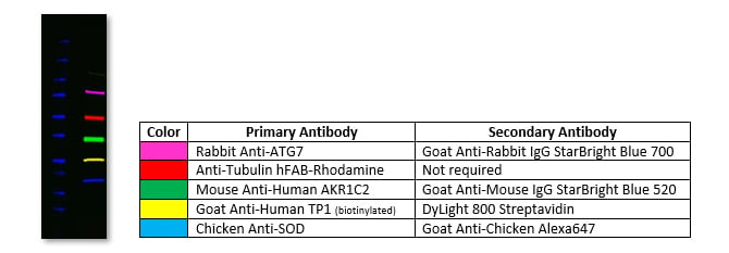
Fig. 2. Western blot of HeLa cell lysate probed with five different antibodies.
Instructions for use, tips, troubleshooting and further information are available in the hFAB Rhodamine Antibodies instruction manual and the Fluorescent Western Blotting Antibodies - Experience Unmatched Sensitivity brochure.
hFAB Rhodamine Antibody Range
# Catalog |
Description |
|---|---|
|
Anti-Actin hFAB Rhodamine Antibody, 200 µl |
|
|
Anti-Actin hFAB Rhodamine Antibody, 40 µl |
|
|
Anti-Tubulin hFAB Rhodamine Antibody, 200 µl |
|
|
Anti-Tubulin hFAB Rhodamine Antibody, 40 µl |
|
|
Anti-GAPDH hFAB Rhodamine Antibody, 200 µl |
|
|
Anti-GAPDH hFAB Rhodamine Antibody, 40 µl |
Imaging with ChemiDoc MP Imaging System and Image Lab Touch Software
ChemiDoc imagers offer visible light (RGB), far red/near infrared (FR/NIR) fluorescence and chemiluminescence detection. Stain-free imaging enables immediate visualization and verification of proteins without the need for gel staining. Image Lab Software is a family of software packages for the acquisition and analysis of gels and images.
Stain Free Technology
Stain free technology allows the visualization of proteins in SDS-PAGE gels without the need for staining or destaining of gels. Trihalo compounds within the gel are covalently linked to tryptophan residues in proteins when activated by UV light. This allows the proteins to be visualized in gels or on PVDF membranes multiple times throughout the electrophoresis and blotting process for validation in a western blotting workflow.
Stain free technology therefore allows the normalization of western blot band intensities against total protein levels from the sample loaded onto the gel enabling you to perform subsequent relative quantification analysis. Mini and midi sized stain free precast gels, containing trihalo compounds, are available. View a workflow example of a stain free technology in an IP experiment using TidyBlot Western Blot Detection Reagent.
Best in-class performance from the ChemiDoc MP Imaging System:
- Sensitive detection with low fluorescence background
- Chemiluminescence sensitivity matching X-ray film
- Easy-to-use software
- Multiplex fluorescence detection
Learn more about the imaging system and software, including full product specification, user guides and resources.
Products for Western Blotting
Clarity and Clarity Max Western ECL Blotting Substrates
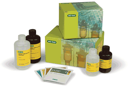
Clarity family of ECL substrates provides high performance for all your western chemiluminescence needs.


