StarBright Violet Dyes

- On This Page
- StarBright Violet Dye Range
- StarBright Resources
Excitable by the 405 nm laser, with a broad range of maximal emissions from 440 nm to 790 nm, StarBright Violet (SBV) Dyes are designed to be the ideal choice when building multicolor panels using the 405 laser in conventional and full spectrum flow cytometry.
They are bright, photostable, compatible with most buffers, and have narrow excitation and emission spectra.
Visit StarBright Dyes to discover all the benefits of these dyes.
Table 1 below shows all the available SBV Dyes.
SBV Dye Range
StarBright Dye |
Max Ex, nm |
Max Em, nm |
Brightness (1–5) |
ZE5 Cell Analyzer Optimal Filter |
Comparison Dye |
Flier |
|---|---|---|---|---|---|---|
|
383 |
436 |
5 |
460/22 |
Super Bright (SB) 436 , Pacific Blue, eFluor 450 |
||
|
405 |
479 |
4 |
525/50 |
Brilliant Violet (BV) 480 |
||
|
402 |
516 |
4 |
525/50 |
BV510, Amethyst Orange |
||
|
404 |
571 |
4 |
615/24 |
BV570 |
||
|
403 |
607 |
4 |
615/24 |
BV605, SB600 |
||
|
401 |
667 |
5 |
670/30 |
BV650, SB645 |
||
|
402 |
713 |
5 |
720/60 |
BV711, SB702 |
||
|
403 |
754 |
5 |
750LP |
BV750 |
|
|
|
402 |
782 |
4 |
750LP |
BV785, SB780 |
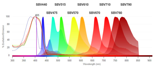
Fig. 1. Excitation and emission spectra of StarBright Violet Dyes.
 Technical Note: A Study Comparing the Benefits of Using StarBright Ultraviolet, Violet, and Blue Dye–Conjugated Antibodies to Commonly Used Fluorophores for Human Immunophenotyping
StarBright Violet Dyes in Focus
Below we provide more in-depth information about each individual dye, including usage information, conventional spectra visuals (wavelength and channel) and brightness comparison data.
Simply click on the “+” to reveal your desired information.
StarBright Violet 440 Dye
StarBright Violet 440 (SBV440) Dye is a perfect replacement for fluorophores that have emission maxima between 420 and 480 nm, this includes dyes such as BV421, SB436, Pacific Blue, V450, vioBlue, and Alexa Fluor 405. Excitable by the violet 405 nm laser and emitting at 436 nm, it can be detected using the 460/22 filter on the ZE5 Cell Analyzer or a similar filter (e.g.450/50) on other instruments. This gives more choice and flexibility when detecting markers which have low expression, as dim fluorophores are not suitable.
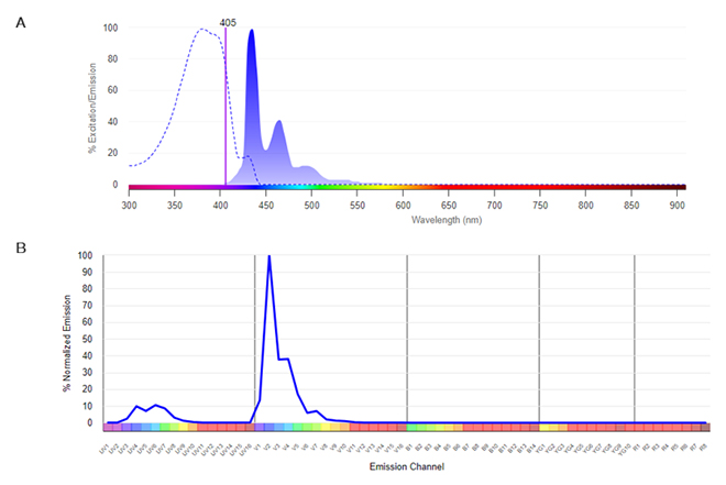
Fig. 2. SBV440. A, Conventional spectra. Excitation by the violet laser (dotted line) with emission shown in the shaded histogram. B, Normalized dye signature. Full emission spectrum showing the emission at all wavelengths. Data were collected on a 5-L Cytek Aurora Flow Cytometry System using SpectroFlo Software.
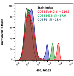
Fig. 3. Brightness comparison. Mouse peripheral blood was stained with CD4SBV440 (MCA2691SBV440) (red), CD4 Pacific Blue (blue), or CD4 SuperBright 436 (green) and analyzed on the ZE5 Cell Analyzer detected using the 460/22 filter. All antibodies were titrated prior to use to determine the optimal concentration.
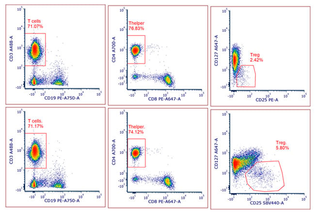
Fig 4. Superior performance of SBV440. In a 6-color panel identifying B cells and T cell populations in human peripheral blood, SBV440 gives better resolution of CD127lo CD25+ Tregs than using PE. Antibodies used: CD19PE-750 (MCA1940P750), CD3A488 (MCA463A488), CD127A647, CD4A700 (MCA1267A700), CD8PE-A647,CD25PE (MCA2127PE) or CD25SBV440 (MCA2125SBV440).
StarBright Violet 475 Dye
StarBright Violet 475 (SBV475) Dye is excitable by the violet 405 nm laser and emits at 479 nm, making it easily detectable using standard filters available on most instruments. StarBright Violet 475 Dye is also compatible with spectral flow cytometry and has a unique profile, allowing incorporation into new and existing flow cytometry panels, giving greater flexibility and increasing multiplex capabilities. The unique profile of SBV475 allows you to combine this dye with StarBright Violet 515 (SBV515) Dye and Brilliant Violet 510 (BV510) in a panel when using full spectrum flow cytometry.
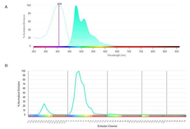
Fig. 5. SBV475. A, Conventional spectra. Excitation by the violet laser (dotted line) with emission shown in the shaded histogram. B, Normalized dye signature. Full emission spectrum showing the emission at all wavelengths. Data were collected on a 5-L Cytek Aurora Flow Cytometry System using SpectroFlo Software.
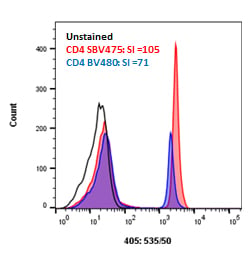
Fig. 6. Brightness comparison. Human peripheral blood was stained with CD4SBV475 (MCA1267SBV475) (red), or CD4 BV480 (blue) and analyzed on the ZE5 Cell Analyzer detected using the 525/50 filter. All antibodies were titrated prior to use to determine the optimal concentration.
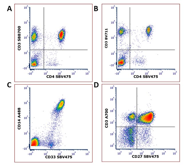
Fig. 7. Examples of SBV475 Dye staining.
Human peripheral blood was stained with A, CD3SBB700 (MCA463SBB700) and CD4SBV475 (MCA1267SBV475); B, CD3BV711 (BD) and CD4SBV475 (MCA1267SBV475); C, CD14A488 (MCA1568A488) and CD33SBV475 (MCA1271SBV475); and D, CD3A700 (MCA463A700) and CD27SBV475 (MCA755SBV475). All staining was performed at
4°C
in PBS containing 1% BSA after blocking with 10% human serum apart from (D) which was stained in Brilliant Buffer. Cells were analyzed on the ZE5 Cell Analyzer.
StarBright Violet 515 Dye
Excitable by the violet 405 nm laser and emitting at 515 nm, StarBright Violet 515 (SBV515) Dye is a perfect replacement for fluorophores that have emission maxima between 500 and 550 nm, such as Pacific Orange, Krome Orange, vioGreen, and Brilliant Violet 510, which can all be detected with a 525/50 filter (or similar) in your flow cytometry experiments.
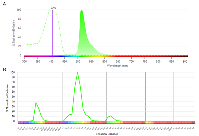
Fig. 8. SBV515. A, Conventional spectra. Excitation by the violet laser (dotted line) with emission shown in the shaded histogram. B, Normalized dye signature. Full emission spectrum showing the emission at all wavelengths. Data were collected on a 5-L Cytek Aurora Flow Cytometry System using SpectroFlo Software.
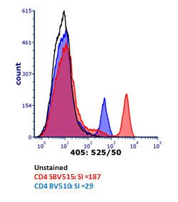
Fig. 9. Brightness comparison. Murine peripheral blood was stained with CD4SBV515 (MCA2691SBV515) (red), or CD4BV510 (blue) and analyzed on the ZE5 Cell Analyzer detected using the 525/50 filter. All antibodies were titrated prior to use to determine the optimal concentration.
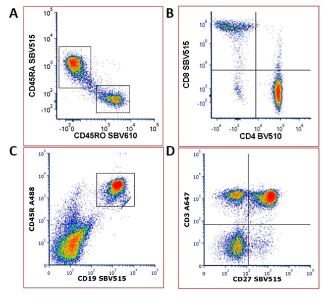
Fig. 10. Examples of SBV515 Dye staining. Human or mouse peripheral blood was stained with A, CD45RASBV515 (MCA88SBV515) and CD45ROSBV610 (MCA461SBV610) B, CD8SBV515 (MCA1226SBV515) and CD4BV510 C, CD19SBV515 (MCA1439SBV515) and CD45RA488 (MCA1258SBV515) and D, CD27SBV515 (MCA755SBV515) and CD3A647 (MCA463A647). All staining was performed at
4°C
in PBS containing 1% BSA after blocking with 10% serum. Cells were analyzed on the ZE5 Cell Analyzer, apart from B which was analyzed on a Cytek Aurora.
StarBright Violet 570 Dye
StarBright Violet 570 (SBV570) Dye is excitable by the violet 405 nm laser and emits at 570 nm. It is brighter than dyes with similar excitation and emission profiles such as Brilliant Violet 570 (BV570) with reduced spillover into PE. Although SBV570 can be detected using the ZE5 Cell Analyzer, SBV570 is detected optimally on the S3e Cell Sorter fitted with a 405 nm laser as this has a 586/25 or 593/40 filter, depending on the configuration. It is also suitable to be used on any cell analyzer or sorter with a similar filter and in spectral flow cytometry due to its unique spectral profile.
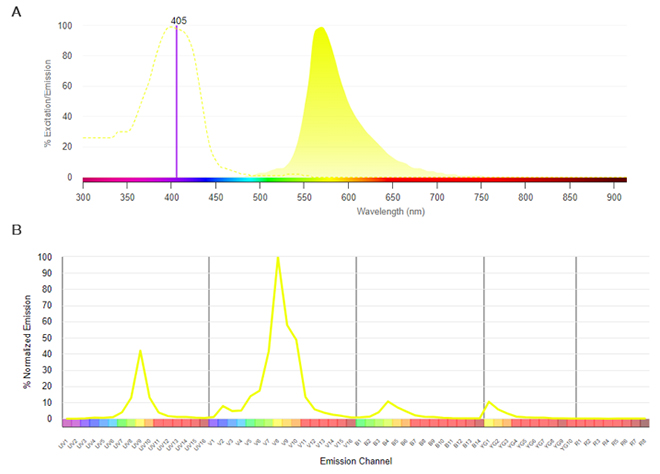
Fig. 11. SBV570. A, Conventional spectra. Excitation by the violet laser (dotted line) with emission shown in the shaded histogram. B, Normalized dye signature. Full emission spectrum showing the emission at all wavelengths. Data were collected on a 5-L Cytek Aurora Flow Cytometry System using SpectroFlo Software.
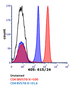
Fig. 12. Brightness comparison. Human peripheral blood was stained with CD4SBV570 (MCA1267SBV570) (red), or CD4BV570 (blue) and analyzed on the ZE5 Cell Analyzer detected using the 615/24 filter. All antibodies were titrated prior to use to determine the optimal concentration.
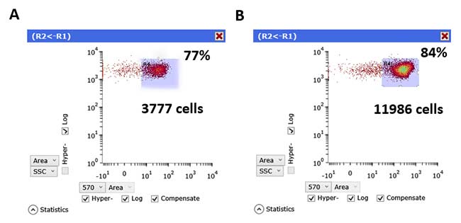
Fig. 13. Sorting purity and recovery is improved with StarBright Dyes. Human PBMCs were stained with CD3 FITC (MCA463F) and either A, CD8BV570 or B, CD8SBV570 (MCA1226SBV570). The number of cells recovered and the purity (%) is shown on the plots.
StarBright Violet 610 Dye
StarBright Violet 610 (SBV610) Dye is excitable by the violet 405 nm laser and maximally emits at 607 nm. It is optimally detected using the 615/24 filter on the ZE5 Cell Analyzer or similar filters on other instruments. StarBright Violet 610 Dye is compatible with spectral flow cytometry and has a unique profile allowing incorporation into new and existing flow cytometry panels.
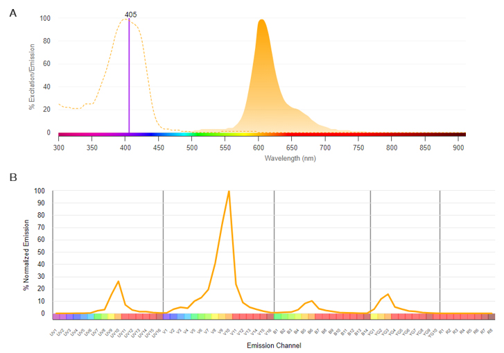
Fig. 14. SBV610. A, Conventional spectra. Excitation by the violet laser (dotted line) with emission shown in the shaded histogram. B, Normalized dye signature. Full emission spectrum showing the emission at all wavelengths. Data were collected on a 5-L Cytek Aurora Flow Cytometry System using SpectroFlo Software.
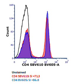
Fig. 15. Brightness comparison. Human peripheral blood was stained with CD4SBV610 (MCA1267SBV610) (red), or CD4 BV605 (blue) and analyzed on the ZE5 Cell Analyzer detected using the 615/24 filter. All antibodies were titrated prior to use to determine the optimal concentration.
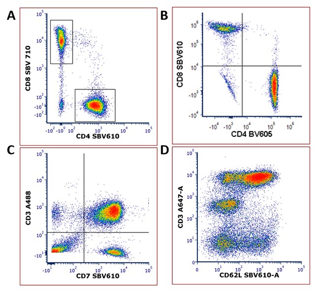
Fig. 16. Examples of SBV610 Dye staining. Human peripheral blood was stained with A, CD4SBV610 (MCA1267SBV610) and CD8SBV710 (MCA1226SBV710) B, CD8SBV610 (MCA1226SBV610) and CD4BV605 (BD) C, CD7 Biotin (MCA1197B) and Streptavidin SBV610 (STAR210SBV610) and D, CD62LSBV610 (MCA1076SBV610) and CD3A647 (MCA463A647). All staining was performed at 4°C in PBS containing 1% BSA after blocking with 10% serum. Cells were analyzed on the ZE5 Cell Analyzer, apart from B which was analyzed on a Cytek Aurora.
StarBright Violet 670 Dye
StarBright Violet 670 (SBV670) Dye is excitable by the violet 405 nm laser and maximally emits at 667 nm. It can be detected using the 670/30 filter on the ZE5 Cell Analyzer or similar filters on other instruments with reduced spillover into the PE-A674 and A647 channels. SBV670 Dye is also compatible with spectral flow cytometry and has a unique profile allowing incorporation into new and existing flow cytometry panels.
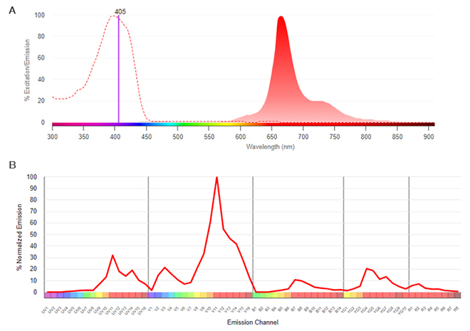
Fig. 17. SBV670. A, Conventional spectra. Excitation by the violet laser (dotted line) with emission shown in the shaded histogram. B, Normalized dye signature. Full emission spectrum showing the emission at all wavelengths. Data were collected on a 5-L Cytek Aurora Flow Cytometry System using SpectroFlo Software.
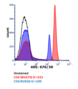
Fig. 18. Brightness comparison. Human peripheral blood was stained with CD4SBV670 (MCA1267SBV670) (red), or CD4BV650 (blue) and analyzed on the ZE5 Cell Analyzer detected using the 670/30 filter. All antibodies were titrated prior to use to determine the optimal concentration.
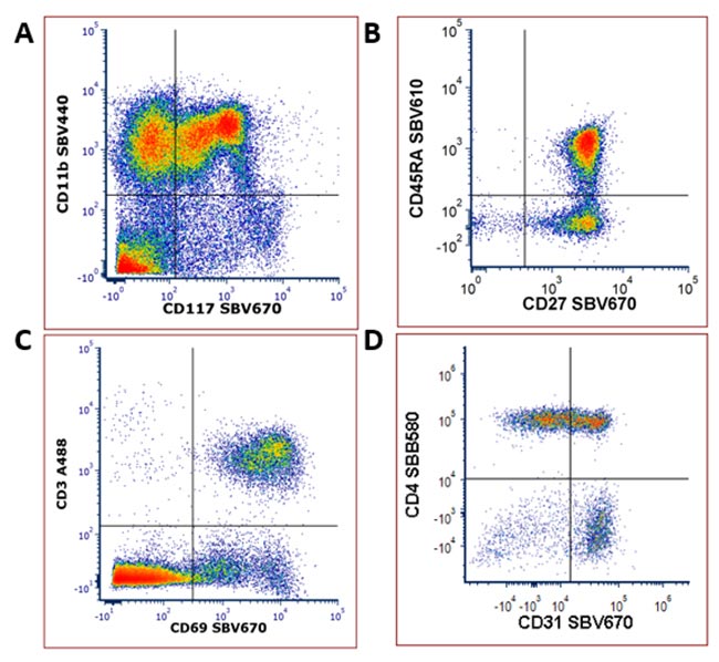
Fig. 19. Examples of SBV670 Dye staining. Mouse bone marrow (A) or human peripheral blood (B-D) was stained with A, CD11bSBV440 (MCA711SBV440) and CD117SBV670 (MCA1365SBV670) B, CD45RASBV610 (MCA88SBV610) and CD27SBV670 (MCA755SBV670) C, CD3A488 (MCA463A488) and CD69SBV670 (MCA2806SBV670) D, CD4SBB580 (Coming soon) and CD31SBV670 (MCA1738SBV670). All staining was performed at
4°C
in PBS containing 1% BSA after blocking with 10% serum. Cells were analyzed on the ZE5 Cell Analyzer, apart from D which was analyzed on a Cytek Aurora.
StarBright Violet 710 Dye
StarBright Violet 710 (SBV710) Dye is excitable by the violet 405 nm laser and maximally emits at 713 nm. It can be detected using the 720/60 filter on the ZE5 Cell Analyzer or similar filters on other instruments with reduced spillover, particularly into the A700 channel. SBV710 Dye is also compatible with spectral flow cytometry and has a unique profile allowing incorporation into new and existing flow cytometry panels.
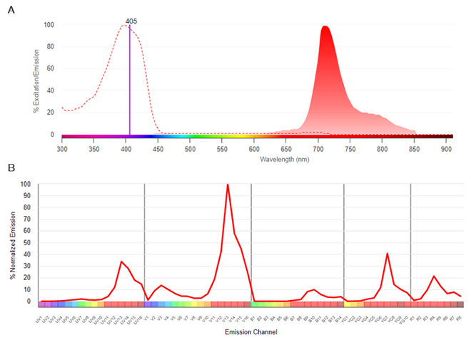
Fig. 20. SBV710. A, Conventional spectra. Excitation by the violet laser (dotted line) with emission shown in the shaded histogram. B, Normalized dye signature. Full emission spectrum showing the emission at all wavelengths. Data were collected on a 5-L Cytek Aurora Flow Cytometry System using SpectroFlo Software.
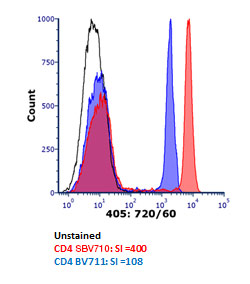
Fig. 21. Brightness comparison. Human peripheral blood was stained with CD4SBV710 (MCA1267SBV710) (red), or CD4 BV711 (blue) and analyzed on the ZE5 Cell Analyzer detected using the 720/60 filter. All antibodies were titrated prior to use to determine the optimal concentration.
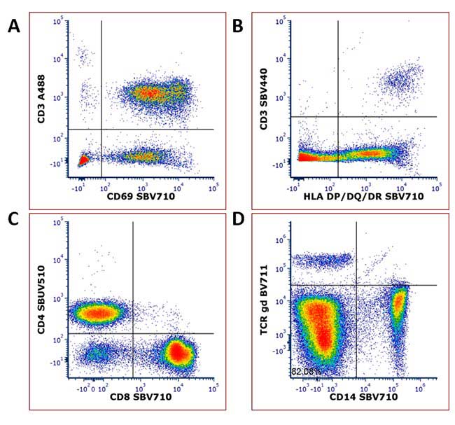
Fig. 22. Examples of SBV710 Dye staining. Human peripheral blood was stained with A, CD3A488 (MCA463A488) and CD69SBV710 (MCA2806SBV710) B, CD3SBV440 (MCA463SBV440) and HLA DP/DQ/DRSBV710 (MCA477SBV710) C, CD4SBUV510 (MCA1267SBUV510) and CD8SBV710 (MCA1226SBV710) D, TCRgdBV711 and CD14SBV710 (MCA1568SBV710). All staining was performed at
4°C
in PBS containing 1% BSA after blocking with 10% serum. Cells were analyzed on the ZE5 Cell Analyzer, apart from D which was analyzed on a Cytek Aurora.
StarBright Violet 760 Dye
StarBright Violet 760 (SBV760) Dye is excitable by the violet 405 nm laser and maximally emits at 760 nm. It can be detected using the 750LP filter on the ZE5 Cell Analyzer or similar filters on other instruments. SBV760 Dye has a similar spillover profile to other dyes with similar emission max but is much brighter giving excellent resolution between positive and negative populations.
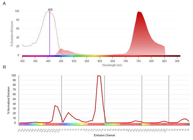
Fig. 23. SBV760. A, Conventional spectra. Excitation by the violet laser (dotted line) with emission shown in the shaded histogram. B, Normalized dye signature. Full emission spectrum showing the emission at all wavelengths. Data were collected on a 5-L Cytek Aurora Flow Cytometry System using SpectroFlo Software.
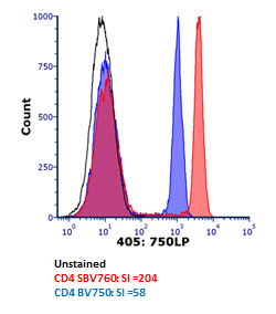
Fig. 24. Brightness comparison. Human peripheral blood was stained with CD4SBV760 (MCA1267SBV760) (red), or CD4 BV750 (blue) and analyzed on the ZE5 Cell Analyzer detected using the 750LP filter. All antibodies were titrated prior to use to determine the optimal concentration.
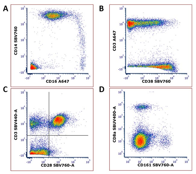
Fig. 25. Examples of SBV760 Dye staining. Human peripheral blood (A-C) or mouse peripheral blood (D) was stained with A, CD16A647 (MCA2537A647) and CD14SBV760 (MCA1568SBV760) B, CD3A647 (MCA463A647) and CD38 (MCA1039SBV760) C, CD3SBV440 (MCA463SBV440) and CD28SBV760 (MCA709SBV760) D, CD8aSBUV400 (MCA609SBUV400) and CD161SBV760 (MCA1266SBV760). All staining was performed at
4°C
in PBS containing 1% BSA after blocking with 10% serum. Cells were analyzed on the ZE5 Cell Analyzer.
StarBright Violet 790 Dye
StarBright Violet 790 (SBV790) Dye is excitable by the violet 405 nm laser and maximally emits at 782 nm. It can be detected using the 750LP filter on the ZE5 Cell Analyzer or similar filters on other instruments. SBV790 Dye is also compatible with spectral flow cytometry and has a unique profile allowing incorporation into new and existing flow cytometry panels.
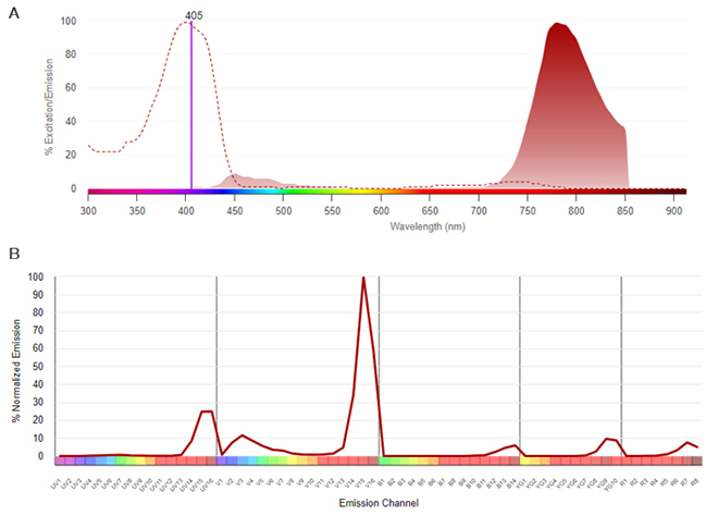
Fig. 26. SBV790. A, Conventional spectra. Excitation by the violet laser (dotted line) with emission shown in the shaded histogram. B, Normalized dye signature. Full emission spectrum showing the emission at all wavelengths. Data were collected on a 5-L Cytek Aurora Flow Cytometry System using SpectroFlo Software.
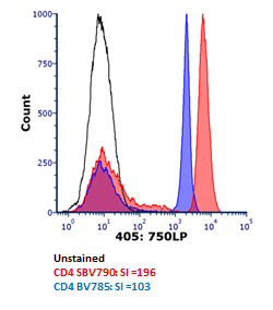
Fig. 27. Brightness comparison. Human peripheral blood was stained with CD4SBV790 (MCA1267SBV790) (red), or CD4 BV785 (blue) and analyzed on the ZE5 Cell Analyzer detected using the 670/30 filter. All antibodies were titrated prior to use to determine the optimal concentration
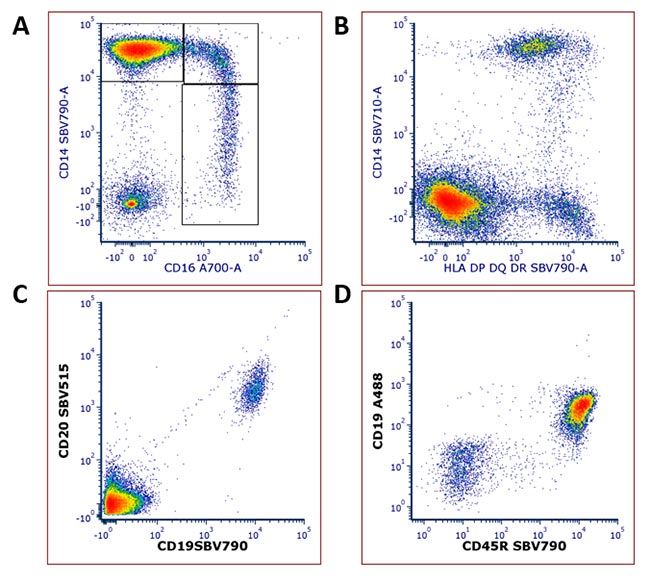
Fig 28. Examples of SBV790 Dye staining. Human peripheral blood (A-C) or mouse bone marrow (D) was stained with A, CD16A700 (MCA2537A700) and CD14SBV790 (MCA1568SBV790) B, CD14SBV710 (MCA1568SBV710) and HLA DP/DQ/DRSBV790 (MCA477SBV790) C, CD20SBV515 (MCA1710SBV515) and CD19SBV790 (MCA1910SBV790) D, CD19A488 (MCA1258A488) and CD45RSBV790 (MCA1439SBV790). All staining was performed at
4°C
in PBS containing 1% BSA after blocking with 10% serum. Cells were analyzed on the ZE5 Cell Analyzer.
For more information on how the StarBright Dye range can fit into your flow cytometry experiments, please take a look at our StarBright Dyes page.
For more information about fluorophores and immunophenotyping, refer to our flow cytometry resources.
Click on the links below to find more in-depth information on each topic and view our popular flow cytometry basics guide.
- StarBright Dyes posters
- StarBright Dye webinars
- Spectral flow cytometry with StarBright Dyes
- An introduction to spectral flow cytometry
- Sample preparation
- Flow controls
- Multicolor panel building tips
- Common flow cytometry protocols
- Cell frequency in common samples
- Gating in flow cytometry
- Fluorophores poster (downloadable)
- Flow cytometry pocket guide (downloadable)
- Fluorescent spectraviewer
- Flow cytometry poster (downloadable)
