Unlabeled Antibodies requiring Conjugation Products, Resources, and Tools
- On This Page
- Products
- Protocol
- Resources
s


If an antibody isn’t available in your preferred fluorophore, conjugation kits can provide a simple solution. Bio-Rad offers a wide range of conjungation kits that require minimal hands-on time. Learn more about these kits and how to use them.
Products
Antibody Conjugation Kits
Fluorophores, Enzymes, and Biotin/Streptavidin Conjugations
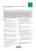
 conjugated to ReadiLink 700/713 Antibody Labeling Kit (1351008) to detect T cells. Data acquired on the ZE5™ Cell Analyzer.")
Fig. 1. Peripheral blood lymphocytes were stained with CD3 (MCA463) conjugated to ReadiLink 700/713 Antibody Labeling Kit (1351008) to detect T cells. Data acquired on the ZE5 Cell Analyzer.
Conjugation of proteins or antibodies of interest with various molecular labels is often necessary in biological research to facilitate detection or quantification of the labeled protein, its binding partners, or antigens in the case of labeled antibodies. Molecular labels such as fluorophores or biotin are covalently conjugated to proteins or antibodies through reactions with thiol or amine groups.
With the LYNX Rapid and Rapid Plus Conjugation Kits and ReadiLink Antibody Conjugation Kits, Bio-Rad offers two complementary, fast, and easy to use antibody conjugations systems that cover a wide variety of labels including the common fluorophores as well as enzymes and biotin conjugations. Save valuable time by quickly conjugating your label of choice using these kits with minimal hands-on time.
LYNX Kits
LYNX Rapid and Rapid Plus Conjugation Kits are available in a wide range of options including common fluorophores, enzymes, and biotin/streptavidin conjugations, suitable for use in flow cytometry, ELISA, western blotting, immunohistochemistry, and immunofluorescence. Identical protocols for small and large scale (up to milligram quantities of antibody) conjugations allow for fast one-step conjugation of proteins and antibodies using a method that is superior to traditional kits and techniques. In contrast to conventional conjugation, LYNX Conjugation Kits offer consistent results with no post-conjugation clean-up process required; therefore, the antibodies are ready to use immediately.
Conjugate your antibodies in 3 easy steps with only 30 seconds hands-on time and no purification required. The total conjugation time is fifteen minutes with the LYNX Rapid Plus Conjugation Kits or 3 hours with the LYNX Rapid Conjugation Kits, however the reaction may run overnight without any adverse effects.
ReadiLink Kits
ReadiLink Antibody Conjugation Kits offer ten unique fluorescence conjugation options for small volumes. Quickly label your antibody of interest in two easy steps in just over an hour with the unique chemistry of ReadiLink Dyes. The excitation wavelengths and narrow emissions spectra of the fluorophores ranging from violet to far red are ideal for multicolor flow cytometry. The infrared excitable conjugations are suitable for westerns, ELISA, and in vivo imaging.
Our Full Conjugation Kit Range
| Description | Target | Format | Clone | Applications | Citations | Code |
|---|
Degree of Labeling (DOL) Calculation
 Want to know how much fluorophore is conjugated to your antibody?
Easily calculate the fluorophore:protein (F:P) ratio using these 4 steps.
You may need to know the amount of fluorophore conjugated to your antibody for control purposes. To determine the fluorophore:protein ratio you will need to calculate the molar concentrations of both the fluorophore and the protein based on absorbance at known wavelengths in a spectrophotometer and then express this as a ratio. This ratio will be an indication of the average number of dye molecules conjugated to your antibody in your solution.
- Remove any excess fluorophore. Excess fluorophore will interfere with the absorbance measurement.
-
Determine the concentration of protein. Measure the absorbance of the protein at A280 nm. Divide this value by the molar extinction coefficient for your protein.
(e.g. mouse IgG has an extinction coefficient of 203,000 M-1 cm-1). -
Determine the fluorophore concentration. Measure the absorbance of the fluorophore at the wavelength of maximal excitation (e.g. for FITC use 495 nm wavelength). Divide this value by the molar extinction coefficient for the dye.
(e.g. FITC has an extinction coefficient of 75,000 M-1 cm-1). - Determine the labeling. Divide the fluorophore concentration by the protein concentration to calculate the fluorophore:protein ratio.
Protocol
Direct Immunofluorescence Staining of Cells and Blood
Applicable where the fluorophore is directly linked to the primary antibody e.g. RPE, FITC and Alexa Fluor® conjugates. RPE conjugates should always be handled in the dark.
This method provides a general procedure for use with the majority of Bio-Rad reagents. In some cases specific recommendations are provided on product datasheets, and these methods should always be used in conjunction with the product and batch specific information provided with each vial. A certain level of technical skill and immunological knowledge is required for the successful design and implementation of these techniques; these are guidelines only and may need to be adjusted for particular applications. Specific methodology for blood appears in [ ] brackets.
Reagents
- Phosphate buffered saline (BUF036A) containing 1% bovine serum albumin (PBS/BSA).
- PBS.
- Erythrolyse red blood cell lysing buffer (BUF04).
- Anticoagulant (Note: for basic staining any appropriate anticoagulant, such as heparin, EDTA, or acid citrate dextrose, may be used. In some instances specific anticoagulants may be required)
- Optional: 0.5% (w/v) paraformaldehyde in PBS (Note: dissolve on heated stirrer and cool before use)
Method
-
Prepare cells appropriately; refer to protocol FC1. 'Preparation of cells for flow cytometry' for further information. Adjust the cell suspension to a concentration of 1 x 10⁷ cells/ml with cold (4oC) PBS/BSA buffer.
[Whole blood samples may be used undiluted unless the cell count is high, e.g. as in leukemia. Use appropriate anticoagulants].
- Aliquot 100 μl of the cell suspension [or whole blood] into as many test tubes as required.
-
Add antibody at the vendor-recommended dilution. Mix well and incubate at 4oC for at least
30 min, avoiding direct light. -
Wash cells with 2 ml cold (4oC) PBS/BSA, centrifuge at 300-400 g for 5 min at 4oC, and discard the resulting supernatant.
[To the blood suspension add 2 ml freshly prepared erythrolyse red cell lysing buffer and mix well. Incubate for 10 min at room temperature. Centrifuge at 300-400 g for 5 min at room temperature and discard the supernatant. Wash with 2 ml room temperature PBS/BSA, centrifuge at 300-400 g for 5 min and discard the supernatant. Proceed to step 5].
- Re-suspend cells in 200 μl cold (4oC) PBS or with 200 μl of 0.5% paraformaldehyde in PBS if required.
- Acquire data by flow cytometry. Analyze fixed cells within 24 hours.
Notes
-
The following should be considered when designing your flow cytometry experiments:
-
To avoid nonspecific binding, you also need to block Fc receptors on cell types such as spleen cells, with FcR blocking reagents e.g. Bio-Rad’s Mouse Seroblock reagent (BUF041).
-
Appropriate controls should always be carried out, for flow cytometry the following should be considered for inclusion;
-
Isotype controls used to determine if the staining is specific
-
Unstained cells should always be included in the experimental set up to monitor autofluorescence.
-
-
-
For all multi-color flow cytometry experiments it is advisable to include compensation controls and fluorescence minus one (FMO) controls, which assist with identifying gating boundaries.
Resources
Choosing Fluorophores for Flow Cytometry

Fluorophores and Viability Dyes for Flow Cytometry
Reference Postcard
Flow cytometry generally requires bright fluorophores that are excitable by one laser with a narrow emission wavelength to allow multicolor panel building. Below is a handy guide to the range of fluorophores Bio-Rad offers, including our new StarBright™ Dyes, that are suitable for flow cytometry, grouped by the laser used to excite them.
Additional fluorophore information can be found on specific webpages organized by the laser that excites them. This includes the maximal emission wavelength, relative brightness, ZE5 Cell Analyzer filter used for detection, similar or incompatible fluorophores, and our antibodies available conjugated to them, helping you select the right fluorophore for your experiment.
For more information on which fluorophores are the most suitable for your experiment, try our fluorescence spectraviewer or download our poster showing common fluorophores used in flow cytometry.
Fluorophores excited by the 355 nm (Ultraviolet) Laser
- StarBright UltraViolet 400
- StarBright UltraViolet 445
- StarBright UltraViolet 510
- StarBright UltraViolet 575
- StarBright UltraViolet 605
- StarBright UltraViolet 665
- StarBright UltraViolet 740
-
StarBright UltraViolet 795

Fluorophores Excited by the 405 nm (Violet) Laser
- Pacific Blue
- StarBright Violet 440
- StarBright Violet 475
- StarBright Violet 515
- Amethyst Orange
- StarBright Violet 570
- StarBright Violet 610
- StarBright Violet 670
- StarBright Violet 710
- StarBright Violet 760
-
StarBright Violet 790

Fluorophores Excited by the 488 nm (Blue) Laser
- FITC
- Alexa Fluor 488
- PE
- PE-Alexa Fluor647
- PE-Cy5
- PE-Cy5.5
- PE-Alexa Fluor750
- PE-Cy7
- PerCP
- StarBright Blue 580
- StarBright Blue 615
- StarBright Blue 675
- StarBright Blue 700
- StarBright Blue 765
-
StarBright Blue 810

Fluorophores Excited by the 561 nm (Yellow) Laser
- PE
- PE-Alexa Fluor647
- PE-Cy5
- PE-Cy5.5
- PE-Alexa Fluor750
- PE-Cy7
- StarBright Yellow 575
- StarBright Yellow 605
- StarBright Yellow 665
- StarBright Yellow 720
-
StarBright Yellow 800

Fluorophores Excited by the 640 nm (Red) Laser
- Alexa Fluor 647
- APC
- Alexa Fluor 700
- StarBright Red 670
- StarBright Red 715
- StarBright Red 775
-
StarBright Red 815


Antigen Density of Common Human and Murine Markers
Quick Guide
Antigen Density
Tutorials for building multicolor flow cytometry panels always highlight the importance of antigen density — but why is it so important?
The number of antigens, or target molecules that a cell carries directly correlates to the intensity of the positive population and will determine the optimal fluorophore you should use for each marker. As a general rule you should pair bright fluorophores (e.g. PE) with low expressing markers and dimmer fluorophores (e.g. Pacific Blue) with highly expressed markers. Careful choice of fluorophores will help with the resolution of your cell populations. It is possible for bright fluorophores to be paired with highly expressed antigens, but better to avoid pairing dim fluorophores with low abundance antigens.
The guide provides advice on how to measure antigen density and lists the antigen density of some common markers.

Essentials for Multicolor Panel Building Poster, incl. tips on antibody titration.
Antibody Titration in Flow Cytometry
To optimize staining in flow cytometry antibody titration is recommended. While antibodies will bind to high affinity targets on a cell, if they are present in excess they will also bind to low affinity targets. This results in an increase in background fluorescence and consequently a reduction in your ability to resolve populations, especially if there are subtle differences. Furthermore, if the antibody concentration is too high, it may result in a false negative prozone effect.
It is therefore important to use the antibodies at the right concentration. Although antibodies are sold with a recommended dilution and it is a good starting point, it may not be optimal for your cell type or protocol, therefore titration is an essential step to optimize your staining.
Basic Multicolor Panel Design Rules
Before you build your panels it is critically important to have as clear an idea as possible of what is required to identify your population of interest. For example, are you looking at one cell type or is it a subset, are you looking at an activation marker or a change in cell frequency, are the markers co-expressed or do you even know the expression pattern?
Ideally, when building multicolor panels, it is best to separate fluorophore excitation across lasers, and where possible, the emission across the detectors. This will minimize the amount of spillover and therefore compensation you will need to do. It will also reduce the effect that fluorescence spread will have on your data. However, as you increase the number of fluorophores in your panel, this will not always be possible. Therefore other considerations need to be included in your design.
Webinar: Multicolor Panel Building in Flow Cytometry
Presented by: Dr Sebastian Hedlund, Flow Cytometry Specialist at Bio-Rad Laboratories
Multicolor flow cytometry is the analysis of multiple fluorescent parameters in one sample. Building large flow cytometry panels can be daunting because each additional fluorophore you add to your panel has the potential to influence another fluorophore.
Join our 45 minute webinar to learn which fluorophores are compatible with each other, how they interact and can affect your staining and how to obtain optimal resolution of signal. Furthermore, we will show you how dump channels, fluorophore brightness, antigen density, marker expression patterns and instrument configuration can all contribute to improving your panel design.
This webinar is recommended for both novices and researchers with some experience of flow cytometry.
Speaker Biography
 Sebastian obtained his degree from the Institute of Technology, Linköping University, Sweden, followed by an MSc in Medical Biology from the same institute. He then moved to Freiburg, Germany where he obtained his PhD in Immunology and Molecular Biology at the Max Planck Institute of Immunobiology and Epigenetics.
The focus of his thesis was the role of microRNA in innate immunity and immunopathology. He then left academia and started work as a dedicated flow cytometry specialist, before joining Bio-Rad in 2016 as the flow cytometry specialist for Scandinavia, Eastern Europe, Israel, and South Africa.
Multicolor Panel Builder

Build multicolor flow cytometry panels in just a few simple steps
- Start your design by first selecting your instrument from the drop down menu, or create a customized instrument, to build a personalized panel
- Select the target and species you want to identify. Keep adding until you have all the markers you want
- Choose antibodies by clone, isotype, and format. If there is not an antibody available in the format you require, select the fluorophore as a placeholder and continue
- Include a viability dye or dump channel for more complexity
The ability to select antigen density and fluorophores based on brightness, combined with automatic exclusion of incompatible fluorophores, will help you build larger panels with confidence.
Interactive Flow Cytometry, Spectral Analyzer, Microscopy, and Western Blot Fluorescence Spectraviewer

Find the right fluorophore for your application using our new interactive, fluorescence spectraviewer with hundreds of fluorophores to choose from.
Bio-Rad’s new spectraviewer is the only spectraviewer to allow a multi-laser view and support three applications in a single tool.
- Supports flow cytometry, fluorescence microscopy, and western blotting
- Displays the excitation and emission profiles of the fluorophores available to you, essential when building multicolor panels for flow cytometry
- Allows full customization: select any fluorophore, laser, and filter combination
- Provides more information than any other spectraviewer: the traditional view, showing the fluorescence spectra, is complemented by the multi-laser view visualizing the spillover and cross-laser excitation and emission
- Saves time with the pre-loaded Bio-Rad instrument settings
The ability to view in a multi-laser format allows you to check fluorophore compatibility, including potential issues due to cross-laser excitation, and predict compensation.
Bio-Rad has over 4,000 antibodies validated for flow cytometry as well as helpful resources to guide you through your flow cytometry experiments. These resources as well as listings of antibodies, kits, and controls can be accessed from our dedicated flow cytometry page.