p53 antibody | DO-1
Mouse anti p53 (aa20-25)
- Product Type
- Monoclonal Antibody
- Clone
- DO-1
- Isotype
- IgG2a
- Format
- Purified
- Specificity
- p53
- Region
- (aa20-25)


KO Validated
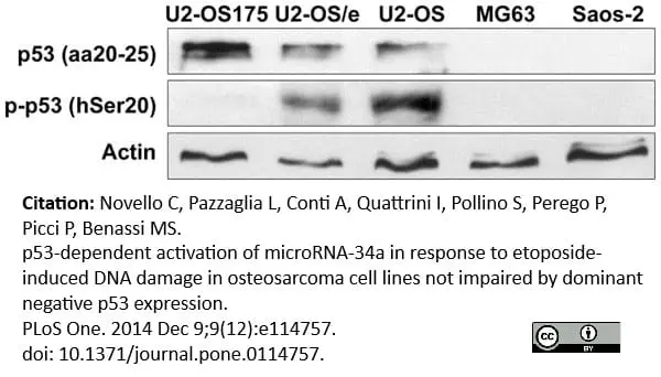
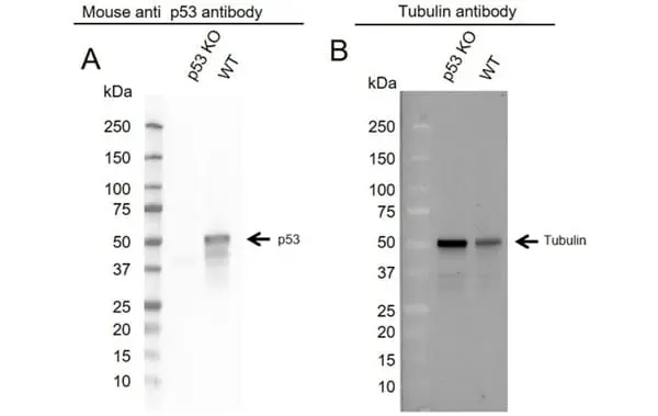
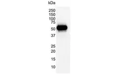
Mouse anti p53 (aa20-25) antibody, clone DO-1 (MCA1701) used to evaluate DO-1 expression in U2-OS cells by western blotting.
Image caption:
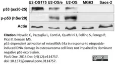
p53 protein expression in OS cells. wt-p53 U2-OS, U2-OS transfected with empty vector (U2-OS/e) and p53-impaired U2-OS175 cells were positive to anti-p53 that binds the transactivation site of N-terminal domain (aa20-25), with increased expression in U2-OS175 cells. U2-OS and U2-OS/e also presented accumulation of p53 phosphorylated at Ser20 residue (p-p53). MG63 and Saos-2 were negative to both antibodies. Actin was used as loading control.
From: Novello C, Pazzaglia L, Conti A, Quattrini I, Pollino S, et al. (2014)
p53-Dependent Activation of microRNA-34a in Response to Etoposide-Induced DNA Damage in Osteosarcoma Cell Lines Not Impaired by Dominant Negative p53 Expression.
PLoS ONE 9(12): e114757.
doi: 10.1371/journal.pone.0114757.
This image is from an open access article distributed under the terms of the Creative Commons Attribution License.
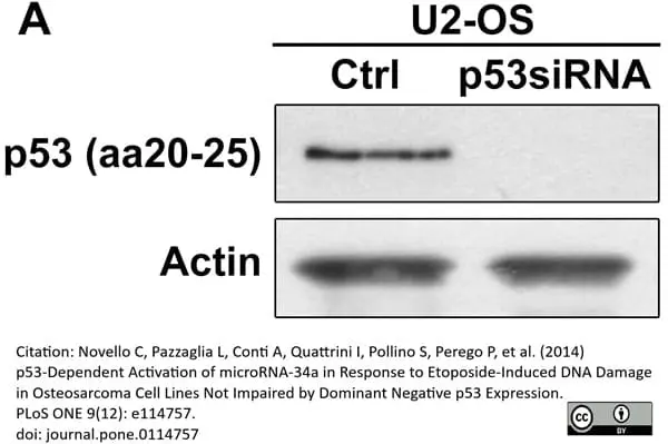
Mouse anti Human p53 antibody, clone DO-1 (MCA1701) used for p53 evaluation in western blotting.
Image caption:
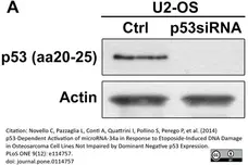
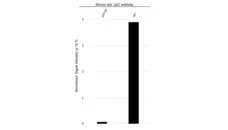
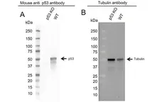
Response of p53siRNA U2-OS cells to etoposide.
(A) Inhibition of p53 expression in p53siRNA U2-OS. Actin was used as loading control.
From: Novello C, Pazzaglia L, Conti A, Quattrini I, Pollino S, et al. (2014)
p53-Dependent Activation of microRNA-34a in Response to Etoposide-Induced DNA Damage in Osteosarcoma Cell Lines Not Impaired by Dominant Negative p53 Expression.
PLoS ONE 9(12): e114757.
doi: 10.1371/journal.pone.0114757.
This image is from an open access article distributed under the terms of the Creative Commons Attribution License.

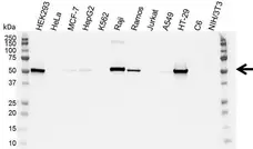
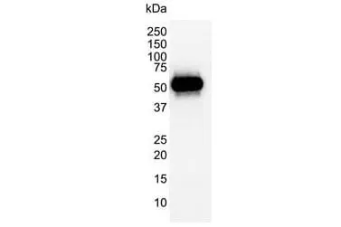
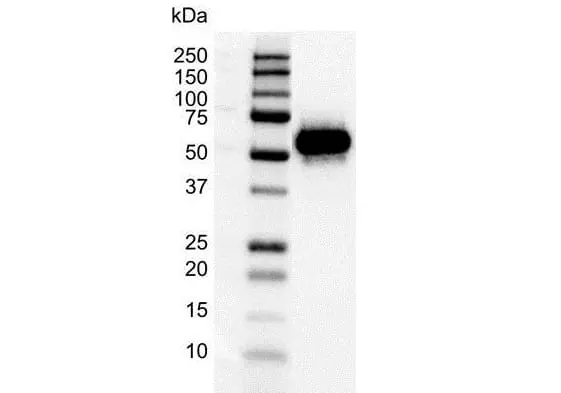
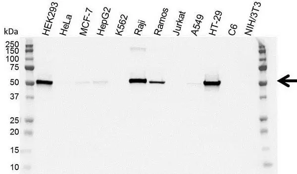
Arrow points to p53 (molecular weight 53 kDa.
Filter by Application:
WB Reset| Mouse anti Human p53 antibody, clone DO-1 recognizes the human p53 tumor suppressor protein, also known as cellular tumor antigen p53 or NY-CO-13. Clone DO-1 binds to both wild type and mutant forms of the p53 protein found in various malignancies (Kern et al. 1992). p53 is important in multicellular organisms, where it regulates cell cycle progression to allow DNA repair or apoptosis in the case of irreparably damaged cells (Haupt et al. 2003) and thus functions as a tumor suppressor that is involved in preventing cancer. Mutations in the p53 gene are found in about half the cases of human cancer (Joerger and Fersht 2007) Mouse anti Human p53 antibody, clone DO-1 recognizes an epitope at the N-terminal end of p53 between amino acids 20-25,common to isoforms 1-3 of p53. |
|
- Target Species
- Human
- Species Cross-Reactivity
-
Target Species Cross Reactivity Mouse Rat Bovine Cat Horse Green Monkey - N.B. Antibody reactivity and working conditions may vary between species.
- Product Form
- Purified IgG - liquid
- Preparation
- Purified IgG prepared by affinity chromatography on Protein A from tissue culture supernatant
- Buffer Solution
- Phosphate buffered saline
- Preservative Stabilisers
- 0.09% sodium azide (NaN3)
- Carrier Free
- Yes
- Immunogen
- Recombinant human p53.
- Approx. Protein Concentrations
- IgG concentration 1.0 mg/ml
- Fusion Partners
- Spleen cells from immunized BALB/c mice were fused with cells of the mouse X63Ag8.653 myeloma cell line.
- Regulatory
- For research purposes only
- Guarantee
- 12 months from date of despatch
This product is shipped at ambient temperature. It is recommended to aliquot and store at -20°C on receipt. When thawed, aliquot the sample as needed. Keep aliquots at 2-8°C for short term use (up to 4 weeks) and store the remaining aliquots at -20°C.
Avoid repeated freezing and thawing as this may denature the antibody. Storage in frost-free freezers is not recommended.
Avoid repeated freezing and thawing as this may denature the antibody. Storage in frost-free freezers is not recommended.
This product has been reported to work in the following applications. This information is derived from testing within our laboratories, peer-reviewed publications or personal communications from the originators. Please refer to references indicated for further information. For general protocol recommendations, please visit the antibody protocols page.
| Application Name | Verified | Min Dilution | Max Dilution |
|---|---|---|---|
| ELISA |  |
||
| Immunohistology - Frozen | |
||
| Immunohistology - Paraffin 1 | |
1/25 | |
| Immunoprecipitation | |
||
| Western Blotting | |
1/1000 |
- 1 This product requires antigen retrieval using heat treatment prior to staining of paraffin sections. Sodium citrate buffer pH 6.0 is recommended for this purpose.
The PrecisionAb label is reserved for antibodies that meet the defined performance criteria within Bio-Rad's ongoing antibody validation programme. Learn about how we validate our PrecisionAb range. Where this product has not been tested for use in a particular technique this does not necessarily exclude its use in such procedures. Further optimization may be required dependent on sample type.
- Histology Positive Control Tissue
- Normal human colon or breast carcinoma
References for p53 antibody
-
Vojtěsek B et al. (1992) An immunochemical analysis of the human nuclear phosphoprotein p53. New monoclonal antibodies and epitope mapping using recombinant p53.
J Immunol Methods. 151 (1-2): 237-44. -
Levesque, M.A. et al. (1995) Time-resolved immunofluorometric assay of p53 protein.
Clin Chem. 41 (12 Pt 1): 1720-9. -
Sironi, G. et al. (1999) p53 protein expression in conjunctival squamous cell carcinomas of domestic animals.
Vet Ophthalmol. 2 (4): 227-231. -
Hietanen, S. et al. (2000) Activation of p53 in cervical carcinoma cells by small molecules.
Proc Natl Acad Sci U S A. 97 (15): 8501-6. -
Phelps, M. et al. (2003) p53-independent activation of the hdm2-P2 promoter through multiple transcription factor response elements results in elevated hdm2 expression in estrogen receptor alpha-positive breast cancer cells.
Cancer Res. 63: 2616-23. -
Carvalho, T. et al. (2009) Immunohistochemical evaluation of vascular urinary bladder tumors from cows with enzootic hematuria.
Vet Pathol. 46 (2): 211-21. -
Bergman, L.M. et al. (2009) CtBPs promote cell survival through the maintenance of mitotic fidelity.
Mol Cell Biol. 29: 4539-51. -
Phillips, A. et al. (2010) HDMX-L is expressed from a functional p53-responsive promoter in the first intron of the HDMX gene and participates in an autoregulatory feedback loop to control p53 activity.
J Biol Chem. 285 (38): 29111-27.
MCA1701
If you cannot find the batch/lot you are looking for please contact our technical support team for assistance.
View more products with p53 specificity
Please Note: All Products are "FOR RESEARCH PURPOSES ONLY"
View all Anti-Human ProductsAlways be the first to know.
When we launch new products and resources to help you achieve more in the lab.
Yes, sign me up