Macrophages antibody | BA4D5
Mouse anti Pig Macrophages
- Product Type
- Monoclonal Antibody
- Clone
- BA4D5
- Isotype
- IgG2b
- Specificity
- Macrophages


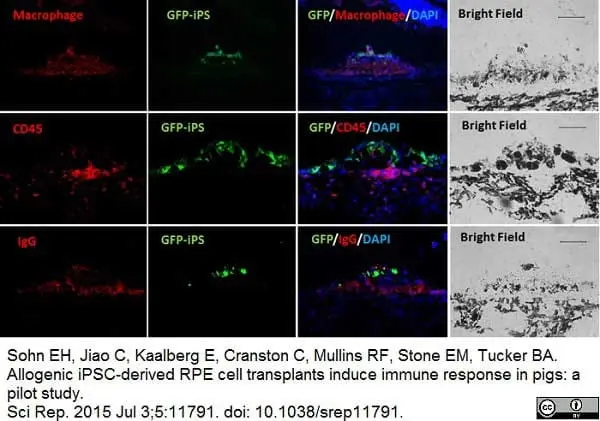
Mouse anti Porcine macrophages antibody, clone BA4D5 (MCA2317GA) used for the demonstration of macrophages in the subretinal space of pigs by immunofluorescence.
Image caption:
Immunolabeling of iPSC-RPE bolus injections in the subretinal space where the GFP-iPSC panels indicate transplanted cells (labeled in green).
The merged panels demonstrate presence of accompanying GFP-negative, non-iPSC-derived cells that are positive for macrophage (top panels), CD45 markers (middle panels), and IgG (lowest panels). Corresponding bright field panels are on the far right column. Scale bar = 100 μm.
From: Sohn EH, Jiao C, Kaalberg E, Cranston C, Mullins RF, Stone EM, Tucker BA.
Allogenic iPSC-derived RPE cell transplants induce immune response in pigs: a pilot study.
Sci Rep. 2015 Jul 3;5:11791.
doi: 10.1038/srep11791
This image is from an open access article distributed under terms of a Creative Commons Attribution License.
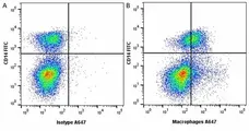
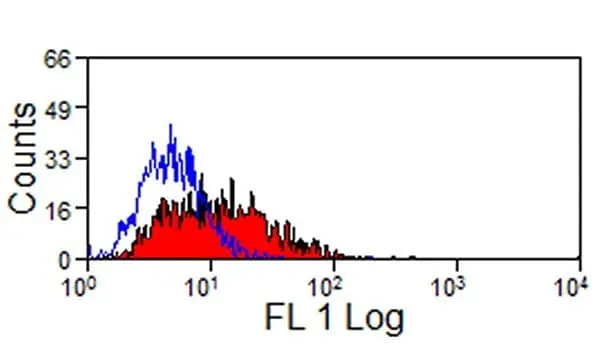
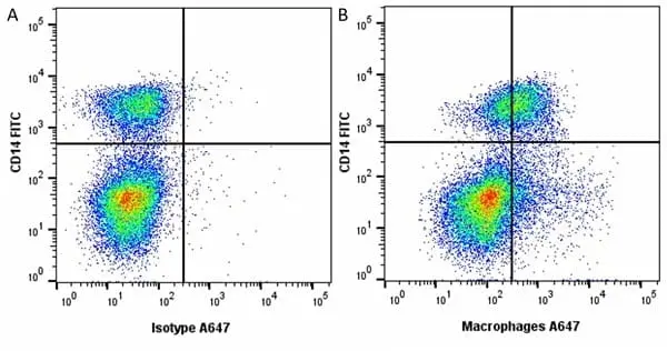
Figure B. FITC conjugated Mouse anti Pig CD14 antibody, clone MIL2 (MCA1218F) and Alexa Fluor® 647 conjugated Mouse anti Pig macrophages antibody, clone BA4D5 (MCA2317A647).
All experiments performed on frozen porcine blood gated on single mononuclear cells, in the presence of 10% pig serum. Data acquired on the ZE5 Cell analyser.
Filter by Application:
F IF Reset| Mouse anti Pig Macrophages antibody, clone BA4D5 recognizes porcine cells of the monocyte/macrophage lineage. Expression of the antigen is increased with maturation, with higher expression on peritoneal and alveolar macrophages. Some expression has also been observed on peripheral blood lymphocytes. The antigen recognized by clone BA4D5 has a broad tissue distribution and this antibody stains macrophages in a range of tissues, including the thymus, spleen periarteriolar lymphoid sheath (PALS), spleen red pulp and the Peyer's patches. Expression has also been reported on some non-heamatopoietic cells including endothelial cells. It is believed that clone BA4D5 may be specific for porcine CD68 (Poulsen et al. 2016) although the protein recognized by this antibody has not yet been fully characterized. The protein is expressed on the cell surface, although it is most abundantly expressed in the cytoplasm. |
- Target Species
- Pig
- Product Form
- Purified IgG - liquid
- Preparation
- Purified IgG prepared by affinity chromatography on Protein A from tissue culture supernatant
- Buffer Solution
- Phosphate buffered saline
- Preservative Stabilisers
- 0.09% sodium azide (NaN3)
- Carrier Free
- Yes
- Immunogen
- Porcine alveolar macrophages.
- Approx. Protein Concentrations
- IgG concentration 1.0 mg/ml
- Fusion Partners
- Spleen cells from immunized BALB/c mice were fused with cells of the mouse SP2/0 mouse myeloma cell line.
- Regulatory
- For research purposes only
- Guarantee
- 12 months from date of despatch
This product is shipped at ambient temperature. It is recommended to aliquot and store at -20°C on receipt. When thawed, aliquot the sample as needed. Keep aliquots at 2-8°C for short term use (up to 4 weeks) and store the remaining aliquots at -20°C.
Avoid repeated freezing and thawing as this may denature the antibody. Storage in frost-free freezers is not recommended.
Avoid repeated freezing and thawing as this may denature the antibody. Storage in frost-free freezers is not recommended.
This product has been reported to work in the following applications. This information is derived from testing within our laboratories, peer-reviewed publications or personal communications from the originators. Please refer to references indicated for further information. For general protocol recommendations, please visit the antibody protocols page.
| Application Name | Verified | Min Dilution | Max Dilution |
|---|---|---|---|
| Flow Cytometry 1 |  |
1/50 | 1/100 |
| Immunofluorescence | |
||
| Immunohistology - Frozen | |
||
| Immunoprecipitation | |
||
| Western Blotting 2 | |
- 1 Membrane permeabilization is required for this application. The use of Leucoperm (Product Code BUF09) is recommended for this purpose.
- 2 BA4D5 recognizes a 105kDa antigen in pig macrophage lysates under non-reducing conditions.
Where this product has not been tested for use in a particular technique this does not necessarily exclude its use in such procedures. Suggested working dilutions are given as a guide only. It is recommended that the user titrates the product for use in their own system using appropriate negative/positive controls.
- Flow Cytometry
- Use 10μl of the suggested working dilution to label 1x10⁶ cells in 100μl
| Description | Product Code | Applications | Pack Size | List Price | Your Price | Quantity | |
|---|---|---|---|---|---|---|---|
| Mouse IgG2b Negative Control | MCA691 | F | 100 Tests |

|
Log in | ||
| List Price | Your Price | ||||||
|
|
Log in | ||||||
| Description | Mouse IgG2b Negative Control | ||||||
References for Macrophages antibody
-
Luechtenborg, B. et al. (2008) Function of scavenger receptor class A type I/II is not important for smooth muscle foam cell formation.
Eur J Cell Biol. 87: 91-9. -
Ezquerra, A. et al. (2009) Porcine myelomonocytic markers and cell populations.
Dev Comp Immunol. 33 (3): 284-98. -
Muscari C et al. (2010) Comparison between Culture Conditions Improving Growth and Differentiation of Blood and Bone Marrow Cells Committed to the Endothelial Cell Lineage.
Biol Proced Online. 12 (1): 9023. -
Fujita M et al. (2013) Technique of endoscopic biopsy of islet allografts transplanted into the gastric submucosal space in pigs.
Cell Transplant. 22 (12): 2335-44. -
Sohn, E.H. et al. (2015) Allogenic iPSC-derived RPE cell transplants induce immune response in pigs: a pilot study.
Sci Rep. 5: 11791. -
Liu, G. et al. (2015) Influenza A Virus Panhandle Structure is Directly Involved in RIG-I Activation and IFN Induction.
J Virol. pii: JVI.00232-15. -
Poulsen, C.B. et al. (2016) Treatment with a human recombinant monoclonal IgG antibody against oxidized LDL in atherosclerosis-prone pigs reduces cathepsin S in coronary lesions.
Int J Cardiol. 215: 506-515. -
Rayat, G.R. et al. (2016) First update of the International Xenotransplantation Association consensus statement on conditions for undertaking clinical trials of porcine islet products in type 1 diabetes - Chapter 3: Porcine islet product manufacturing and release testing criteria.
Xenotransplantation. 23 (1): 38-45.
View The Latest Product References
-
Wang, L. et al. (2017) Porcine alveolar macrophage polarization is involved in inhibition of porcine reproductive and respiratory syndrome virus (PRRSV) replication.
J Vet Med Sci. 79 (11): 1906-15. -
Porras, A.M. et al. (2018) Creation of disease-inspired biomaterial environments to mimic pathological events in early calcific aortic valve disease.
Proc Natl Acad Sci U S A. 115 (3): E363-E371. -
Maciag, S.S. et al. (2022) On the influence of the source of porcine colostrum in the development of early immune ontogeny in piglets.
Sci Rep. 12 (1): 15630. -
dos Santos, M.C. et al. (2023) Effect of yeast extracted β-glucans on the immune response and reproductive performance of gilts in the adaptation, gestation, and lactation periods
Livestock Science. 275: 105289. -
Haach, V. et al. (2023) A polyvalent virosomal influenza vaccine induces broad cellular and humoral immunity in pigs.
Virol J. 20 (1): 181. -
Petitpas, K. et al. (2022) Genetic modifications designed for xenotransplantation attenuate sialoadhesin-dependent binding of human erythrocytes to porcine macrophages.
Xenotransplantation. 29 (6): e12780. -
Forner, R. et al. (2021) Distribution difference of colostrum-derived B and T cells subsets in gilts and sows.
PLoS One. 16 (5): e0249366.
Further Reading
-
Piriou-Guzylack, L. (2008) Membrane markers of the immune cells in swine: an update.
Vet Res. 39: 54.
MCA2317GA
If you cannot find the batch/lot you are looking for please contact our technical support team for assistance.
Request a different product with this specificity
Please Note: All Products are "FOR RESEARCH PURPOSES ONLY"
View all Anti-Pig ProductsAlways be the first to know.
When we launch new products and resources to help you achieve more in the lab.
Yes, sign me up