Leucoperm
Leucoperm
- Product Type
- Accessory Reagent
- Specificity
- Leucoperm


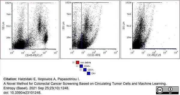
Leucoperm Reagent kit (BUF09) used to fix and permeablize human peripheral blood cells prior to flow cytometric staining for cytokeratins.
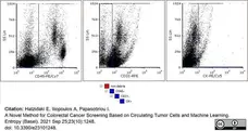
Image caption:
Gating strategy for the identification of CTCs in PBMCs. First on the left plot shows exclusion of CD45-positive cells (hematopoietic); second plot shows exclusion of CD31-positive cells (epithelial); third plot shows selection of pan-CK-positive cells.
From: Hatzidaki E, Iliopoulos A, Papasotiriou I.
A Novel Method for Colorectal Cancer Screening Based on Circulating Tumor Cells and Machine Learning.
Entropy (Basel). 2021 Sep 25;23(10):1248.
doi: 10.3390/e23101248.
This image is from an open access article distributed under terms of a Creative Commons Attribution License.
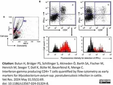
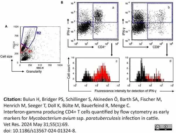
Image caption:
Strategy to analyse flow cytometrically acquired data. A Representative scatter plot with gates for lymphocytes (R1) and for lymphoblasts (R2). B gating strategy to analyse IFN-γ in CD4+ and CD8+ T cells, respectively. Representative dot plots of gated lymphoblasts to detect IFN-γ+, CD4+ and CD8+cells, respectively (a, b). Representative histograms depict the mean fluorescence intensity (MFI) of the IFN-γ signal in CD4+ and CD8+ lymphoblasts, respectively [c, d]. Black curves represent MFI data of unstimulated (medium control) CD4+ (c) and CD8+ (d) lymphoblasts. Red curves represent fluorescence intensity data of WCSj stimulated CD4+ (c) and CD8+ (d) lymphoblasts
From: Bulun H, Bridger PS, Schillinger S, Akineden Ö, Barth SA, Fischer M, Henrich M, Seeger T, Doll K, Bülte M, Bauerfeind R, Menge C.
Interferon-gamma producing CD4+ T cells quantified by flow cytometry as early markers for Mycobacterium avium ssp. paratuberculosis infection in cattle.
Vet Res. 2024 May 31;55(1):69.
doi: 10.1186/s13567-024-01324-8.
This image is from an open access article distributed under terms of a Creative Commons Attribution License.
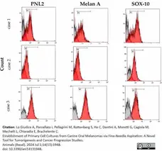
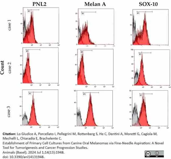
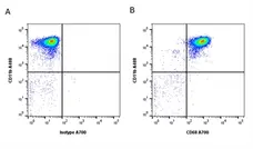
Leucoperm (BUF09B) used to fix and permablize canine melanoma aspirate cells prior to phenotype determination by flow cytometry.
Image caption:
Flow cytometric analysis of cases 1, 2, and 3. The histograms show the different fluorescence intensity patterns for the tested markers: PNL2, Melan-A, and Sox-10. The red peaks represent the positive cells, while the grey peaks represent the negative ones. The sign #1 indicates the name of the gate.
From: Lo Giudice A, Porcellato I, Pellegrini M, Rottenberg S, He C, Dentini A, Moretti G, Cagiola M, Mechelli L, Chiaradia E, Brachelente C.
Establishment of Primary Cell Cultures from Canine Oral Melanomas via Fine-Needle Aspiration: A Novel Tool for Tumorigenesis and Cancer Progression Studies.
Animals (Basel). 2024 Jul 1;14(13):1948.
doi: 10.3390/ani14131948.
This image is from an open access article distributed under terms of a Creative Commons Attribution License.
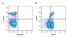
Leucoperm (BUF09) used to permablise cell membranes prior to analysis of interleukin expression in peripheeral mononuclear cells by flow cytometry.
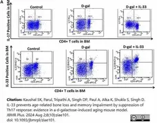
Image caption:
Effects of IL-33 on Th-17 cell proliferation and Treg cells. (A) FACS was used to detect the percentage of IL-17– and IL-33–positive cells in bone marrow (BM) PBMCs across all groups.
From: Kaushal SK, Parul, Tripathi A, Singh DP, Paul A, Alka K, Shukla S, Singh D.
IL-33 prevents age-related bone loss and memory impairment by suppression of Th17 response: evidence in a d-galactose-induced aging mouse model.
JBMR Plus. 2024 Aug 2;8(10):ziae101.
doi: 10.1093/jbmrpl/ziae101.
This image is from an open access article distributed under terms of a Creative Commons Attribution License.
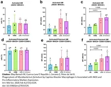
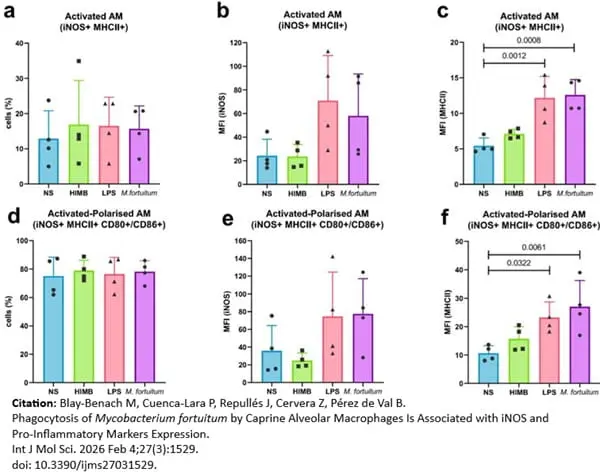
Alveolar macrophages treated with Leucoperm (BUF09B) prior to flow cytometric analysis of iNOS expression.
Image caption:
Flow cytometry analysis of activation and polarisation profiles of stimulated Ams. (a) Percentage of activated AMs (iNOS+ MHCII+). (b) MFI of intracellular iNOS in activated cells. (c) MFI of surface MHCII in activated cells. (d) Percentage of activated-polarised AMs (iNOS+ MHCII+ CD80+/CD86+). (e) MFI of iNOS in active-polarised AMs. (f) MFI of MHCII in active-polarised AMs. Each dot presents an independent macrophage batch. Results are represented as Mean ± SD. AMs: alveolar macrophages; MFI: mean fluorescent intensity; NS: non-stimulated.
From: Blay-Benach M, Cuenca-Lara P, Repullés J, Cervera Z, Pérez de Val B.
Phagocytosis of Mycobacterium fortuitum by Caprine Alveolar Macrophages Is Associated with iNOS and Pro-Inflammatory Markers Expression.
Int J Mol Sci. 2026 Feb 4;27(3):1529.
doi: 10.3390/ijms27031529.
This image is from an open access article distributed under terms of a Creative Commons Attribution License.
Filter by Application:
CE F Reset| Flow cytometric analyses with monoclonal antibodies have been restricted primarily to cell surface molecules. Intracellular structures such as cytoplasmic or nuclear enzymes, oncoproteins, cytokines, immunoglobulins etc. were largely excluded from such assays. Also excluded from flow cytometric assays were cytoplasmic localizations of well established membrane molecules such as CD3 and CD22. LEUCOPERM reagents allow intracellular antigen analysis with the same ease as surface antigens. The only prerequisite is the availability of suitable antibody conjugates. Most commercially available monoclonal antibody conjugates can be used with LEUCOPERM reagents. Some determinants are sensitive, however, to the fixation step involved. This and the optimal fixation time may have to be determined experimentally for each antibody conjugate. |
- Product Form
- Reagent A - Fixation medium
Reagent B - Permeabilisation medium - Preservative Stabilisers
- Formaldehyde in Reagent A
- Regulatory
- For research purposes only
- Guarantee
- 12 months from date of despatch
This product is shipped at ambient temperature.
LEUCOPERM Cell Permeabilisation reagents should be stored and used at room temperature. DO NOT FREEZE. Do not use reagents if a precipitate forms or discolouration occurs.
LEUCOPERM Cell Permeabilisation reagents should be stored and used at room temperature. DO NOT FREEZE. Do not use reagents if a precipitate forms or discolouration occurs.
This product has been reported to work in the following applications. This information is derived from testing within our laboratories, peer-reviewed publications or personal communications from the originators. Please refer to references indicated for further information. For general protocol recommendations, please visit the antibody protocols page.
| Application Name | Verified | Min Dilution | Max Dilution |
|---|---|---|---|
| Flow Cytometry |  |
LEUCOPERM reagents are intended for fixing cells in suspension with Reagent A and then permeabilizing the cells with Reagent B. The specific formulations reduce background staining and allow simultaneous addition of permeabilization medium and fluorochrome labeled antibodies.
- Instructions For Use
-
BUF09C, BUF09B, BUF09
For the detection of cell cycle antigens such as Ki-67, PCNA and BrdU, methanol modification is recommended - see protocol #F5.
1. Prepare cells in the appropriate manner. Adjust cell suspension to a concentration of 1 x 10⁷ cells/ml in PBS/BSA. Whole blood samples may also be used. Bio-Rad recommend the use of EDTA anti-coagulant in these circumstances, although satisfactory results may be obtained using heparin or acid-citrate dextrose.
2. Add 100ul of cell suspension to the appropriate number of test tubes.
If required, perform staining of cell surface antigens at this stage. Following staining for the recommended period, wash cells once in PBS/BSA and discard supernatant.
3. Add 100ul of Reagent A (fixation medium, stored at room temperature).
4. Incubate for 15 minutes at room temperature.
5. Add 3ml PBS/BSA and centrifuge for 5 minutes at 300 x g. Remove supernatant.
6. Re-suspend cells in 100ul of Reagent B (Permeabilization Medium).
7. Immediately add recommended volume of the appropriate directly conjugated antibody. Vortex and incubate for 30 minutes at room temperature.
If using an unconjugated primary antibody, wash in 3ml of PBS/BSA (as per step 5) and then repeat step 7 using an appropriate secondary antibody. There is no requirement to add further Leucoperm.
8. Wash once in PBS/BSA. Remove supernatant and resuspend cells in sheath fluid for immediate analysis or resuspend cells in 0.25ml of 0.5% formaldehyde and store them at 2-8oC in the dark. Analyse fixed cells within 24 hours.
References for Leucoperm
-
Chiu, W.C. et al. (2009) Effects of dietary fish oil supplementation on cellular adhesion molecule expression and tissue myeloperoxidase activity in hypercholesterolemic mice with sepsis.
J Nutr Biochem. 20: 254-60. -
Grundy, M. et al. (2010) The FLT3 internal tandem duplication mutation is a secondary target of the aurora B kinase inhibitor AZD1152-HQPA in acute myelogenous leukemia cells.
Mol Cancer Ther. 9: 661-72. -
Taylor, L. et al. (2010) The effect of acute hypoxia on heat shock protein 72 expression and oxidative stress in vivo.
Eur J Appl Physiol. 109 (5): 849-55. -
Bairey, O. et al. (2010) Arsenic-trioxide-induced apoptosis of chronic lymphocytic leukemia cells.
Int J Lab Hematol. 32 (1 Pt 1): e77-85. -
Myles, A. et al. (2011) Expression of Toll-like receptors 2 and 4 is increased in peripheral blood and synovial fluid monocytes of patients with enthesitis-related arthritis subtype of juvenile idiopathic arthritis.
Rheumatology (Oxford). 50: 481-8. -
Osorio, Y. et al. (2011) Identification of small molecule lead compounds for visceral leishmaniasis using a novel ex vivo splenic explant model system
PLoS Negl Trop Dis. 5:e962. -
Suradhat, S. et al. (2015) A novel DNA vaccine for reduction of PRRSV-induced negative immunomodulatory effects: A proof of concept.
Vaccine. 33 (32): 3997-4003. -
Parry, D.A. et al. (2016) A homozygous STIM1 mutation impairs store-operated calcium entry and natural killer cell effector function without clinical immunodeficiency.
J Allergy Clin Immunol. 137 (3): 955-7.e8.
View The Latest Product References
-
Dishon, S. et al. (2017) Inhibition of Myeloid Differentiation Factor 88 Reduces Human and Mouse T-Cell Interleukin-17 and IFN&gamma Production and Ameliorates Experimental Autoimmune Encephalomyelitis Induced in Mice.
Front Immunol. 8: 615. -
Nie, H. et al. (2017) Phenotypic switch in lung interstitial macrophage polarization in an ovalbumin-induced mouse model of asthma.
Exp Ther Med. 14 (2): 1284-92. -
Jiang, W.J. et al. (2017) Structure-activity relationship of the inhibitory effects of flavonoids on nitric oxide production in RAW264.7 cells.
Bioorg Med Chem. 25 (2): 779-788. -
Kliminski, V. et al. (2017) Popdc1/Bves Functions in the Preservation of Cardiomyocyte Viability While Affecting Rac1 Activity and Bnip3 Expression.
J Cell Biochem. 118 (6): 1505-17. -
Arrieta-Villegas, C. et al. (2020) Immunogenicity and Protection against Mycobacterium caprae Challenge in Goats Vaccinated with BCG and Revaccinated after One Year.
Vaccines (Basel). 8 (4): 751. -
Alhuthali, H.M. et al. (2020) The natural alkaloid Jerantinine B has activity in acute myeloid leukemia cells through a mechanism involving c-Jun.
BMC Cancer. 20 (1): 629. -
Martelli, P. et al. (2021) Immune B cell responsiveness to single-dose intradermal vaccination against Mycoplasma hyopneumoniae..
Res Vet Sci. 141: 66-75. -
Hatzidaki, E. et al. (2021) A Novel Method for Colorectal Cancer Screening Based on Circulating Tumor Cells and Machine Learning.
Entropy (Basel). 23 (10): 1248. -
Martelli, P. et al. (2021) Immune B cell responsiveness to single-dose intradermal vaccination against Mycoplasma hyopneumoniae..
Res Vet Sci. 141: 66-75. -
Cequier, A. et al. (2022) Equine Mesenchymal Stem Cells Influence the Proliferative Response of Lymphocytes: Effect of Inflammation, Differentiation and MHC-Compatibility.
Animals (Basel). 12 (8) 984. -
Sanchez-Pino, M.D. (2022) Detection of Circulating and Tissue Myeloid-Derived Suppressor Cells (MDSC) by Flow Cytometry.
Methods Mol Biol. 2422: 247-61. -
Franzoni, G. et al. (2022) Analyses of the Impact of Immunosuppressive Cytokines on Porcine Macrophage Responses and Susceptibility to Infection to African Swine Fever Viruses.
Pathogens. 11 (2): 166. -
Jeong, E.M. et al. (2022) Targeting RUNX1 as a novel treatment modality for pulmonary arterial hypertension.
Cardiovasc Res. 118 (16): 3211-24. -
Matralis, D.T. et al. (2023) Intracellular IFN-γ and IL-4 levels of CD4 + and CD8 + T cells in the peripheral blood of naturally infected (Leishmania infantum) symptomatic dogs before and following a 4-week treatment with miltefosine and allopurinol: a double-blinded, controlled and cross-sectional study.
Acta Vet Scand. 65 (1): 2. -
Liu, Y. et al. (2024) Porous PLGA/MBG scaffold enhanced bone regeneration through osteoimmunomodulation
Composites Part B: Engineering. 272: 111202. -
Gordon, H. et al. (2024) Human Intestinal Dendritic Cells Can Overcome Retinoic Acid Signaling to Generate Proinflammatory CD4 T Cells with Both Gut and Skin Homing Properties.
J Immunol. 212 (1): 96-106. -
Takeuchi, T. et al. (2024) Potential Effects of Ischemic Postconditioning and Changes in Heat Shock Protein 72 in Patients with Acute Myocardial Infarction without Prodromal Angina.
Int Heart J. 65 (3): 395-403. -
Bulun, H. et al. (2024) Interferon-gamma producing CD4+ T cells quantified by flow cytometry as early markers for Mycobacterium avium ssp. paratuberculosis infection in cattle.
Vet Res. 55 (1): 69. -
Gordon, H. et al. (2024) Human Intestinal Dendritic Cells Can Overcome Retinoic Acid Signaling to Generate Proinflammatory CD4 T Cells with Both Gut and Skin Homing Properties.
J Immunol. 212 (1): 96-106. -
McCarthy, N.E. et al. (2021) Patients with gastrointestinal irritability after TGN1412-induced cytokine storm displayed selective expansion of gut-homing αβ and γδT cells.
Cancer Immunol Immunother. 70 (4): 1143-53. -
Lo Giudice, A. et al. (2024) Establishment of Primary Cell Cultures from Canine Oral Melanomas via Fine-Needle Aspiration: A Novel Tool for Tumorigenesis and Cancer Progression Studies.
Animals (Basel). 14 (13): 1948. -
Kaushal, S.K. et al. (2024) IL-33 prevents age-related bone loss and memory impairment by suppression of Th17 response: evidence in a d-galactose-induced aging mouse model.
JBMR Plus. 8 (10): ziae101. -
Rogato, F. et al. (2024) Leukemia cutis as a prominent clinical sign in a dog with acute myeloid leukemia.
Vet Clin Pathol. 53 (4): 448-57. -
Wójtowicz, A. et al. (2025) Th1 and Th2 cells in equine endometrosis and their interactions with endometrial fibroblasts.
Sci Rep. 15 (1): 36263. -
Blay-Benach, M. et al. (2026) Phagocytosis of Mycobacterium fortuitum by Caprine Alveolar Macrophages Is Associated with iNOS and Pro-Inflammatory Markers Expression.
Int J Mol Sci. 27 (3): 1529. -
Arribas-Rodríguez, E. et al. (2026) Type 1 and CD103(+) Type 2 Conventional Dendritic Cells Are Decreased in Active Patients with Ulcerative Colitis but Not with Crohn's Disease.
Eur J Immunol. 56 (2): e70118.
Request a different product with this specificity
Please Note: All Products are "FOR RESEARCH PURPOSES ONLY"
Always be the first to know.
When we launch new products and resources to help you achieve more in the lab.
Yes, sign me up