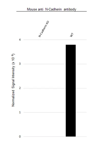
N-Cadherin antibody | 13A9
Mouse anti Human N-Cadherin
- Product Type
- Monoclonal Antibody
- Clone
- 13A9
- Isotype
- IgG1
- Format
- Purified
- Specificity
- N-Cadherin


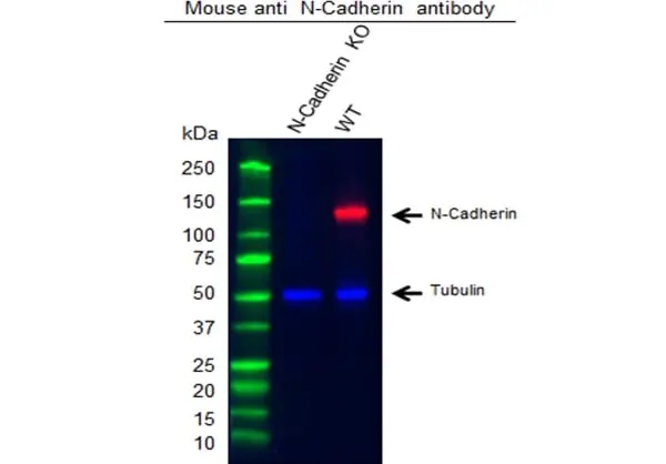
KO Validated
Filter by Application:
P WB Reset| Mouse anti Human N-cadherin antibody, clone 13A9 recognizes neural cadherin, otherwise known as CD325, a calcium dependent cell-cell adhesion glycoprotein, and member of the cadherin superfamily, which links to the actin cytoskeleton via catenins, and plays a role in cell-matrix adhesion, cell growth and differentiation, and the establishment of left-right asymmetry. N-cadherin is expressed by neurons, endothelial cells, muscle cells, and stem cells, and is one of the primary cadherins recruited to the site of neuronal synapse formation. N-cadherin is directly involved in the differentiation of early hematopoietic progenitors, and is commonly expressed by cancer cells, playing a role in transendothelial migration and metastasis, through the up-regulation of the src kinase pathway, and subsequent failure of the intercellular connection between two adjacent endothelial cells. Mouse anti Human N-cadherin antibody, clone 13A9 does not recognize E-cadherin, M-cadherin or P-cadherin (Knudsen et al. 1995). Mouse anti Human N-cadherin antibody, clone 13A9 is a reliable marker for the differential diagnosis of pleural mesotheliomas and lung adenocarcinomas, when used in conjunction with E-cadherin (Han et al. 1997). |
|
- Target Species
- Human
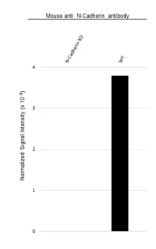
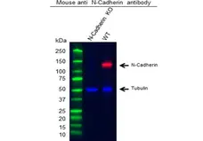
- Western Blotting
- MCA5698 detects a band of approximately 135-140kDa in human HeLa cell lysates.
- Species Cross-Reactivity
-
Target Species Cross Reactivity Rat - N.B. Antibody reactivity and working conditions may vary between species.
- Product Form
- Purified IgG - liquid
- Preparation
- Purified IgG prepared by affinity chromatography on Protein A from tissue culture supernatant
- Buffer Solution
- Phosphate buffered saline
- Preservative Stabilisers
- 0.09% Sodium Azide (NaN3)
- Carrier Free
- Yes
- Immunogen
- Recombinant MBP fusion protein containing the entire cytoplasmic domain of human N-cadherin.
- Approx. Protein Concentrations
- IgG concentration 1.0mg/ml
- Regulatory
- For research purposes only
- Guarantee
- 12 months from date of despatch
- Acknowledgements
- PrecisionAb is a trademark of Bio-Rad Laboratories
This product is shipped at ambient temperature. It is recommended to aliquot and store at -20°C on receipt. When thawed, aliquot the sample as needed. Keep aliquots at 2-8°C for short term use (up to 4 weeks) and store the remaining aliquots at -20°C.
Avoid repeated freezing and thawing as this may denature the antibody. Storage in frost-free freezers is not recommended.
Avoid repeated freezing and thawing as this may denature the antibody. Storage in frost-free freezers is not recommended.
This product has been reported to work in the following applications. This information is derived from testing within our laboratories, peer-reviewed publications or personal communications from the originators. Please refer to references indicated for further information. For general protocol recommendations, please visit the antibody protocols page.
| Application Name | Verified | Min Dilution | Max Dilution |
|---|---|---|---|
| Immunofluorescence |  |
||
| Immunohistology - Frozen | |
||
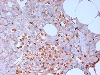
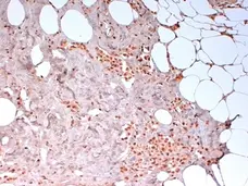
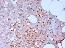
| Immunohistology - Paraffin 1 | |
||
| Immunoprecipitation | |
||
| Western Blotting | |
1/100 | 1/1000 |
- 1 This product requires antigen retrieval using steam heat treatment prior to staining of paraffin sections. Sodium citrate buffer pH 6.0 is recommended for this purpose.
The PrecisionAb label is reserved for antibodies that meet the defined performance criteria within Bio-Rad's ongoing antibody validation programme. Learn about how we validate our PrecisionAb range. Where this product has not been tested for use in a particular technique this does not necessarily exclude its use in such procedures. Further optimization may be required dependent on sample type.
References for N-Cadherin antibody
-
Sacco, P.A. et al. (1995) Identification of plakoglobin domains required for association with N-cadherin and alpha-catenin.
J Biol Chem. 270: 20201-6. -
Peralta Soler, A. et al. (1995) The differential expression of N-cadherin and E-cadherin distinguishes pleural mesotheliomas from lung adenocarcinomas.
Hum Pathol. 26 (12): 1363-9. -
Knudsen, K.A. et al. (1995) Interaction of alpha-actinin with the cadherin/catenin cell-cell adhesion complex via alpha-catenin.
J Cell Biol. 130 (1): 67-77. -
Islam, S. et al. (1996) Expression of N-cadherin by human squamous carcinoma cells induces a scattered fibroblastic phenotype with disrupted cell-cell adhesion.
J Cell Biol. 135 (6 Pt 1): 1643-54. -
Han, A.C. et al. (1997) Differential expression of N-cadherin in pleural mesotheliomas and E-cadherin in lung adenocarcinomas in formalin-fixed, paraffin-embedded tissues.
Hum Pathol. 28 (6): 641-5. -
Peralta Soler, A. et al. (1999) P-cadherin expression in breast carcinoma indicates poor survival.
Cancer. 86: 1263-72. -
Nieman, M.T. et al. (1999) N-cadherin promotes motility in human breast cancer cells regardless of their E-cadherin expression.
J Cell Biol. 147 (3): 631-44. -
Machell, N.H. et al. (2000) Developmental expression and distribution of N- and E-cadherin in the rat ovary.
Biol Reprod. 63 (3): 797-804.
View The Latest Product References
-
Van Aken, E.H. et al. (2002) Structure and function of the N-cadherin/catenin complex in retinoblastoma.
Invest Ophthalmol Vis Sci. 43 (3): 595-602. -
Wahl, J.K. 3rd et al. (2003) N-cadherin-catenin complexes form prior to cleavage of the proregion and transport to the plasma membrane.
J Biol Chem. 278 (19): 17269-76. -
Shintani, Y. et al. (2006) Phosphoinositide-3 kinase-Rac1-c-Jun NH2-terminal kinase signaling mediates collagen I-induced cell scattering and up-regulation of N-cadherin expression in mouse mammary epithelial cells.
Mol Biol Cell. 17: 2963-75. -
Theisen, C.S. et al. (2007) NHERF links the N-cadherin/catenin complex to the platelet-derived growth factor receptor to modulate the actin cytoskeleton and regulate cell motility.
Mol Biol Cell. 18: 1220-32. -
Tian, G. et al. (2009) Clarin-1, encoded by the Usher Syndrome III causative gene, forms a membranous microdomain: possible role of clarin-1 in organizing the actin cytoskeleton.
J Biol Chem. 284: 18980-93.
- Synonyms
- CD325
- RRID
- AB_11152772
- UniProt
- P19022
- Q9Z1Y3
- Entrez Gene
- CDH2
- Cdh2
- GO Terms
- GO:0005509 calcium ion binding
- GO:0016021 integral to membrane
- GO:0008013 beta-catenin binding
- GO:0016342 catenin complex
- GO:0034329 cell junction assembly
- GO:0034332 adherens junction organization
- GO:0042692 muscle cell differentiation
- GO:0045294 alpha-catenin binding
- GO:0045295 gamma-catenin binding
- View More GO Terms
- GO:0051149 positive regulation of muscle cell differentiation
- GO:0005916 fascia adherens
- GO:0007416 synapse assembly
- GO:0016339 calcium-dependent cell-cell adhesion
- GO:0019901 protein kinase binding
- GO:0031641 regulation of myelination
- GO:0032403 protein complex binding
- GO:0032880 regulation of protein localization
- GO:0035023 regulation of Rho protein signal transduction
- GO:0044456 synapse part
- GO:0050770 regulation of axonogenesis
- GO:0051291 protein heterooligomerization
MCA5698
If you cannot find the batch/lot you are looking for please contact our technical support team for assistance.
Request a different product with this specificity
Please Note: All Products are "FOR RESEARCH PURPOSES ONLY"
View all Anti-Human ProductsAlways be the first to know.
When we launch new products and resources to help you achieve more in the lab.
Yes, sign me up