Macrophages/Monocytes/Granulocytes antibody | MAC387
Mouse anti Human Macrophages
- Product Type
- Monoclonal Antibody
- Clone
- MAC387
- Isotype
- IgG1
- Specificity
- Macrophages/Monocytes/Granulocytes


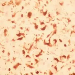
Mouse anti Human macrophages antibody, clone MAC387 (MCA874G) used for the identification of canine macrophages in skin by immunohistochemistry on formalin fixed, paraffin embedded tissue sections.
Image caption:
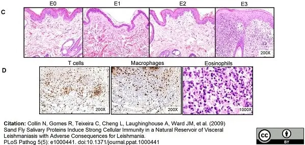
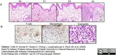
Dogs develop a strong humoral and cellular immune response to bites of Lu. longipalpis sand flies. Dogs (n = 9) were exposed for 10 min to bites of 20 sand flies three times at one week intervals (first exposure, E1; second exposure, E2; third exposure, E3). (C) Representative H&E staining of biopsies taken from sand fly bite sites prior to exposure (E0) and 48 h after each of three sand fly exposures (E1–E3). Note marked cellular infiltrate within dermis and thickening of epidermis in E3. (D) Immunohistochemical labeling of tissue sections from E3 demonstrating the presence of abundant CD3+T cells (CD3), macrophages (Mac387) and eosinophil granules (Luna stain).
From: Collin N, Gomes R, Teixeira C, Cheng L, Laughinghouse A, et al. (2009)
Sand Fly Salivary Proteins Induce Strong Cellular Immunity in a Natural Reservoir of Visceral Leishmaniasis with Adverse Consequences for Leishmania.
PLoS Pathog 5(5): e1000441.
This image is from an open access article distributed under terms of a Creative Commons Attribution License.

Mouse anti Human macrophages antibody, clone MAC387 (MCA874G) used for the identification of macrophages in canine skin by immunohistochemistry on formalin fixed, paraffin embedded tissue sections.
Image caption:
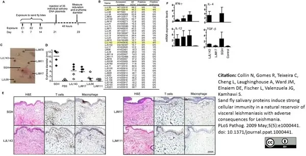
Identification of salivary proteins from Lu. longipalpis that produce a cellular immune response in dogs. (A) A schematic representation of the reverse antigen screening approach based on the intradermal injection of DNA plasmids in dogs previously exposed to sand fly bites (first exposure, E1; second exposure, E2; third exposure, E3). (B–F) Dogs pre-exposed to sand fly bites were challenged intradermally with DNA plasmids and one pair of salivary gland homogenate (SGH) and PBS (positive and negative controls, respectively) and investigated 48 h post-injection. (B) The number of dogs showing local induration and/or erythema at the site of injection for 35 DNA plasmids coding for secreted salivary molecules. Yellow bars highlight the response of dogs to LJM17 and LJL143. (C) Photograph to demonstrate specificity of the cellular reaction to DNA plasmids and SGH. (D) The diameter of erythema in the absence (◇) or presence (◆) of induration for each dog at the site of injection of SGH, PBS, LJL143 and LJM17 (reactive plasmids) and LJL04 and LJM111 (intermediate and non-reactive plasmids, respectively). (E–F) Skin biopsies (6mm) obtained from injection sites were cut in half and processed for histology and RNA extraction. (E) Representative H&E staining and immunohistochemical labeling of dermal T cells (anti-CD3) and macrophages (Mac387) at the injection sites of SGH, LJL143, LJM17 and LJM111. Note marked dermal infiltrates of inflammatory cells characterized as CD3+ T cells and scattered macrophages (Mac387) in the SGH, LJL143 and LJM17. There is no inflammation with LJM111. (F) Reverse-transcriptase quantitative PCR showing the expression levels of IFN-γ, IL-12, IL-4 and TGF-β for LJL143, LJM17, a pair of SGH and control (a mix of PBS and empty plasmid) 48 h post-injection. Error bars represent means±S.E.
From: Collin N, Gomes R, Teixeira C, Cheng L, Laughinghouse A, et al. (2009)
Sand Fly Salivary Proteins Induce Strong Cellular Immunity in a Natural Reservoir of Visceral Leishmaniasis with Adverse Consequences for Leishmania.
PLoS Pathog 5(5): e1000441.
This image is from an open access article distributed under terms of a Creative Commons Attribution License.
Mouse anti Human macrophages antibody, clone MAC387 (MCA874G) used for the identification of macrophages in canine skin by immunohistochemistry on formalin fixed, paraffin embedded tissue sections.
Image caption:
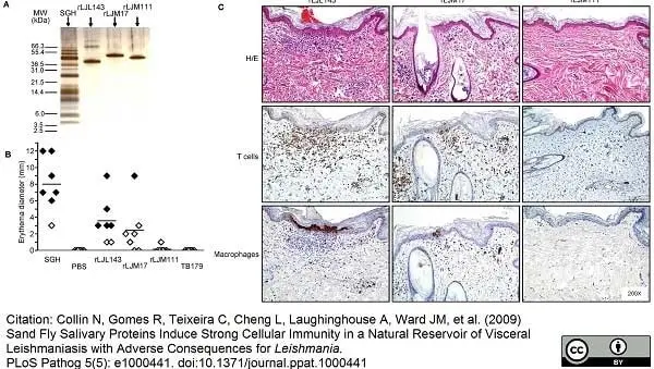
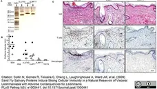
Sand fly salivary recombinant proteins produce a DTH response in dogs previously exposed to sand flies. (A) Purity of the recombinant salivary proteins produced by HEK-293F mammalian cells and purified by a HPLC nickel trap column. (B) The diameter of erythema in the absence (◇) or presence (◆) of induration for each dog at the site of injection 48 h after challenge with salivary gland homogenate (SGH), PBS, recombinant proteins rLJL143 and rLJM17 (reactive), rLJM111 (non-reactive) and a non-related tick recombinant protein TB179. (C) Representative H&E staining and immunohistochemical labeling of T cells (anti-CD3) and macrophages (Mac387) at the injection sites of rLJL143, rLJM17 and rLJM111. Note marked dermal infiltrates of inflammatory cells characterized as CD3+ T cells and scattered macrophages (Mac387) with rLJL143 and rLJM17; rLJM111 is negative.
From: Collin N, Gomes R, Teixeira C, Cheng L, Laughinghouse A, et al. (2009)
Sand Fly Salivary Proteins Induce Strong Cellular Immunity in a Natural Reservoir of Visceral Leishmaniasis with Adverse Consequences for Leishmania.
PLoS Pathog 5(5): e1000441.
This image is from an open access article distributed under terms of a Creative Commons Attribution License.

Mouse anti Human macrophages antibody, clone MAC387 (MCA874G used for the identification of macrophages in canine skin by immunohistochemistry on formalin fixed, paraffin embedded tissue sections.
Image caption:
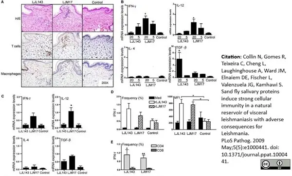
Bites of Lu. longipalpis sand flies induce a strong focal and systemic adaptive cellular immune response in dogs immunized with LJL143 or LJM17. (A–C) Dogs were exposed to uninfected and infected sand flies for 10 min one month after the final immunization with either LJM17, LJL143 or the empty plasmid (control). (A–C) Skin biopsies (6mm) obtained from bite sites 48 h post challenge with 20 and five uninfected and 10 infected sand flies were cut in half and processed for histology and RNA extraction. (A) Representative H&E staining and immunohistochemical labeling of T cells (anti-CD3) and macrophages (Mac387) at the bite sites of 20 uninfected sand flies in LJL143- and LJM17-immunized and control dogs. (B) Reverse-transcriptase quantitative PCR showing the expression levels of IFN-γ, IL-12, IL-4 and TGF-β at the bite sites of 20 or five uninfected sand flies in LJL143- and LJM17-immunized and control dogs (for control dogs RNA was combined from sites of 20 and 5 uninfected sand fly bites). (C) Same as (B) using 10 infected sand flies. Histological sections from bite sites of five uninfected and 10 infected sand flies are provided as Figure S1 and Figure S2, respectively. (D–E) PBMC from LJL143- and LJM17-immunized and control dogs obtained one week after exposure to sand flies. (D) Frequency and mean fluorescence intensity (MFI) of CD3+ T cells following stimulation with medium, rLJL143 or rLJM17. (E) Frequency of CD4+ and CD8+ T cells expressing IFN-γ in PBMC from LJL143- and LJM17-immunized dogs. Error bars represent means±S.E. * P<0.05, ** P<0.01.
From: Collin N, Gomes R, Teixeira C, Cheng L, Laughinghouse A, et al.
(2009) Sand Fly Salivary Proteins Induce Strong Cellular Immunity in a Natural Reservoir of Visceral Leishmaniasis with Adverse Consequences for Leishmania.
PLoS Pathog 5(5): e1000441.
This image is from an open access article distributed under terms of a Creative Commons Attribution License.
Mouse anti Human macrophage antibody, clone MAC387 (MCA874G) used for the detection of macrophages in rabbit skin by immunohistochemistry on formalin fixed, paraffin embedded tissue sections.
Image caption:
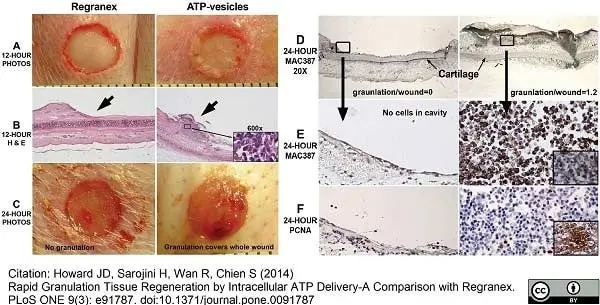
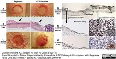
A photomontage of wounds and microscopic changes occurring within 24 hours after surgery. When ATP-vesicles are used, granulation starts to appear within 12 hours (A), and H&E staining indicates a rich cellular component (B). Granulation tissue growth continues and covers the whole wound at 24 hours (C). Granulation tissue shows positive anti-MAC387 staining (D), which is further confirmed by CD163 staining (inset) (E). PCNA staining indicates very active proliferation of these cells (F), which is further confirmed by BrdU antibody staining (inset). Wounds treated with Regranex do not display this rapid growth.
From: Howard JD, Sarojini H, Wan R, Chien S (2014)
Rapid Granulation Tissue Regeneration by Intracellular ATP Delivery-A Comparison with Regranex.
PLoS ONE 9(3): e91787.
This image is from an open access article distributed under terms of a Creative Commons Attribution License.

Mouse anti Human macrophage antibody, clone MAC387 (MCA874G) used for the detection of macrophages in rabbit skin by immunohistochemistry on formalin fixed, paraffin embedded tissue sections.
Image caption:
Macrophage accumulation in early days. When the wounds are treated with ATP-vesicles, solid granulation occurs at day 3 (B). The growth is filled with macrophages, which occur not only in wound cavity, but also underneath the ear cartilage (D). The wounds treated with Regranex do not have similar growth (A, C).
From: Howard JD, Sarojini H, Wan R, Chien S (2014)
Rapid Granulation Tissue Regeneration by Intracellular ATP Delivery-A Comparison with Regranex.
PLoS ONE 9(3): e91787.
This image is from an open access article distributed under terms of a Creative Commons Attribution License.
Mouse anti Human macrophage antibody, clone MAC387 (MCA874G) used for the detection of macrophages in rabbit skin by immunohistochemistry on formalin fixed, paraffin embedded tissue sections.
Image caption:
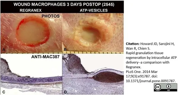
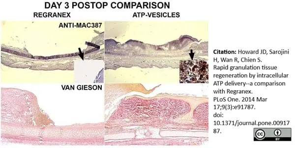
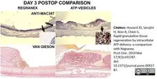
Comparison of Regranex and ATP-vesicle treated wounds 3 days postoperatively. Rich collections of macrophages are stained dark brown to black by Anti-Mac387 (top panels), and more collagen is shown by van Gieson stain (low panels) in the wounds treated with ATP-vesicles. Regranex treated wounds do not show a similar effect.
From: Howard JD, Sarojini H, Wan R, Chien S (2014)
Rapid Granulation Tissue Regeneration by Intracellular ATP Delivery-A Comparison with Regranex.
PLoS ONE 9(3): e91787.
This image is from an open access article distributed under terms of a Creative Commons Attribution License.
Mouse antiHuman macrophages antibody, clone MAC387 (MCA874) used for the demonstration of lesion associated macrophages in Burkholderia pseudomallei infected marmosets by immunohistochemistry on formalin fixed, paraffin embedded tissue sections.
Image caption:
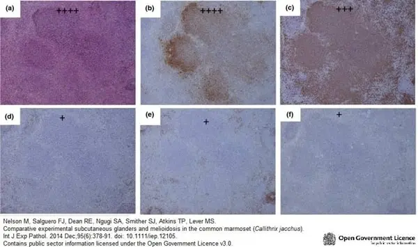
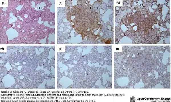
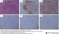
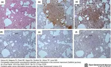
Representative H&E and IHC stained tissue sections from the spleen of a marmoset humanely euthanised at 48 h p.c. challenged with 1.85 × 10² ± 57 cfu of Burkholderia pseudomallei by the subcutaneous route. (a) H & E showing multifocal lesion with severe necrosis (++++), (b) Burkholderia pseudomallei antigen IHC showing abundant bacteria associated with the lesion (++++), (c) IHC staining showing a moderate number of macrophages associated with the lesion (+++), (d) IHC staining showing very few T cells associated with the lesion (+), (e) IHC staining showing very few B cells associated with the lesion (+), (f) IHC staining showing very few inducible Nitric oxide synthase (iNOS) producing cells associated with the lesion (+).
From: Nelson M, Salguero FJ, Dean RE, Ngugi SA, Smither SJ, Atkins TP, Lever MS.
Comparative experimental subcutaneous glanders and melioidosis in the common marmoset (Callithrix jacchus).
Int J Exp Pathol. 2014 Dec;95(6):378-91.
Contains public sector information licensed under the Open Government Licence v3.0.
Mouse anti Human macrophages antibody, clone MAC387 (MCA874G) used for the demonstration of lesion associated macrophages in Burkholderia pseudomallei infected marmosets by immunohistochemistry on formalin fixed, paraffin embedded tissue sections.
Image caption:
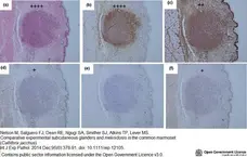
Representative H&E and IHC stained tissue sections from the inoculation site of a marmoset humanely euthanised at 36 h p.c. challenged with 1.85 × 10² ± 57 cfu of Burkholderia pseudomallei by the subcutaneous route. (a) H & E showing multifocal lesion with severe necrosis (++++), (b) B. pseudomallei antigen IHC showing abundant bacteria associated with the lesion (++++), (c) IHC staining showing a small number of macrophages associated with the lesion (++), (d) IHC staining showing very few T cells associated with the lesion (+), (e) IHC staining showing no B cells associated with the lesion, (f) IHC staining showing very few inducible Nitric oxide synthase (iNOS) producing cells associated with the lesion (+).
From: Nelson M, Salguero FJ, Dean RE, Ngugi SA, Smither SJ, Atkins TP, Lever MS.
Comparative experimental subcutaneous glanders and melioidosis in the common marmoset (Callithrix jacchus).
Int J Exp Pathol. 2014 Dec;95(6):378-91.
doi: 10.1111/iep.12105.
Contains public sector information licensed under the Open Government Licence v3.0.
Mouse anti Human macrophages antibody, clone MAC387 (MCA874G) used for the demonstration of lesion associated macrophages in Burkholderia pseudomallei infected marmosets by immunohistochemistry on formalin fixed, paraffin embedded tissue sections.
Image caption:
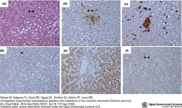
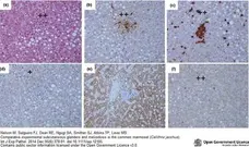
Representative H&E and IHC stained tissue sections from the liver of a marmoset humanely euthanised at 144 h p.c. challenged with 1.79 × 10² ± 20 cfu of Burkholderia mallei by the subcutaneous route. (a) H&E showing non-necrotic multifocal solid lesions (++), (b) Burkholderia mallei antigen IHC showing a small number of bacteria associated with the lesion (++), (c) IHC staining showing a small number macrophages associated with the lesion (++), (d) IHC staining showing very few T cells associated with the lesion (+), (e) IHC staining showing no B cells associated with the lesion, (f) IHC staining showing a small number inducible Nitric oxide synthase (iNOS) producing cells associated with the lesion (++).
From: Nelson M, Salguero FJ, Dean RE, Ngugi SA, Smither SJ, Atkins TP, Lever MS.
Comparative experimental subcutaneous glanders and melioidosis in the common marmoset (Callithrix jacchus).
Int J Exp Pathol. 2014 Dec;95(6):378-91.
Contains public sector information licensed under the Open Government Licence v3.0.
Mouse antiHuman macrophages antibody, clone MAC387 (MCA874G) used for the demonstration of lesion associated macrophages in Burkholderia pseudomallei infected marmosets by immunohistochemistry on formalin fixed, paraffin embedded tissue sections.
Image caption:
Representative H&E and IHC stained tissue sections from the lungs of a marmoset humanely euthanised at 144 h p.c. challenged with 1.79 × 10² ± 20 cfu of Burkholderia mallei by the subcutaneous route. (a) H & E showing multifocal lesion with severe necrosis (++++), (b) B. mallei antigen IHC showing abundant bacteria associated with the lesion (++++), (c) IHC staining showing abundant macrophages associated with the lesion (++++), (d) IHC staining showing a small number of T cells associated with the lesion (++), (e) IHC staining showing very few B cells associated with the lesion (+), (f) IHC staining showing a small number inducible Nitric oxide synthase (iNOS) producing cells associated with the lesion (++).
From: Nelson M, Salguero FJ, Dean RE, Ngugi SA, Smither SJ, Atkins TP, Lever MS.
Comparative experimental subcutaneous glanders and melioidosis in the common marmoset (Callithrix jacchus).
Int J Exp Pathol. 2014 Dec;95(6):378-91.
Contains public sector information licensed under the Open Government Licence v3.0.
Mouse anti Human macrophages antibody, clone MAC387 (MCA874G) used for the identification of macrophages in canine skin by immunohistochemistry on formalin fixed, paraffin embedded tissue sections.
Image caption:
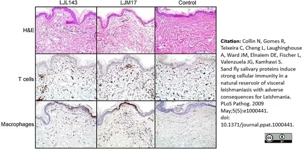
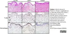
Bites of Lu. longipalpis sand flies induce a strong focal cellular immune response in dogs immunized with LJL143 or LJM17. Dogs were exposed to 5 uninfected sand flies for 10 min one month after the final immunization with either LJM17, LJL143 or empty plasmid (control). Skin biopsies (6mm) obtained from bite sites 48 h post challenge were processed for histology. Representative H&E staining and immunohistochemical labeling of T cells (anti-CD3) and macrophages (Mac387) at the bite sites in LJL143- and LJM17-immunized and control dogs.
From: Collin N, Gomes R, Teixeira C, Cheng L, Laughinghouse A, et al. (2009)
Sand Fly Salivary Proteins Induce Strong Cellular Immunity in a Natural Reservoir of Visceral Leishmaniasis with Adverse Consequences for Leishmania.
PLoS Pathog 5(5): e1000441.
This image is from an open access article distributed under terms of a Creative Commons Attribution License.
Mouse anti Human macrophages antibody, clone MAC387 (MCA874G) used for the identification of macrophages in canine skin by immunohistochemistry on formalin fixed, paraffin embedded tissue sections.
Image caption:
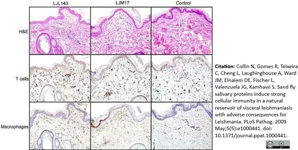
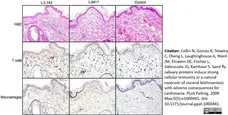
Bites of L. i. chagasi infected sand flies induce a strong focal cellular immune response in dogs immunized with LJL143 or LJM17. Dogs were exposed to ten L. i. chagasi infected sand flies for 10 min one month after the final immunization with either LJM17, LJL143 or empty plasmid (control). Skin biopsies (6mm) obtained from bite sites 48 h post challenge were processed for histology. Representative H&E staining and immunohistochemical labeling of T cells (anti-CD3) and macrophages (Mac387) at the bite sites in LJL143- and LJM17-immunized and control dogs.
From: Collin N, Gomes R, Teixeira C, Cheng L, Laughinghouse A, et al. (2009)
Sand Fly Salivary Proteins Induce Strong Cellular Immunity in a Natural Reservoir of Visceral Leishmaniasis with Adverse Consequences for Leishmania.
PLoS Pathog 5(5): e1000441.
This image is from an open access article distributed under terms of a Creative Commons Attribution License.
Mouse anti Human macrophages antibody, clone MAC387 (MCA874GA) used to label macrophages in ovine tissues by immunohistochemistry of formalin fixed, paraffin embedded tissue sections following proteinase K mediated antigen retrieval.
Image caption:
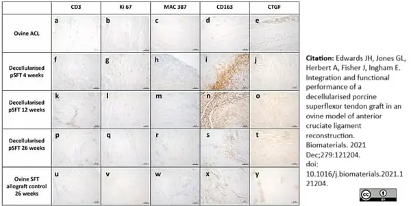
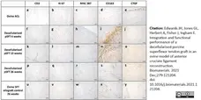
Representative images of sections of the native ovine ACL and intra-articular graft portion stained with antibodies to CD3, Ki-67, MAC 387; CD163 and CTGF.
A-E Native ovine ACL; F-J decellularised pSFT explanted at 4 weeks; K–O; decellularised pSFT explanted at 12 weeks; P-T decellularised pSFT explanted at 26 weeks; U–Y ovine SFT (allograft control) explanted at 26 weeks. Images captured at 10× magnification unless otherwise stated. Scale bars show 100 μm.
From: Edwards JH, Jones GL, Herbert A, Fisher J, Ingham E.
Integration and functional performance of a decellularised porcine superflexor tendon graft in an ovine model of anterior cruciate ligament reconstruction.
Biomaterials. 2021 Oct 21;279:121204.
doi: 10.1016/j.biomaterials.2021.121204
This image is from an open access article distributed under terms of a Creative Commons Attribution License.
Mouse anti Human macrophages, clone MAC387 (MCA874GA) used to label macrophages in ovine pulmonary root tissues by immunohistochemistry on formalin fixed cryosections.
Image caption:
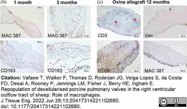
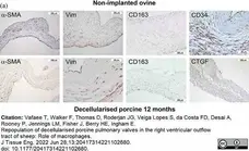
Representative images of sections of explanted pulmonary root leaflet tissues stained with antibodies to α-SMA, vimentin, MAC 387, CD163, CD34 or CTGF. (a) Upper panel: native ovine pulmonary root leaflet tissues. Images captured at 10× magnification (scale bars 100 μm) except CD34 (20× magnification; scale bar 50 μm). Lower panel: decellularised porcine pulmonary wall tissues explanted after 12 months in vivo. Images captured at 10× magnification (scale bars 100 μm) except vimentin (20× magnification; scale bar 50 μm). Images show that the distribution of cells expressing α-SMA, vimentin and CD163 was similar, although sparser, in the decellularised porcine pulmonary root leaflet tissues compared to native ovine tissue, however cells in the decellularised porcine pulmonary root leaflets did not express CD34 and there were high levels of expression of CTGF.
From: Vafaee T, Walker F, Thomas D, Roderjan JG, Veiga Lopes S, da Costa FD, Desai A, Rooney P, Jennings LM, Fisher J, Berry HE, Ingham E.
Repopulation of decellularised porcine pulmonary valves in the right ventricular outflow tract of sheep: Role of macrophages.
J Tissue Eng. 2022 Jun 28;13:20417314221102680.
doi: 10.1177/20417314221102680.
This image is from an open access article distributed under terms of a Creative Commons Attribution License.
Mouse anti Human macrophages, clone MAC387 (MCA874GA) used to label macrophages in ovine pulmonary root tissues by immunohistochemistry on formalin fixed cryosections.
Image caption:
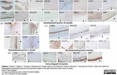
Representative images of sections of explanted pulmonary root wall tissues stained with antibodies to α-SMA, vimentin, MAC 387, CD163, CD34, CD271, vWF and CTGF. (a) Upper panel: native ovine pulmonary artery wall tissues. Images captured at 10× magnification (scale bars 100 μm) unless otherwise stated. α-SMA (scan 2.5× magnification; scale bar 2000 μm), CD34 and vWF (20× magnification; scale bars 50 μm). Lower panel: decellularised porcine pulmonary wall tissues explanted after 12 months. Images captured at 10× magnification (scale bars 100 μm) unless otherwise stated. α-SMA (2.5× magnification; scale bar 500 μm), vimentin and vWF (20× magnification; scale bars 50 μm). Black arrows indicate the intima. (b) Images of the distal decellularised porcine pulmonary artery wall tissues explanted at 1 month. Images captured at 2.5× magnification (scale bars 500 μm). Images show MAC 387+ and CD163+ cells at the interface between cellular and acellular tissue with α-SMA+ and CD271+ cells populating the adventitia and media. Black arrows: intima. (c) Upper panel: images of the same region of central area of a decellularised porcine pulmonary artery wall tissue explanted at 3 months captured at 2.5× magnification (scale bars 500 μm). Images show the ‘front’ (dashed line) of MAC 387+ and CD 163+ cells between the media and intimal regions with α-SMA+ cells in the central media. Black arrows: intima. Lower panel: images of sequential sections of the central area of a decellularised porcine pulmonary artery wall tissue explanted at 3 months captured at 20× magnification (scale bars 50 μm) clearly showing distinct populations of MAC 387+/ CD163low; CD163+/ MAC 387+; CD34+ and CD271+ cells. Red circle: vessel present in all images. The dashed lines demarcate cellular and acellular tissue. (d) Ovine allograft pulmonary artery wall tissues explanted at 12 months. Images captured at 10× magnification (scale bars 100 μm) except α-SMA (2.5× magnification; scale bar 500 μm). Images show the presence of α-SMA+, CD163+ and CD34+ cells in the media with an amorphous intimal region and the presence of MAC 387+ cell foci at the intima with high levels of expression of CTGF and vWF. Black arrows: intima.
From: Vafaee T, Walker F, Thomas D, Roderjan JG, Veiga Lopes S, da Costa FD, Desai A, Rooney P, Jennings LM, Fisher J, Berry HE, Ingham E.
Repopulation of decellularised porcine pulmonary valves in the right ventricular outflow tract of sheep: Role of macrophages.
J Tissue Eng. 2022 Jun 28;13:20417314221102680.
doi: 10.1177/20417314221102680.
This image is from an open access article distributed under terms of a Creative Commons Attribution License.
Mouse anti Human macrophages, clone MAC387 (MCA874GA) used to label macrophages in ovine pulmonary root tissues by immunohistochemistry on formalin fixed cryosections.
Image caption:
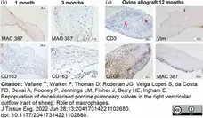
(b) Images of explanted decellularised porcine root leaflets stained with antibodies to MAC 387 and CD163 at 1 (images captured at 10× magnification; scale bars 100 μm) and 3 months (images captured at 20× magnification; scale bars 50 μm). At 1 month MAC387+ cells populated the surfaces of the leaflets distally and at 3 months, the body of the leaflets. CD163+ cells populated the basal regions of the leaflets at 1 and 3 months. (c) Images of explanted ovine allograft leaflet tissues at 12 months. Images captured at 10× magnification (scale bars 100 μm) except vimentin (20× magnification; scale bar 50 μm). Images show inflammatory infiltrate with CD3+ and CTGF+ cells (two of four explants) and paucity of vimentin+ cells with MAC 387+ cell infiltrate on leaflet surface (two of four explants).
From: Vafaee T, Walker F, Thomas D, Roderjan JG, Veiga Lopes S, da Costa FD, Desai A, Rooney P, Jennings LM, Fisher J, Berry HE, Ingham E.
Repopulation of decellularised porcine pulmonary valves in the right ventricular outflow tract of sheep: Role of macrophages.
J Tissue Eng. 2022 Jun 28;13:20417314221102680.
doi: 10.1177/20417314221102680.
This image is from an open access article distributed under terms of a Creative Commons Attribution License.
Mouse anti Human macrophages/monocytes/granulocytes antibody, clone MAC387 (MCA874G) used to label cells in crypt lumen of pigs.
Image caption:
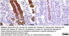
Crypt abscesses in pig with post-weaning diarrhoea. B Cytokeratin stain demonstrating red/brown epithelial lining cells of the crypt abscess (40×/0.75 NA, scale bar, 120 µm). C MAC387 stain demonstrating red MAC-positive cells within the lumen of a crypt (40×/0.75 NA, scale bar, 120 μm)
From: Blirup-Plum SA, Jensen HE, Nielsen SS, Pankoke K, Hansen MS, Pedersen KS, Eriksen EØ, Nielsen JP, Olsen JE, Kudirkiene E, Larsen LE, Goecke NB, Barington K.
Gastro-intestinal lesions are not relatable to diarrhoea or specific pathogens in post-weaning diarrhoea (PWD) in pigs.
Acta Vet Scand. 2023 Jul 3;65 (1):30.
doi: 10.1186/s13028-023-00693-y.
This image is from an open access article distributed under terms of a Creative Commons Attribution License.
Mouse anti Human macrophages, clone MAC387 (MCA874G) used to label macrophages and granulocytes in marmoset formalin fixed, paraffin embedded lung sections by immubohistochemistry.
Image caption:
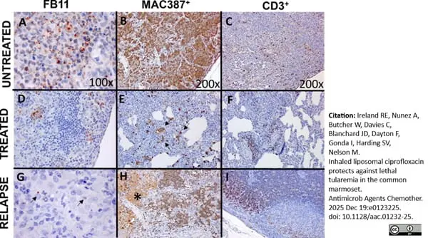
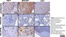
Representative stained lung section images from marmosets challenged by the inhalational route with F. tularensis and then administered placebo or antibiotics at the onset of fever. Lung tissue from placebo-treated animals was stained with FB11 (antibody used to detect the LPS of F. tularensis) (A), MAC387 antibody (macrophages/neutrophils) (B), and an anti-CD3 antibody (T cells) (C). Brown coccobacilli were located intracytoplasmically and extracellularly with abundant labeling of bacteria, neutrophils, macrophages, and T cells in areas of acute inflammation. Extracellular staining of neutrophils, macrophages, and T cells was also abundant in areas of exudation. Less bacterial staining was observed in the lung tissue of animals administered antibiotics (D, ciprofloxacin-treated animal) with MAC387+ (E, ciprofloxacin-treated animal) and CD3+ cells (F, Apulmiq-treated animal) and in areas of septal thickening and cellular infiltration. The arrows indicate stained clusters of macrophages. The animal that received Apulmiq and relapsed had some bacterial staining (G), with the arrows highlighting individual bacterial cells. Neutrophils and macrophages were observed in areas of acute inflammation (H), with areas of diffuse extracellular labeling (*) likely due to presence of cellular components and debris in the exudate. CD3+ cells were observed in the cortex (I).
From: Ireland RE, Nunez A, Butcher W, Davies C, Blanchard JD, Dayton F, Gonda I, Harding SV, Nelson M.
Inhaled liposomal ciprofloxacin protects against lethal tularemia in the common marmoset.
Antimicrob Agents Chemother. 2025 Dec 19:e0123225.
doi: 10.1128/aac.01232-25.
This image is from an open access article distributed under terms of a Creative Commons Attribution License.
Filter by Application:
F P FLA C Reset| Mouse anti Human macrophages, clone MAC387 recognizes the L1 or Calprotectin molecule, an intracytoplasmic antigen comprised of a 12 kDa alpha chain and a 14 kDa beta chain. Although originally described as binding to epitopes common to both the alpha and beta chains (Flavell et al. 1987) subsequent evidence indicates that the antibody detects an epitope exclusively expressed on the beta chain (Goebeler et al. 1994) demonstrated by immunofluorescent and western blotting on both naturally expressing and transfected targets. In addition, Mouse anti Human macrophages, clone MAC387 detects the beta chain in complex with the alpha. The antigen recognized by Mouse anti Human macrophages, clone MAC387 is expressed by granulocytes, monocytes and by tissue macrophages. Variable results have been reported for staining brain macrophages and microglia. The epitope recognized appears to be well conserved and the antibody is routinely used for the detection of myeloid cells in a wide range of species. |
- Target Species
- Human
- Species Cross-Reactivity
-
Target Species Cross Reactivity Horse Pig Dog Rabbit Baboon Bovine Guinea Pig Rat Cat Cynomolgus monkey Rhesus Monkey Goat Fallow deer Pygmy hippopotamus Mink Marmoset - N.B. Antibody reactivity and working conditions may vary between species.
- Product Form
- Purified IgG - liquid
- Preparation
- MCA874GT, MCA874G: Purified IgG prepared by affinity chromatography on Protein A from tissue culture supernatant
- MCA874GA: Purified IgG prepared by affinity chromatography on Protein A from tissue culture supernatant.
- Buffer Solution
- MCA874GT, MCA874G: Phosphate buffered saline
- MCA874GA: Phosphate buffered saline.
- Preservative Stabilisers
0.09% Sodium Azide - Carrier Free
- Yes
- Immunogen
- Human monocytes.
- Approx. Protein Concentrations
- IgG concentration 1.0 mg/ml
- Fusion Partners
- Spleen cells from immunized BALB/c mice were fused with cells of the mouse NS1 myeloma cell line.
- Regulatory
- For research purposes only
- Guarantee
- MCA874GT, MCA874G: 12 months from date of despatch
- MCA874GA: 12 months from date of despatch.
This product is shipped at ambient temperature. It is recommended to aliquot and store at -20°C on receipt. When thawed, aliquot the sample as needed. Keep aliquots at 2-8°C for short term use (up to 4 weeks) and store the remaining aliquots at -20°C.
Avoid repeated freezing and thawing as this may denature the antibody. Storage in frost-free freezers is not recommended.
Avoid repeated freezing and thawing as this may denature the antibody. Storage in frost-free freezers is not recommended.
This product has been reported to work in the following applications. This information is derived from testing within our laboratories, peer-reviewed publications or personal communications from the originators. Please refer to references indicated for further information. For general protocol recommendations, please visit the antibody protocols page.
| Application Name | Verified | Min Dilution | Max Dilution |
|---|---|---|---|
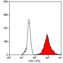
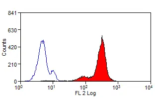
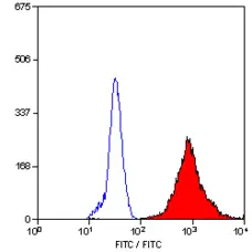
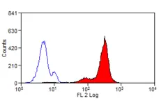
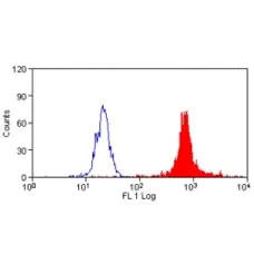
| Flow Cytometry 1 |  |
1/50 | 1/100 |
| Immunohistology - Frozen | |
1/100 | 1/200 |
| Immunohistology - Paraffin 2 | |
1/100 | 1/200 |
- 1 Membrane permeabilization is required for this application. The use of Leucoperm (Product Code BUF09) is recommended for this purpose.
- 2This product requires protein digestion pre-treatment of paraffin sections e.g. trypsin or pronase.
Where this antibody has not been tested for use in a particular technique this does not necessarily exclude its use in such procedures. Suggested working dilutions are given as a guide only. It is recommended that the user titrates the antibody for use in their own system using appropriate negative/positive controls.
- Flow Cytometry
- Use 10ul of the suggested working dilution to label 1x10⁶ cells in 100ul.
- Histology Positive Control Tissue
- Human Spleen
| Description | Product Code | Applications | Pack Size | List Price | Your Price | Quantity | |
|---|---|---|---|---|---|---|---|
| Mouse IgG1 Negative Control | MCA928 | F | 100 Tests |

|
Log in | ||
| List Price | Your Price | ||||||
|
|
Log in | ||||||
| Description | Mouse IgG1 Negative Control | ||||||
Source Reference
-
Flavell, D.J. et al. (1987) Identification of tissue histiocytes on paraffin sections by a new monoclonal antibody.
J Histochem Cytochem. 35 (11): 1217-26.
Antibody Characterization Reference
-
Brandtzaeg, P. et al. (1988) Mac 387 antibody and detection of formalin resistant myelomonocytic L1 antigen.
J Clin Pathol. 41 (9): 963-70.
References for Macrophages/Monocytes/Granulocytes antibody
-
Burudi, E.M. et al. (2002) Regulation of indoleamine 2,3-dioxygenase expression in simian immunodeficiency virus-infected monkey brains.
J Virol. 76: 12233-41. -
Ueland, T. et al. (2009) Dickkopf-1 enhances inflammatory interaction between platelets and endothelial cells and shows increased expression in atherosclerosis.
Arterioscler Thromb Vasc Biol. 29: 1228-34 -
Brandtzaeg, P. et al. (1992) The leucocyte protein L1 (calprotectin): usefulness as an immunohistochemical marker antigen and putative biological function.
Histopathology. 21: 191-6. -
Gutierrez, M. et al. (1999) The detection of CD2+, CD4+, CD8+, and WC1+ T lymphocytes, B cells and macrophages in fixed and paraffin embedded bovine tissue using a range of antigen recovery and signal amplification techniques.
Vet Immunol Immunopathol. 71 (3-4): 321-34. -
Ramsay, A.D. et al. (1991) Phenotypic analysis of malignant lymphoma in simian immunodeficiency virus infection using anti-human antibodies.
J Pathol. 164 (4): 321-8. -
Christgau, M. et al. (1998) Characterization of immunocompetent cells in the diseased canine periodontium.
J Histochem Cytochem. 46 (12): 1443-54. -
Pérez, J. et al. (1999) Immunohistochemical study of the inflammatory infiltrate associated with equine squamous cell carcinoma.
J Comp Pathol. 121 (4): 385-97. -
Nanney, L.B. et al. (2008) Calreticulin enhances porcine wound repair by diverse biological effects.
Am J Pathol. 173: 610-30.
View The Latest Product References
-
Poncelet, L. et al. (2008) Detection of antigenic heterogeneity in feline coronavirus nucleocapsid in feline pyogranulomatous meningoencephalitis.
Vet Pathol. 45: 140-53. -
Sethi, R.S. et al. (2010) Immunolocalization of pulmonary intravascular macrophages, TLR4, TLR9 and IL-8 in normal and Pasteurella multocida-infected lungs of water buffalo (Bubalus bubalis).
J Comp Pathol. 144: 135-44. -
Sanchez, J. et al. (2011) Microscopical and immunological features of tuberculoid granulomata and cavitary pulmonary tuberculosis in naturally infected goats.
J Comp Pathol. 145 (2-3): 107-17. -
Isling, L.K. et al. (2010) Pyelonephritis in slaughter pigs and sows: morphological characterization and aspects of pathogenesis and aetiology.
Acta Vet Scand. 52: 48. -
Vranckx, K. et al. (2012) Vaccination reduces macrophage infiltration in bronchus-associated lymphoid tissue in pigs infected with a highly virulent Mycoplasma hyopneumoniae strain.
BMC Vet Res. 8: 24. -
Campuzano, O. et al. (2012) Arrhythmogenic right ventricular cardiomyopathy: severe structural alterations are associated with inflammation.
J Clin Pathol. 65 (12): 1077-83. -
García-Jiménez, W.L. (2012) Histological and immunohistochemical characterisation of Mycobacterium bovis induced granulomas in naturally infected fallow deer (Dama dama).
Vet Immunol Immunopathol. 149: 66-75. -
Santana, C.H. et al. (2016) Relationship between the inflammatory infiltrate and the degree of differentiation of the canine cutaneous squamous cell carcinoma.
Vet Anim Sci. 1-2: 4-8. -
Masure, D. et al. (2013) A Role for Eosinophils in the Intestinal Immunity against Infective Ascaris suum Larvae.
PLoS Negl Trop Dis. 2013 Mar;7(3): e2138. -
Tellez, A. et al. (2014) Experimental evaluation of efficacy and healing response of everolimus-eluting stents in the familial hypercholesterolemic swine model: a comparative study of bioabsorbable versus durable polymer stent platforms.
Coron Artery Dis. 25 (3): 198-207. -
Collin, N. et al. (2009) Sand fly salivary proteins induce strong cellular immunity in a natural reservoir of visceral leishmaniasis with adverse consequences for Leishmania.
PLoS Pathog. 5(5):e1000441. -
McCurdy, P. et al. (2014) Acute lymphoblastic leukemia in a pygmy hippopotamus (Hexaprotodon liberiensis).
J Zoo Wildl Med. 45 (4): 906-10. -
Marcaccini, A. et al. (2008) Pseudorabies virus infection in mink: a host-specific pathogenesis.
Vet Immunol Immunopathol. 124 (3-4): 264-73. -
Romero-Palomo, F. et al. (2017) Immunopathologic Changes in the Thymus of Calves Pre-infected with BVDV and Challenged with BHV-1.
Transbound Emerg Dis. 64 (2): 574-84. -
Rossi, C.N. et al. (2016) In situ Cutaneous cellular immune response in dogs naturally infected by visceral leishmaniasis.
Rev Inst Med Trop Sao Paulo. 58: . -
Vrolyk, V. et al. (2017) Lung Inflammation Associated With Clinical Acute Necrotizing Pancreatitis in Dogs.
Vet Pathol. 54 (1): 129-40. -
Nelson, M. et al. (2014) Comparative experimental subcutaneous glanders and melioidosis in the common marmoset (Callithrix jacchus).
Int J Exp Pathol. 95 (6): 378-91. -
Amarilla, S.P. et al. (2016) Thymic depletion of lymphocytes is associated with the virulence of PRRSV-1 strains.
Vet Microbiol. 188: 47-58. -
García-Jiménez, W.L. et al. (2013) Immunopathology of granulomas produced by Mycobacterium bovis in naturally infected wild boar.
Vet Immunol Immunopathol. 156 (1-2): 54-63. -
Zhao, L. et al. (2020) Reducing macrophage numbers alleviates temporomandibular joint ankylosis.
Cell Tissue Res. 379 (3): 521-36. -
Lai, H.Y. et al. (2017) CCAAT/enhancer-binding protein delta promotes intracellular lipid accumulation in M1 macrophages of vascular lesions.
Cardiovasc Res. 113 (11): 1376-88. -
Wacinski, P. et al. (2021) Anti-Inflammatory Effect of Very High Dose Local Vessel Wall Statin Administration: Poly(L,L-Lactide) Biodegradable Microspheres with Simvastatin for Drug Delivery System (DDS).
Int J Mol Sci. 22 (14): 7486. -
Edwards, J.H. et al. (2021) Integration and functional performance of a decellularised porcine superflexor tendon graft in an ovine model of anterior cruciate ligament reconstruction.
Biomaterials. 279: 121204. -
Bertolo, P.H.L. et al. (2022) Influence of serum progesterone levels on the inflammatory response of female dogs with visceral leishmaniosis.
Vet Parasitol. 302: 109658. -
do Prado Duzanski, A. et al. (2022) Cell-mediated immunity and expression of MHC class I and class II molecules in dogs naturally infected by canine transmissible venereal tumor: Is there complete spontaneous regression outside the experimental CTVT?
Research in Veterinary Science. 145: 193-204. -
Vafaee, T. et al. (2022) Repopulation of decellularised porcine pulmonary valves in the right ventricular outflow tract of sheep: Role of macrophages.
J Tissue Eng. 13: 20417314221102680. -
Roux, H.M. et al. (2023) DNA ultra-sensitive quantification, a technology for studying HIV unintegrated linear DNA.
Cell Rep Methods. 3 (4): 100443. -
Agerholm, J.S. et al. (2023) Actinobacillus lignieresii‐associated myocellulitis of the nasal planum in a Jersey cow
Veterinary Record Case Reports. 11 (4) [Epub ahead of print]. -
Blirup-Plum, S.A. et al. (2023) Gastro-intestinal lesions are not relatable to diarrhoea or specific pathogens in post-weaning diarrhoea (PWD) in pigs.
Acta Vet Scand. 65 (1): 30. -
Anderson, S.L. et al. (2021) Depletion of pulmonary intravascular macrophages rescues inflammation-induced delayed neutrophil apoptosis in horses.
Am J Physiol Lung Cell Mol Physiol. 320 (1): L126-L136. -
Chen, L.Y. et al. (2024) Anti-oxidative and anti-inflammatory effects of Ginkgo biloba extract (EGb761) on hindlimb skeletal muscle ischemia-reperfusion injury in rats.
Physiol Rep. 12 (11): e16050. -
Hong, S. et al. (2024) Impact of an Injectable Trace Mineral Supplement on the Immune Response and Outcome of Mannheimia haemolytica Infection in Feedlot Cattle.
Biol Trace Elem Res. Jun 10 [Epub ahead of print]. -
Anderson, S.L. et al. (2021) Depletion of pulmonary intravascular macrophages rescues inflammation-induced delayed neutrophil apoptosis in horses.
Am J Physiol Lung Cell Mol Physiol. 320 (1): L126-L136. -
Rodrigues, A. et al. (2024) Kupffer Cells and Hepatocytes: A Key Relation in the Context of Canine Leishmaniasis
Microorganisms 12 (9) 1887. -
Rogato, F. et al. (2024) Leukemia cutis as a prominent clinical sign in a dog with acute myeloid leukemia.
Vet Clin Pathol. 53 (4): 448-57. -
Ireland, R.E. et al. (2025) Inhaled liposomal ciprofloxacin protects against lethal tularemia in the common marmoset.
Antimicrob Agents Chemother. : e0123225. -
Mushati, K.A. et al. (2026) Modulation of uterine responses in dogs: The divergent effects of semen and embryonal signals on steroidogenic receptors and selected immune system-related factors.
Theriogenology. 250: 117644.
Further Reading
-
Burk, J. et al. (2013) Equine cellular therapy--from stall to bench to bedside?
Cytometry A. 83 (1): 103-13. -
Piriou-Guzylack, L. (2008) Membrane markers of the immune cells in swine: an update.
Vet Res. 39: 54.
- Synonyms
- Calprotectin
- RRID
- AB_321963
- UniProt
- P06702
- Entrez Gene
- S100A9
- GO Terms
- GO:0005886 plasma membrane
- GO:0005515 protein binding
- GO:0005509 calcium ion binding
- GO:0005576 extracellular region
- GO:0004871 signal transducer activity
- GO:0005634 nucleus
- GO:0005737 cytoplasm
- GO:0005856 cytoskeleton
- GO:0006935 chemotaxis
- View More GO Terms
- GO:0006954 inflammatory response
- GO:0007267 cell-cell signaling
Request a different product with this specificity
Please Note: All Products are "FOR RESEARCH PURPOSES ONLY"
View all Anti-Human ProductsAlways be the first to know.
When we launch new products and resources to help you achieve more in the lab.
Yes, sign me up