Insulin Receptor Signaling Pathway
- On This Page
- Overview
- Interactive poster
- Validated PrecisionAb Antibodies
- Insulin receptor signaling pathway antibody range
- References
s

Insulin Receptor Signaling Overview
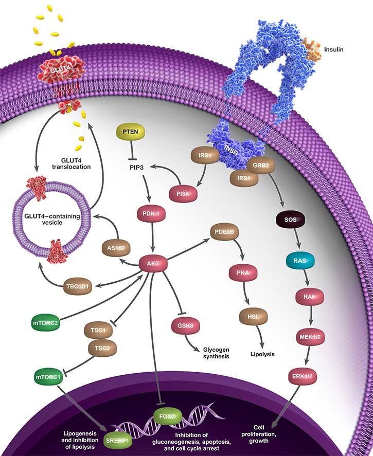
Insulin is a key hormone that regulates human metabolism. It binds to and activates the insulin receptor (IR). IR phosphorylates and recruits adaptor proteins including the IRS family. The p85 subunit of phosphoinositide 3-kinase (PI3K), bound to phosphorylated IRS, activates the catalytic subunit p110. Phosphorylation of PI3K phosphatidylinositol-4, 5-bisphophaste (PIP2) by PI3K to phosphatidylinositol-3, 4, 5-trisphophaste (PIP3), activates 3-phosphoinositide dependent protein kinase-1 (PDK1). PDK1 phosphorylates AKT on Thr308. Then, the mammalian target of rapamycin complex (mTORC2) fully activates AKT through phosphorylation on Ser473 (Poloz and Stambolic 2015). AKT phosphorylates the hamartin (TSC1) and tuberin (TSC2) complex that regulates the activity of mammalian target of rapamycin complex 1 (mTORC1). mTORC1 phosphorylates the sterol regulatory element-binding protein 1 (SREBP1) inducing lipogenesis and repressing lipolysis (Portstmann et al. 2008).
Insulin activated AKT phosphorylates GTPase AS160 (Sano et al. 2003) and TBC1D1 (TBC domain family, member 1) (Peck et al. 2009), which is required for the glucose transporter GLUT4 translocation. AKT also phosphorylates the forkhead box O transcription factor (FOXO). The inhibitory phosphorylation includes the phosphorylation on Thr32, Ser256, and Ser318 and leads to the nuclear exclusion and degradation of FOXO in the cytoplasm (Arden 2004). Phosphorylation by AKT of the glycogen synthase kinase 3 (GSK3) results in glycogen synthesis (Cohen and Frame 2001).
Insulin receptor pathway signaling is crucial for the regulation of glucose homeostastis and research has been focused around antidiabetic medicines. Interestingly, metformin, the most often given medicine for type 2 diabetes, has been linked to a lower risk of a variety of malignancies, particularly pancreatic and colon cancers (Kasznicki et al. 2014).
Interactive Insulin Receptor Signaling Poster
We have compiled key proteins involved in the insulin receptor signaling pathway into one handy poster you can download for your reference.
Click the pathway components within the images below to discover the key antibodies that Bio-Rad provides.
Validated PrecisionAb Antibodies to Study the Insulin Receptor Signaling
Bio-Rad offers validated antibodies to study the insulin receptor signaling pathway. The featured product VMA00900 shows the quality of these antibodies.
Validated Rabbit AKT pSer473 Antibody (VMA00900)
Rabbit AKT pSer473 Antibody (VMA00900) detects the phosphorylated AKT protein at pSer473. There is an increase in the signal intensity after stimulation with calculin A and a reduction after lambda phosphatase treatment. The phospho-specific antibody does not detect the unphosphorylated AKT. In addition, there is a significant reduction (≥90%) in signal intensity in both simulated and unstimulated lysates following phosphatase treatment.
Applications: Western Blotting, Immunohistology
 Antibody (1/1,000, VMA00900).")
Fig. 1. Western blot analysis of whole cell lysates probed with A, Rabbit Anti-AKT Antibody or B, Rabbit Anti-AKT (pSer473) Antibody (1/1,000, VMA00900). It is followed by detection with HRP Conjugated Goat Anti-Rabbit (1/10,000, 170-5046). Membranes were treated with (+) and without (-) lambda protein phosphatase as indicated and visualized on the ChemiDoc MP Imaging System.
Bio-Rad’s Insulin Receptor Signaling Pathway Antibody Range
Browse Bio-Rad’s range of antibodies against targets involved with the insulin receptor pathway. Please use the filters to sort the attributes in the table below to find the antibody that meets your needs. If you need any further assistance, please do not hesitate to contact us.
Antibodies for Insulin Receptor Signaling
| Description | Target | Format | Clone | Applications | Citations | Code |
|---|
References
- Arden K (2004). FoxO: linking new signaling pathways. Mol Cell 14, 416–418.
- Cohen P and Frame S (2001). The renaissance of GSK3. Nat Rev Mol Cell Biol 2, 769–776.
- Kasznicki J (2014). Metformin in cancer prevention and therapy. Ann Transl Med 2, 57.
- Peck G et al. (2009). Insulin-stimulated phosphorylation of the Rab GTPase-activating protein TBC1D1 regulates GLUT4 translocation. J Biol Chem 284, 30,016–30,023.
- Poloz Y and Stambolic V (2015). Obesity and cancer, a case for insulin signaling. Cell Death & Disease. Accessed August 19, 2021.
- Portstmann T et al. (2008). SREBP activity is regulated by mTORC1 and contributes to Akt-dependent cell growth. Cell Metab 8, 224–236.
- Sano H et al. (2003). Insulin-stimulated phosphorylation of a Rab GTPase-activating protein regulates GLUT4 translocation. J Biol Chem 278, 14,599–14,602.