MHC Class II RT1B antibody | OX-6
Mouse anti Rat MHC Class II RT1B
- Product Type
- Monoclonal Antibody
- Clone
- OX-6
- Isotype
- IgG1
- Specificity
- MHC Class II RT1B


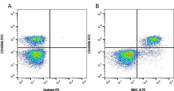
Figure B. FITC conjugated Mouse anti Rat CD45RA antibody, clone OX-33 (MCA340F*) and RPE conjugated Mouse anti Rat MHC-II antibody, clone OX-6 (MCA46PE). All experiments performed on red cell lysed rat blood gated on lymphoid cells in the presence of 10% rat serum.
Data acquired on the ZE5 Cell analyser.
*N.B. FITC format is no longer available. FITC conjugated Mouse anti Rat CD45RA antibody, clone OX-33 is available in both purified and RPE conjugated formats.
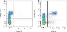
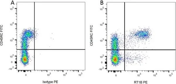
Figure B. FITC conjugated Mouse anti Rat CD45RC antibody, clone OX-22 (MCA53FT) and RPE conjugated Mouse anti Rat RT1B antibody, clone OX-6 (MCA46PE). All experiments performed on red cell lysed rat blood gated on mononuclear cells.
Data acquired on the ZE5 Cell analyser.
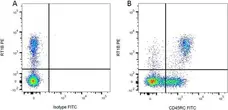
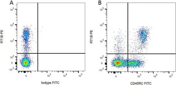
Figure B. RPE conjugated Mouse anti Rat RT1B antibody, clone OX-6 (MCA46PE) and FITC conjugated Mouse anti Rat CD45RC antibody, clone OX-22 (MCA53FT). All experiments performed on red cell lysed rat blood gated on mononuclear cells.
Data acquired on the ZE5 Cell analyser.

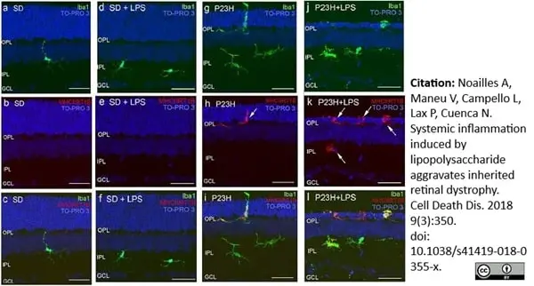
Mouse anti Rat MHC Class II RT1B, clone OX-6 (MCA46R) used for the identification of MHC II expressing microglia in rat retinal sections by immunofluorescence.
Image caption:
Effect of LPS on retinal microglia in SD and P23H rats.
Representative vertical retinal sections immunostained with antibodies against Iba1 (green) and MHC-II (red) in vehicle- and LPS-injected SD rats a–f, or vehicle- and LPS-injected P23H rats g–l. Arrows point microglia MHC-II-immunopositive cells. Nuclei were stained with TO-PRO (blue). All images were taken in the central retina. GCL ganglion cell layer, IPL inner plexiform layer, OPL outer plexiform layer. Scale bar 40μm
From: Noailles A, Maneu V, Campello L, Lax P, Cuenca N.
Systemic inflammation induced by lipopolysaccharide aggravates inherited retinal dystrophy.
Cell Death Dis. 2018 Mar 2;9(3):350.
This image is from an open access article distributed under terms of a Creative Commons Attribution License.
Mouse anti Rat MHC Class II RT1B antibody, clone OX-6 (MCA46G) used for the demonstration of MHC class II expressing cells by immunofluorescence.
Image caption:
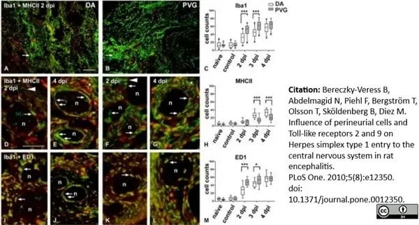
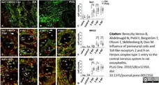
Delayed recruitment of phagocytic cells and increased MHC II activation in the epineurium of DA rats.
Immunofluorescence micrographs illustrating sections of the whiskers area at 2 dpi (A, B, D, F, I and K) and 4 dpi (E, G, J and L). The DA (A, D, E, I and J) and the PVG (B, F, G, K and L) sections were stained with macrophage marker Iba1 (green, A, B, D–G and I–L), MHC II marker (red, A, B and D–G) to show the antigen presenting properties and phago-lysosome marker ED1 (red, I–L). Cellular quantification is represented in C, H and M panels. (A–C) At 2 dpi massive recruitment of Iba1+ cells (green) was present in the whiskers area of both DA (A) and PVG (B) rats at the site of the infection. However, the quantification of infiltrating Iba1+ cells to the epineurium (C) showed that the recruitment of macrophages was delayed and reduced in the epineurium of DA (arrow in A) compared to PVG rats (arrow in B). (D–H) At 2 dpi (D, F, H) the activation of antigen presenting cells detected by MHC II (red) was similar in both strains, but at 3 dpi (H) and 4 dpi (E and H) MHC II increased in DA compared to PVG rats (G and H). At 4 dpi in the whiskers area of DA rats (E) Iba1+/MHC II+ cells (arrows in E) were seen in the perineurium, in a pattern similar to the HSV-1 staining seen in Figure 1. In PVG rats (G), double and single stained Iba1+ and MHC II+ cells were more scattered in the vicinity of nerve fascicles (arrows in G), not delineating the perineurium in the same way as in DA. (I–M) Most Iba1+ cells were also ED1+ in both DA (I and J) and in PVG rats (K and L) indicating their phagocytic activity (arrows). However, there were more ED1+ cells in PVG at 2 dpi and 3 dpi compared to DA (M). At 4 dpi (J and L), Iba1+/ED1+ cells clearly delineated the perineurial cell layer in DA (arrows in J), but not in PVG (arrow in L), where these cells remained in the epineurium. Asterisks (E and G) point to the cellular infiltration into the epineurium; small arrows indicate cells in the perineurium; arrowheads indicate differences in Iba1+ morphology in DA compared to PVG rats; n, nerve fascicles; dots in panel C represent outliers. Scale bar: 50 μm.
From: Bereczky-Veress B, Abdelmagid N, Piehl F, Bergström T, Olsson T, Sköldenberg B, et al. (2010)
Influence of Perineurial Cells and Toll-Like Receptors 2 and 9 on Herpes simplex Type 1 Entry to the Central Nervous System in Rat Encephalitis.
PLoS ONE 5(8): e12350.
This image is from an open access article distributed under terms of a Creative Commons Attribution License.
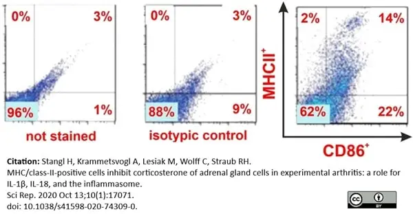
RPE conjugated Mouse anti Rat MHC Class II RT1B antibody, clone OX-6 (MCA46PE) used for the evaluation of MHC Class II expression on rat adrenal cells by flow cytometry.
Image caption:
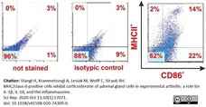
Intraadrenal MHCII+/CD86+ cells during collagen type II induced arthritis (CIA).
Quantification of MHCII and CD86 positive adrenal gland cells (exemplary FACS staining).
From: Stangl H, Krammetsvogl A, Lesiak M, Wolff C, Straub RH.
MHC/class-II-positive cells inhibit corticosterone of adrenal gland cells in experimental arthritis: a role for IL-1β, IL-18, and the inflammasome.
Sci Rep. 2020 Oct 13;10(1):17071.
doi: 10.1038/s41598-020-74309-0.
This image is from an open access article distributed under terms of a Creative Commons Attribution License.
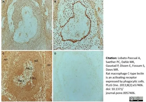
Mouse anti Rat MHC Class II RT1B antibody, clone OX-6 (MCA46G) used for the visualization of MHC class II expressing cells in rat spleen by immunohistochemistry on cryosections.
Image caption:
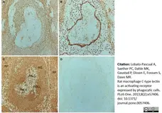
Immunostaining of rat spleen.
Serial-cut frozen sections were stained with mAbs towards A, rat MCL B, rat CD169 C, rat MHC class II D, human MHC class I (negative control) and visualized with peroxidase-conjugated secondary antibody and DAB substrate. RP: red pulp. PALS: periarteriolar lymphoid sheath. FOLL: follicle. MZ: marginal zone.
From: Lobato-Pascual A, Saether PC, Dahle MK, Gaustad P, Dissen E, Fossum S, et al. (2013)
Rat Macrophage C-Type Lectin Is an Activating Receptor Expressed by Phagocytic Cells.
PLoS ONE 8(2): e57406.
This image is from an open access article distributed under terms of a Creative Commons Attribution License.
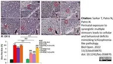
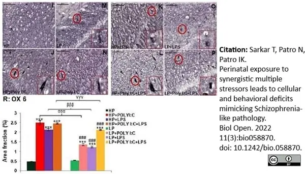
Mouse anti Rat MHC Class II RT1B antibody, clone OX-6 (MCA46G) used to label activated microglia in mouse brain by immunohistochemistry on formalin fixed cryosections.
Image caption:
OX 6 labeled immunohistochemical images and stacked graph showing stress related changes in microglial population: From the OX 6 labeled images it is evident that both single and combined exposure of Poly I:C and LPS to HP and LP animals hyped MHC II expression in the hippocampus of the stressed animals when compared to HP control (I) and LP alone groups (M). HP+Poly I:C (J), HP+LPS (K) and HP+Poly I:C+LPS groups (L) were seen to contain intensely labeled OX 6 positive cells around the CA layer, which was comparatively less prominent in LP groups (N,O P), (n=6 slides from different animals/group, scale bar=100μm). From the quantification data (R) it was seen that, MHC II expression increased in both HP and LP animals, consequent upon Poly I:C and LPS treatment. LP groups however showed comparatively less MHC II expression when compared to similarly treated HP groups. (n=108 images, 6 slides from different animals/group), values are expressed as mean±SEM; ***P≤0.001 with respect to controls; ###P≤0.001 with respect to LP alone group; αααP≤0.001 with respect to HP+Poly I:C and LP+Poly I:C; βββP≤0.05 with respect to HP+LPS and LP+LPS; γγγP≤0.001 with respect to HP+Poly I:C+LPS and LP+Poly I:C+LPS.
From: Tiyasha Sarkar, Nisha Patro, Ishan Kumar Patro;
Perinatal exposure to synergistic multiple stressors lead to cellular and behavioral deficits mimicking Schizophrenia like pathology.
Biol Open 2022; bio.058870
doi:10.1242/bio.058870
This image is from an open access article distributed under terms of a Creative Commons Attribution License.

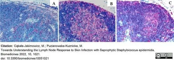
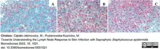
Mouse anti Rat MHC Class II RT1B antibody, clone OX-6 (MCA46R) used to label MHC Class II expressing cells in rat lymph nodes by immunohistochemistry on cryostat sections.
Image caption:
The rat popliteal lymph node with the visible follicle stained for the presence of activated antigen-presenting cells (MHC class II+, red). (A) The node of the control animal receiving 0.9% NaCl (days 1–7, node evaluation on day 8). (B) The node after initial massive S. epidermidis infection (days 1–7) with early node evaluation (day 8). (C) The node after secondary S. epidermidis infection (day 28, node evaluation on day 29). Magnification 200×.
From: Cąkała-Jakimowicz, M.; Puzianowska-Kuznicka, M.
Towards Understanding the Lymph Node Response to Skin Infection with Saprophytic Staphylococcus epidermidis.
Biomedicines 2022, 10, 1021.
doi: 10.3390/biomedicines10051021
This image is from an open access article distributed under terms of a Creative Commons Attribution License.
Mouse anti Rat MHC Class II RT1B antibody, clone OX-6 (MCA46R) used to label MHC Class II expressing cells in rat lymph nodes by immunohistochemistry on cryostat sections.
Image caption:
Paracortex of the rat popliteal lymph node stained for the presence of activated antigen-presenting cells (MHC class II+, red). (A) The node of the control animal receiving 0.9% NaCl (days 1–7, node evaluation on day 8). (B) The node after initial massive S. epidermidis infection (days 1–7) with early node evaluation (day 8). (C) The node after secondary. S. epidermidis infection (day 28, node evaluation on day 29). Magnification 200×.
From: Cąkała-Jakimowicz, M.; Puzianowska-Kuznicka, M.
Towards Understanding the Lymph Node Response to Skin Infection with Saprophytic Staphylococcus epidermidis.
Biomedicines 2022, 10, 1021.
doi: 10.3390/biomedicines10051021
This image is from an open access article distributed under terms of a Creative Commons Attribution License.
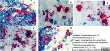
Mouse anti Rat MHC Class II RT1B antibody, clone OX-6 (MCA46R) used to label MHC Class II expressing cells in rat lymph nodes by immunohistochemistry on cryostat sections.
Image caption:
The medulla of the rat popliteal lymph node in initial massive S. epidermidis infection (days 1–7) with early node’s evaluation (day 8). The node stained for: (A) Dendritic cells (OX62+, red). (B) Macrophages and monocytes (CD68+, red). (C) Stem cells, thymocytes and immature B cells (CD90+, red). (D) T helper lymphocytes and monocytes (CD4+, red). (E) Activated antigen-presenting cells (MHC class II+, red). Magnification 1000×.
From: Cąkała-Jakimowicz, M.; Puzianowska-Kuznicka, M.
Towards Understanding the Lymph Node Response to Skin Infection with Saprophytic Staphylococcus epidermidis.
Biomedicines 2022, 10, 1021.
doi: 10.3390/biomedicines10051021
This image is from an open access article distributed under terms of a Creative Commons Attribution License.
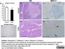
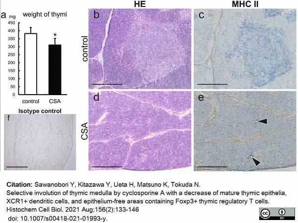
Mouse anti Rat MHC Class II RT1B antibody, clone OX-6 (MCA46G) used to demonstrate MHC Class II expression in rat thymic tissue by immunohistochemistry on cryosections.
Image caption:
Histological overview of CSA-treated thymi. a Weight of thymi. b–f Sections of freshly frozen thymi from control (b, c, f) and CSA (d, e) rats were H&E (b, d) and immunohistologically stained with an anti-MHCII (c, e) or polyclonal mouse IgG isotype control (f) antibodies followed by alkaline phosphatase-conjugated anti-mouse IgG antibody.
From: Sawanobori Y, Kitazawa Y, Ueta H, Matsuno K, Tokuda N.
Selective involution of thymic medulla by cyclosporine A with a decrease of mature thymic epithelia, XCR1+ dendritic cells, and epithelium-free areas containing Foxp3+ thymic regulatory T cells.
Histochem Cell Biol. 2021 Aug;156(2):133-146.
doi: 10.1007/s00418-021-01993-y.
This image is from an open access article distributed under terms of a Creative Commons Attribution License.
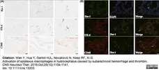
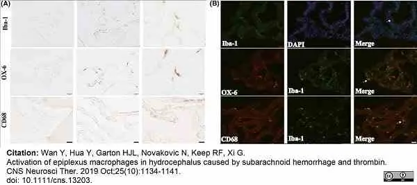
Mouse anti Rat MHC Class II RT1B antibody, clone OX-6 (MCA46R) used to identify MHC Class II expressing cells by immunohistology on formalin fixed cryosections and by immunofluorescence on formalin fixed, paraffin embeddedtissue sections.
Image caption:
A, Iba-1, OX-6, and CD68 immunoreactivity in epiplexus cells of rats at 24 h after sham operation. Scale bar =100 μm in first column, 20 μm in second column, and 10 μm for third column. B, Immunofluorescence staining of Iba-1 and the double labeling of Iba-1 with OX-6 or CD68 in epiplexus cells at 24 h in sham-operated rats. Scale bar =20 μm
From: Wan Y, Hua Y, Garton HJL, Novakovic N, Keep RF, Xi G.
Activation of epiplexus macrophages in hydrocephalus caused by subarachnoid hemorrhage and thrombin.
CNS Neurosci Ther. 2019 Oct;25(10):1134-1141.
doi: 10.1111/cns.13203..
This image is from an open access article distributed under terms of a Creative Commons Attribution License.
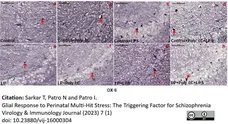
Mouse anti Rat MHC Class II RT1B antibody, clone OX-6 (MCA46G) used to assess MHC class II expression in rat brain by immunohistochemistry on cryostat sections.
Image caption:
The representative images of the CA region at PND 30 showing increase in OX 6 positive cells following stress exposure, depicting an increase in MHC II protein expression.
From: Sarkar T, Patro N and Patro I.
Glial Response to Perinatal Multi-Hit Stress: The Triggering Factor for Schizophrenia.
Virol Immunol J (2023) 7 (1): 1-21.
doi: 10.23880/vij-16000304.
This image is from an open access article distributed under terms of a Creative Commons Attribution License.
Filter by Application:
F IF C Reset| Mouse anti Rat MHC Class II RT1B antibody, clone OX-6 recognizes a monomorphic determinant of the rat RT1B MHC class II antigen present on B lymphocytes, dendritic cells, some macrophages and certain epithelial cells. Rat MHC Class II RT1B antibody, clone OX-6 does not react with the rat BDIX strain due to a defect in RT1B expression (Male et al. 1987). The major histocompatibility complex (MHC) is a cluster of genes that are important in the immune response to infections. In rats, this complex is referred to as the RT1 region. In mice, this complex is referred to as the H-2 region. Mouse anti Rat MHC Class II RT1B antibody, clone OX-6 also cross reacts with a polymorphic determinant on mouse strains of the H-2 haplotypes k and s. Analysis of recombinant mouse strains has mapped the OX-6 determinant to the H-2I-A region (McMaster and Williams 1979 and Male et al. 1987). Mouse anti Rat MHC Class II RT1B antibody, clone OX-6 is routinely tested in flow cytometry on rat splenocytes. |
- Target Species
- Rat
- Species Cross-Reactivity
-
Target Species Cross Reactivity Mouse - N.B. Antibody reactivity and working conditions may vary between species.
- Product Form
- Purified IgG - liquid
- Preparation
- Purified IgG prepared by affinity chromatography on Protein A from tissue culture supernatant
- Buffer Solution
- Phosphate buffered saline
- Preservative Stabilisers
0.09% Sodium Azide - Carrier Free
- Yes
- Immunogen
- Rat thymocyte membrane glycoproteins.
- Approx. Protein Concentrations
- IgG concentration 1.0 mg/ml
- Fusion Partners
- Spleen cells from immunized BALB/c mice were fused with cells from the NS1 mouse myeloma cell line.
- Regulatory
- For research purposes only
- Guarantee
- 12 months from date of despatch
This product is shipped at ambient temperature. It is recommended to aliquot and store at -20°C on receipt. When thawed, aliquot the sample as needed. Keep aliquots at 2-8°C for short term use (up to 4 weeks) and store the remaining aliquots at -20°C.
Avoid repeated freezing and thawing as this may denature the antibody. Storage in frost-free freezers is not recommended.
Avoid repeated freezing and thawing as this may denature the antibody. Storage in frost-free freezers is not recommended.
This product has been reported to work in the following applications. This information is derived from testing within our laboratories, peer-reviewed publications or personal communications from the originators. Please refer to references indicated for further information. For general protocol recommendations, please visit the antibody protocols page.
| Application Name | Verified | Min Dilution | Max Dilution |
|---|---|---|---|
| Flow Cytometry |  |
1/50 | 1/100 |
| Immunofluorescence | |
||
| Immunohistology - Frozen | |
||
| Immunohistology - Paraffin 1 | |
1/50 | 1/100 |
- 1This product requires antigen retrieval using heat treatment prior to staining of paraffin sections.Sodium citrate buffer pH 6.0 is recommended for this purpose. PLP fixation is recommended for optimal results.
Where this antibody has not been tested for use in a particular technique this does not necessarily exclude its use in such procedures. Suggested working dilutions are given as a guide only. It is recommended that the user titrates the antibody for use in their own system using appropriate negative/positive controls.
- Flow Cytometry
- Use 10ul of the suggested working dilution to label 106 cells in 100ul.
| Description | Product Code | Applications | Pack Size | List Price | Your Price | Quantity | |
|---|---|---|---|---|---|---|---|
| Mouse IgG1 Negative Control | MCA1209 | F | 0.1 mg |

|
Log in | ||
| List Price | Your Price | ||||||
|
|
Log in | ||||||
| Description | Mouse IgG1 Negative Control | ||||||
Source Reference
-
McMaster, W.R. & Williams, A.F. (1979) Identification of Ia glycoproteins in rat thymus and purification from rat spleen.
Eur J Immunol. 9 (6): 426-33.
References for MHC Class II RT1B antibody
-
Fernandez, J.L. & Weeks, M. (1986) Genetic monitoring of inbred strains of mice using monoclonal antibodies to major histocompatibility haplotypes and lymphocyte alloantigens.
Lab Anim. 20 (4): 293-7. -
Male, D.K. et al. (1987) Serological evidence for a defect in RT1.B (I-A) expression by the BDIX rat strain.
J Immunogenet. 14 (6): 301-12. -
Charteris, D.G. & Lightman, S.L. (1993) In vivo lymphokine production in experimental autoimmune uveoretinitis.
Immunology. 78 (3): 387-92. -
Whiteland, J.L. et al. (1995) Immunohistochemical detection of T-cell subsets and other leukocytes in paraffin-embedded rat and mouse tissues with monoclonal antibodies.
J Histochem Cytochem. 43 (3): 313-20. -
McKechnie, N.M. et al. (1997) Immunization with the cross-reactive antigens Ov39 from Onchocerca volvulus and hr44 from human retinal tissue induces ocular pathology and activates retinal microglia.
J Infect Dis. 176 (5): 1334-43. -
Burrows, G.G. et al. (1998) Two-domain MHC class II molecules form stable complexes with myelin basic protein 69-89 peptide that detect and inhibit rat encephalitogenic T cells and treat experimental autoimmune encephalomyelitis.
J Immunol. 161 (11): 5987-96. -
Hofmann, N. et al. (2002) Increased expression of ICAM-1, VCAM-1, MCP-1, and MIP-1 alpha by spinal perivascular macrophages during experimental allergic encephalomyelitis in rats.
BMC Immunol. 3:11. -
Banerjee, S. et al. (2003) Development of organised conjunctival leucocyte aggregates after corneal transplantation in rats.
Br J Ophthalmol. 87: 1515-22.
View The Latest Product References
-
Bode, U. et al. (2008) Dendritic cell subsets in lymph nodes are characterized by the specific draining area and influence the phenotype and fate of primed T cells.
Immunology. 123 (4): 480-90. -
King, G.D. et al. (2008) Flt3L in combination with HSV1-TK-mediated gene therapy reverses brain tumor-induced behavioral deficits.
Mol Ther. 16: 682-90. -
Wang, Q. et al. (2009) Pyruvate protects against experimental stroke via an anti-inflammatory mechanism.
Neurobiol Dis. 2009 Oct;36(1):223-31. -
Zilka, N. et al. (2009) Human misfolded truncated tau protein promotes activation of microglia and leukocyte infiltration in the transgenic rat model of tauopathy.
J Neuroimmunol. 209 (1-2): 16-25. -
Baca Jones, C.C. et al. (2009) Rat cytomegalovirus infection depletes MHC II in bone marrow derived dendritic cells.
Virology. 388: 78-90. -
Bereczky-Veress, B. et al. (2010) Influence of perineurial cells and Toll-like receptors 2 and 9 on Herpes simplex type 1 entry to the central nervous system in rat encephalitis.
PLoS One. 5(8): e12350. -
Kawamura, J. et al. (2010) Neuron-immune Interactions in the Sensitized Thalamus Induced by Mustard Oil Application to Rat Molar Pulp.
J Dent Res. 89: 1309-14. -
Calvo, M. et al. (2010) Neuregulin-ErbB signaling promotes microglial proliferation and chemotaxis contributing to microgliosis and pain after peripheral nerve injury.
J Neurosci. 30 (15): 5437-50. -
McClain, J.A. et al. (2011) Adolescent binge alcohol exposure induces long-lasting partial activation of microglia.
Brain Behav Immun. 25 Suppl 1: S120-8. -
Lobato-Pascual, A. et al. (2013) Rat macrophage C-type lectin is an activating receptor expressed by phagocytic cells.
PLoS One. 8: e57406. -
Maneu, V. et al. (2016) Immunosuppression, peripheral inflammation and invasive infection from endogenous gut microbiota activate retinal microglia in mouse models.
Microbiol Immunol. 60 (9): 617-25. -
Takizawa, T. et al. (2017) High-mobility group box 1 is an important mediator of microglial activation induced by cortical spreading depression.
J Cereb Blood Flow Metab. 37 (3): 890-901. -
Liu, M. et al. (2017) Pioglitazone Attenuates Neuroinflammation and Promotes Dopaminergic Neuronal Survival in the Nigrostriatal System of Rats after Diffuse Brain Injury.
J Neurotrauma. 34 (2): 414-22. -
Collins, J.J.P. et al. (2018) Impaired Angiogenic Supportive Capacity and Altered Gene Expression Profile of Resident CD146+ Mesenchymal Stromal Cells Isolated from Hyperoxia-Injured Neonatal Rat Lungs.
Stem Cells Dev. 27 (16): 1109-24. -
Noailles, A. et al. (2018) Systemic inflammation induced by lipopolysaccharide aggravates inherited retinal dystrophy.
Cell Death Dis. 9 (3): 350. -
Lodygin, D. et al. (2019) β-Synuclein-reactive T cells induce autoimmune CNS grey matter degeneration.
Nature. 566 (7745): 503-8. -
Wan, Y. et al. (2019) Activation of epiplexus macrophages in hydrocephalus caused by subarachnoid hemorrhage and thrombin.
CNS Neurosci Ther. 25 (10): 1134-41. -
Stangl, H. et al. (2020) MHC/class-II-positive cells inhibit corticosterone of adrenal gland cells in experimental arthritis: a role for IL-1β, IL-18, and the inflammasome.
Sci Rep. 10 (1): 17071. -
Sinha, S. et al. (2020) Maternal Spirulina supplementation during pregnancy and lactation partially prevents oxidative stress, glial activation and neuronal damage in protein malnourished F1 progeny.
Neurochem Int. 141: 104877. -
Koppe, C. et al. (2021) Local Inflammatory Response after Intramuscularly Implantation of Anti-Adhesive Plasma-Fluorocarbon-Polymer Coated Ti6AI4V Discs in Rats.
Polymers (Basel). 13 (16): 2684. -
Campello, L. et al. (2020) New Nrf2-Inducer Compound ITH12674 Slows the Progression of Retinitis Pigmentosa in the Mouse Model rd10.
Cell Physiol Biochem. 54 (1): 142-59. -
Matsuyama, S. et al. (2021) Properties of macrophages and lymphocytes appearing in rat renal fibrosis followed by repeated injection of cisplatin.
J Vet Med Sci. 83 (9): 1435-42. -
Čepcová, D. et al. (2021) The protective effect of 1-methyltryptophan isomers in renal ischemia-reperfusion injury is not exclusively dependent on indolamine 2,3-dioxygenase inhibition.
Biomed Pharmacother. 135: 111180. -
Sawanobori, Y. et al. (2021) Selective involution of thymic medulla by cyclosporine A with a decrease of mature thymic epithelia, XCR1+ dendritic cells, and epithelium-free areas containing Foxp3+ thymic regulatory T cells.
Histochem Cell Biol. 156 (2): 133-146. -
Silva, B.A. et al. (2022) Understanding the role of the blood brain barrier and peripheral inflammation on behavior and pathology on ongoing confined cortical lesions.
Mult Scler Relat Disord. 57: 103346. -
Sarkar, T. et al. (2022) Perinatal exposure to synergistic multiple stressors leads to cellular and behavioral deficits mimicking Schizophrenia-like pathology.
Biol Open. 11(3):bio058870. -
Pervin, M. et al. (2022) Possible Cytoprotection of Low Dose Lipopolysaccharide in Rat Thioacetamide-Induced Liver Lesions, Focusing on the Analyses of Hepatic Macrophages and Autophagy.
Toxicol Pathol. : 1926233221076758. -
Cąkała-Jakimowicz, M. & Puzianowska-Kuznicka, M. (2022) Towards Understanding the Lymph Node Response to Skin Infection with Saprophytic Staphylococcus epidermidis.
Biomedicines. 10(5):1021. -
Zakani, M. et al. (2023) Paths to hippocampal damage in neuromyelitis optica spectrum disorders
Neuropathol App Neurobiol. 49 (2) e12893 -
Takami, Y. et al. (2023) The effect of lipopolysaccharide on liver homeostasis and diseases based on the mutual interaction of macrophages, autophagy, and damage-associated molecular patterns in male F344/DuCrlCrlj rats.
Vet Pathol. 60 (4): 461-72. -
Sukar, T. et al. (2023) Glial Response to Perinatal Multi-Hit Stress: The Triggering Factor for Schizophrenia
Virology & Immunology Journal. 7 (1): 1-21. -
Pervin, M. et al. (2021) Immunophenotypic analysis of the distribution of hepatic macrophages, lymphocytes and hepatic stellate cells in the adult rat liver.
Anat Histol Embryol. 50 (4): 736-745.
- RRID
- AB_322113
View more products with MHC CLASS II specificity
Please Note: All Products are "FOR RESEARCH PURPOSES ONLY"
View all Anti-Rat ProductsAlways be the first to know.
When we launch new products and resources to help you achieve more in the lab.
Yes, sign me up