CD83 antibody | HB15e
Mouse anti Human CD83
- Product Type
- Monoclonal Antibody
- Clone
- HB15e
- Isotype
- IgG1
- Specificity
- CD83

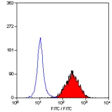


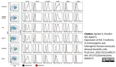
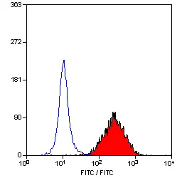
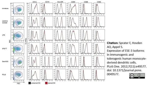
Phycoerythrin conjugated Mouse anti Human CD83 antibody, clone HB15e (MCA1582PE) used for the evaluation of CD83 expression on monocyte derived dendritic cells by flow cytometry.
Image caption:
Phenotypic characterization of immunogenic and tolerogenic moDC populations by flow cytometry. Monocytes were negatively selected from PBMC using magnetic beads. Immature moDC were generated with IL-4 and GM-CSF for 6 days. 15d-PGJ2 (PGJ2 DC) and dexamethasone plus 1α,25-dihydroxyvitamin were added to generate tolerogenic moDC, respectively (PGJ2 DC and Dex/VD3 DC). To generate immunogenic moDC, immature moDC were stimulated for 24 h with LPS, polyI:C and a cytokine cocktail containing TNF-α, IL-1β, IL-6 and PGE2, respectively. The phenotypes of the cells were analyzed by flow cytometry. Live cells were gated according to FSC/SSC. One representative experiment out of three is shown.
From: Sprater F, Hovden A-O, Appel S (2012)
Expression of ESE-3 Isoforms in Immunogenic and Tolerogenic Human Monocyte-Derived Dendritic Cells.
PLoS ONE 7(11): e49577.
doi: 10.1371/journal.pone.0049577
This image is from an open access article distributed under the terms of the Creative Commons Attribution License.
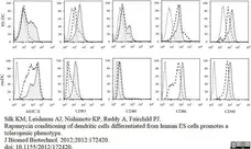
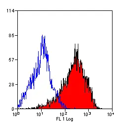
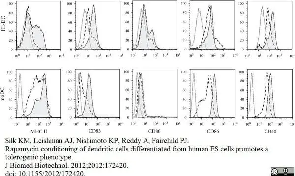
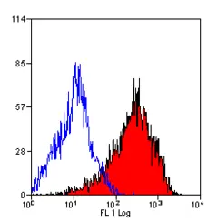
Mouse anti Human CD83 antibody, clone HB15e (MCA1582) used for the evaluation of CD83 expression on cell populations by flow cytometry.
Image caption:
DC differentiation from H1 hESC.
Cells were harvested from cultures at various time points and analysed by flow cytometry for the onset of hematopoiesis and the appearance of DC. (e) Phenotype of immature and mature H1-DCs compared with human moDC. DCs were cultured either in medium alone or medium supplemented with the maturation cocktail and stained for MHC class II, the maturation marker CD83 and classical costimulatory molecules. Dead cells were excluded from the analysis using 7-AAD. Dashed histograms show the phenotype of immature DCs while the filled histograms represent mature DCs. Open histograms depict background staining using isotype-matched controls.
From: Silk KM, Leishman AJ, Nishimoto KP, Reddy A, Fairchild PJ.
Rapamycin conditioning of dendritic cells differentiated from human ES cells promotes a tolerogenic phenotype.
J Biomed Biotechnol. 2012;2012:172420.
doi: 10.1155/2012/172420.
This image is from an open access article distributed under the terms of the Creative Commons Attribution License.
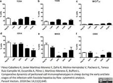
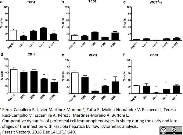
FITC conjugated Mouse anti Human CD83 antibody, clone HB15e (MCA1582F) used for the evaluation of CD83 expression on ovine peritoneal cells by flow cytometry.
Image caption:
Percentage of cell populations in the peritoneal fluid during the early stages of infection. The specific cell populations are shown as follows: (a) TCD4; (b) TCD8; (c) WC1+γδ; (d) CD14; (e) MHCII; (f) CD83. Cell subsets of uninfected (Neg. Ctl) and infected sheep (Group 1) are shown in columns. Mann-Whitney U-test was used to compare data from Groups 1 and 3. Values represent the mean ± standard deviation (SD). Asterisks indicate different levels of significance between groups: *P ≤ 0.05, **P ≤ 0.01
From: Pérez-Caballero R, Javier Martínez-Moreno F, Zafra R, Molina-Hernández V, Pacheco IL, Teresa Ruiz-Campillo M, Escamilla A, Pérez J, Martnez-Moreno Á, Buffoni L.
Comparative dynamics of peritoneal cell immunophenotypes in sheep during the early and late stages of the infection with Fasciola hepatica by flow cytometric analysis.
Parasit Vectors. 2018 11(1):640.
doi: 10.1186/s13071-018-3250-5
This image is from an open access article distributed under the terms of the Creative Commons Attribution License.
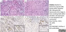
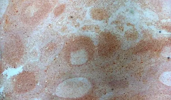
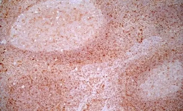
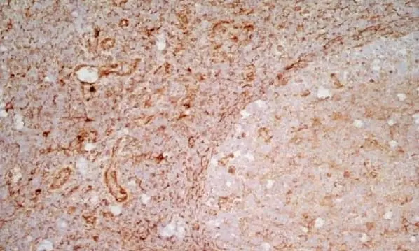
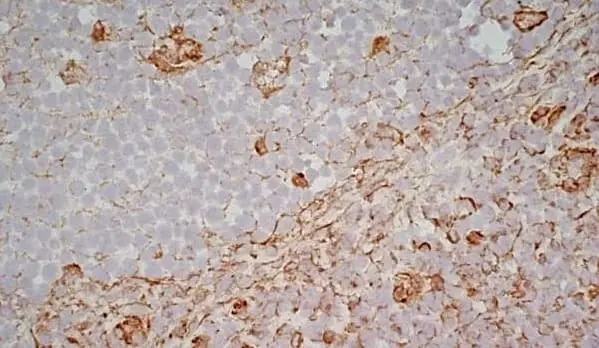
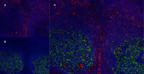
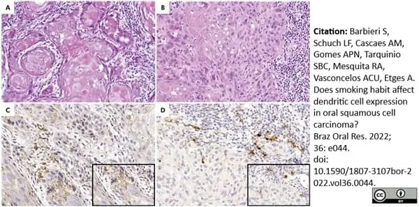
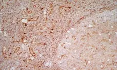
Mouse anti Human CD83 antibody, clone HB15e (MCA1582) used to illustrate mature dendritic cells in human connective tissue by immunohistochemistry on formalin fixed, paraffin embedded tissue sections.
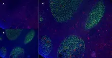
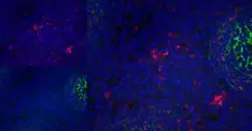
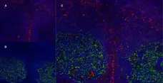
Image caption:
Figure A - Low-grade oral squamous cell carcinoma (OSCC) is represented by sheets and islands of proliferative cells within connective tissue. The proliferative cells show discrete pleomorphism and hyperchromatic nuclei with individual keratinization (H&E, 200X magnification). B - High-grade OSCC is represented by sheets of proliferative cells within the connective tissue. The proliferative cells showed intense pleomorphism and hyperchromatic nuclei without individual keratinization (H&E, 200X magnification). C - Immunostaining for CD1a shows immature dendritic cells located predominantly within the OSSC and with dendritic morphology (box) (immunohistochemistry, 200X magnification). D - Immunostaining of mature CD83+ dendritic cells was observed predominantly in connective tissue in the form of rounded brown structures (box) (immunohistochemistry, 200X magnification).
From: Barbieri S, Schuch LF, Cascaes AM, Gomes APN, Tarquinio SBC, Mesquita RA, Vasconcelos ACU, Etges A.
Does smoking habit affect dendritic cell expression in oral squamous cell carcinoma?
Braz Oral Res. 2022 Mar 14;36:e044.
doi: 10.1590/1807-3107bor-2022.vol36.0044.
This image is from an open access article distributed under terms of a Creative Commons Attribution License.
Filter by Application:
F C IF P Reset| Mouse anti Human CD83 antibody, clone HB15e recognizes the human CD83 cell surface antigen, a 40-45 kDa glycoprotein expressed by peripheral blood dendritic cells. Peripheral lymphocytes can be induced to express very low levels of CD83 after culture in agents such as Con A or PHA. In immunohistology CD83 is shown to be expressed strongly by interfollicular interdigitating reticulum cells and more weakly by cells within germinal centres. CD83 is also expressed by Langerhan's cells in the skin. The CD83 antigen is a 186-amino-acid single-chain glycoprotein. This molecule is a member of the immunoglobulin superfamily and is composed of an extracellular V-type Ig-like single domain, a transmembrane region, and a short, 40-amino-acid cytoplasmic tail. CD83 antigen undergoes extensive post-translational glycosylation, since the determined Mr is twice the predicted size of the core protein (Zhou et al. 1992). However, CD83+ cells have a unique cell surface immuno-phenotype that does not correlate with that of T cells, B cells, NK cells, or cells of the myelomonocytic lineage (Zhou et al. 1995).CD83+ cells co-express the highest levels of MHC class II molecules, when compared with other leucocyte lineages. They also co-express T cell markers (CD2, CD5), B cell markers (CD40, CD78), myeloid cell markers (CD13, CD33, CD36), cytokine receptors as well as other cell surface molecules (Zhou et al.1995) and Zhou and Tedder 1995). |
- Target Species
- Human
- Species Cross-Reactivity
-
Target Species Cross Reactivity Cynomolgus monkey Chimpanzee Baboon Rhesus Monkey Tasmanian Devil Sheep - N.B. Antibody reactivity and working conditions may vary between species.
- Product Form
- Purified IgG - liquid
- Preparation
- MCA1582T, MCA1582GA: Purified IgG prepared by affinity chromatography on Protein A from tissue culture supernatant
- MCA1582: Purified IgG prepared by affinity chromatography on Protein G from tissue culture supernatant
- Buffer Solution
- Phosphate buffered saline
- Preservative Stabilisers
- 0.09% sodium azide (NaN3)
- Carrier Free
- Yes
- Immunogen
- Cos cells transfected with HB15 cDNA.
- Approx. Protein Concentrations
- IgG concentration 1.0 mg/ml
- Fusion Partners
- Spleen cells from immunised BALB/c mice were fused with cells of the mouse NS1 myeloma cell line.
- Regulatory
- For research purposes only
- Guarantee
- 12 months from date of despatch
This product is shipped at ambient temperature. It is recommended to aliquot and store at -20°C on receipt. When thawed, aliquot the sample as needed. Keep aliquots at 2-8°C for short term use (up to 4 weeks) and store the remaining aliquots at -20°C.
Avoid repeated freezing and thawing as this may denature the antibody. Storage in frost-free freezers is not recommended.
Avoid repeated freezing and thawing as this may denature the antibody. Storage in frost-free freezers is not recommended.
This product has been reported to work in the following applications. This information is derived from testing within our laboratories, peer-reviewed publications or personal communications from the originators. Please refer to references indicated for further information. For general protocol recommendations, please visit the antibody protocols page.
| Application Name | Verified | Min Dilution | Max Dilution |
|---|---|---|---|
| Flow Cytometry |  |
1/50 | 1/100 |
| Immunofluorescence | |
||
| Immunohistology - Frozen | |
1/500 | 1/1000 |
| Immunohistology - Paraffin 1 | |
1/50 | 1/50 Pack Size: 20 µg 1/100 Pack Size: 0.1 mg, 0.2 mg |
| Immunoprecipitation | |
- 1 This product requires protein digestion pre-treatment of paraffin sections e.g. trypsin or pronase.
Where this product has not been tested for use in a particular technique this does not necessarily exclude its use in such procedures. Suggested working dilutions are given as a guide only. It is recommended that the user titrates the product for use in their own system using appropriate negative/positive controls.
- Flow Cytometry
- Use 10μl of the suggested working dilution to label 106cells in 100μl
- Histology Positive Control Tissue
- Human tonsil
| Description | Product Code | Applications | Pack Size | List Price | Your Price | Quantity | |
|---|---|---|---|---|---|---|---|
| Mouse IgG1 Negative Control | MCA928 | F | 100 Tests |

|
Log in | ||
| List Price | Your Price | ||||||
|
|
Log in | ||||||
| Description | Mouse IgG1 Negative Control | ||||||
References for CD83 antibody
-
Zhou, L.J. et al. (1992) A novel cell-surface molecule expressed by human interdigitating reticulum cells, Langerhans cells, and activated lymphocytes is a new member of the Ig superfamily.
J Immunol. 149 (2): 735-42. -
Zhou, L.J. & Tedder, T.F. (1995) Human blood dendritic cells selectively express CD83, a member of the immunoglobulin superfamily.
J Immunol. 154 (8): 3821-35. -
Schlossman, S.F., et al. Eds. Engel, P. et al. (1995) 'CD83 Workshop report' in Leucocyte Typing V, White Cell Differentiation Antigens,
Oxford University Press pp. 693-5. -
Hesselink, D.A. et al. (2005) The effects of renal transplantation on circulating dendritic cells.
Clin Exp Immunol. 140: 384-93. -
Walker, J.G. et al. (2007) Characterisation of a dendritic cell subset in synovial tissue which strongly expresses Jak/STAT transcription factors from patients with rheumatoid arthritis.
Ann Rheum Dis. 66: 992-9. -
Ifergan, I. et al. (2008) The blood-brain barrier induces differentiation of migrating monocytes into Th17-polarizing dendritic cells.
Brain. 131: 785-99. -
Denniston, A.K. et al. (2011) Endogenous Cortisol and TGF-{beta} in Human Aqueous Humor Contribute to Ocular Immune Privilege by Regulating Dendritic Cell Function.
J Immunol. 186:305-11. -
Hovden, A.O. et al. (2011) Maturation of monocyte derived dendritic cells with OK432 boosts IL-12p70 secretion and conveys strong T-cell responses.
BMC Immunol. ; 12:2.
View The Latest Product References
-
Van Vré, E.A. et al. (2011) Immunohistochemical characterisation of dendritic cells in human atherosclerotic lesions: possible pitfalls.
Pathology. 43 (3): 239-47. -
Silk, K.M. et al. (2012) Rapamycin conditioning of dendritic cells differentiated from human ES cells promotes a tolerogenic phenotype.
J Biomed Biotechnol. 2012: 172420. -
Sprater, F. et al. (2012) Expression of ESE-3 Isoforms in Immunogenic and Tolerogenic Human Monocyte-Derived Dendritic Cells
PLoS One. 7: e49577. -
Shikotra, A. et al. (2012) Increased expression of immunoreactive thymic stromal lymphopoietin in patients with severe asthma.
J Allergy Clin Immunol. 129: 104-11.e1-9. -
Denniston, A.K. et al. (2012) Aqueous humor suppression of dendritic cell function helps maintain immune regulation in the eye during human uveitis.
Invest Ophthalmol Vis Sci. 53 (2): 888-96. -
Howson, L.J. et al. (2014) Identification of dendritic cells, B cell and T cell subsets in Tasmanian devil lymphoid tissue; evidence for poor immune cell infiltration into devil facial tumors.
Anat Rec (Hoboken). 297: 925-38. -
Eren, U. et al. (2016) The several elements of intestinal innate immune system at the beginning of the life of broiler chicks.
Microsc Res Tech. 79 (7): 604-14. -
Wang, P. et al. (2016) Distribution and expression profiles of dendritic cell subpopulations in human bladder cancer.
Int J Clin Exp Pathol 9(7):7180-7. -
Duan, Y.G. et al. (2016) Characterisation of dendritic cell subsets in chronically inflamed human epididymis.
Andrologia. 48 (4): 431-40. -
Pérez-caballero, R. et al. (2018) Comparative dynamics of peritoneal cell immunophenotypes in sheep during the early and late stages of the infection with Fasciola hepatica by flow cytometric analysis.
Parasit Vectors. 11 (1): 640. -
Arya, S. et al. (2019) Quantitative proteomic changes in LPS-activated monocyte-derived dendritic cells: A SWATH-MS study.
Sci Rep. 9 (1): 4343. -
Yildiz, M. et al. (2019) Histological and immunohistochemical studies of the proximal caecum and caecal tonsils of quail (Coturnix coturnix japonica).
Anat Histol Embryol. 48 (5): 476-85. -
Eren, U. et al. (2022) TLR2 and TLR4 molecules and antigen-presenting cell compositions in cecal tonsils of broiler chicks (Gallus gallus domesticus.) in the first two weeks of the post-hatch period.
Anat Histol Embryol. 51 (1): 125-35. -
Barbieri, S. et al. (2022) Does smoking habit affect dendritic cell expression in oral squamous cell carcinoma?
Braz Oral Res. 36: e044.
- Synonyms
- HB15
- RRID
- AB_321773
- UniProt
- Q01151
- Entrez Gene
- CD83
- GO Terms
- GO:0006952 defense response
- GO:0007165 signal transduction
- GO:0005887 integral to plasma membrane
- GO:0006959 humoral immune response
View more products with CD83 specificity
Please Note: All Products are "FOR RESEARCH PURPOSES ONLY"
View all Anti-Human ProductsAlways be the first to know.
When we launch new products and resources to help you achieve more in the lab.
Yes, sign me up