CD105 antibody | SN6
Mouse anti Human CD105
- Product Type
- Monoclonal Antibody
- Clone
- SN6
- Isotype
- IgG1
- Specificity
- CD105


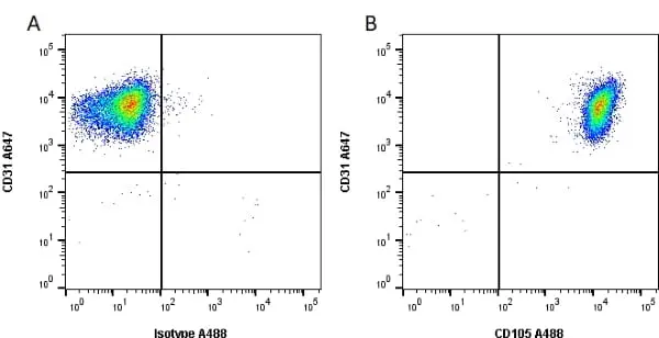
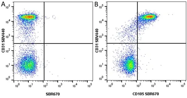
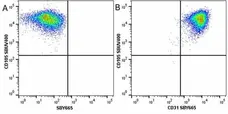
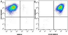
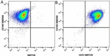
Figure B. Alexa Fluor® 647 conjugated Mouse anti Human CD31 antibody, clone WM59 (MCA1738A647) and Alexa Fluor® 488 conjugated Mouse anti Human CD105 antibody, clone SN6 (MCA1557A488). All experiments performed on HUVECs gated on live single cells, in the presence of 10% human serum.
Data acquired on the ZE5 Cell Analyzer.
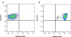
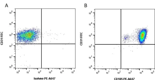
Figure B. FITC conjugated Mouse anti Human CD31 antibody, clone WM59 (MCA1738F) and PE-Alexa Fluor® 647 conjugated Mouse anti Human CD105 antibody, clone SN6 (MCA1557P647). All experiments performed on HUVECs gated on live single cells, in the presence of 10% human serum.
Data acquired on the ZE5 Cell Analyzer.

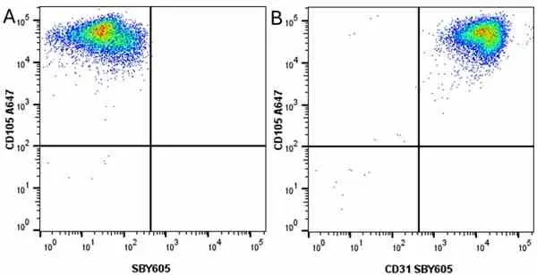
Figure B. FITC conjugated Mouse anti Human CD31 antibody, clone WM59 (MCA1738F) and Alexa Fluor® 647 conjugated Mouse anti Human CD105 antibody, clone SN6 (MCA1557A647). All experiments performed on HUVEC cells gated on live single cells in the presence of 10% human serum.
Data acquired on the ZE5 Cell Analyzer.
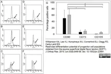
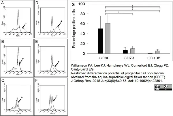
AlexaFlour®488 conjugated Mouse anti Human CD105 antibody, clone SN6 (MCA1557A488) used to evaluate endoglin expression on equine, tendon derived stem/progenitor cells by flow cytometry.
Image caption:
Flow cytometry analysis of cell surface markers CD90, CD73, and CD105 on tendon‐derived stem/progenitor cells isolated by differential adhesion onto substrates precoated with 20 μg/ml fibronectin (f‐TSPCs) and grown in normoxia and 5% hypoxia. A–C: f‐TSPCs (age 1 year) grown in 21% oxygen and incubated with control or antibodies (+) to CD90 (A), CD73 (B), or CD105 (C) for flow cytometry. D–F: f‐TSPCs grown in 5% oxygen and incubated with control or antibodies (+) to CD90 (D), CD73 (E), or CD105 (F) for flow cytometry. a p = 0.024, b p = 0.04, c p = 0.038.
From: Williamson KA, Lee KJ, Humphreys WJ, Comerford EJ, Clegg PD, Canty-Laird EG.
Restricted differentiation potential of progenitor cell populations obtained from the equine superficial digital flexor tendon (SDFT).
J Orthop Res. 2015 Jun;33(6):849-58.
This image is from an open access article distributed under the terms of a Creative Commons Attribution License.
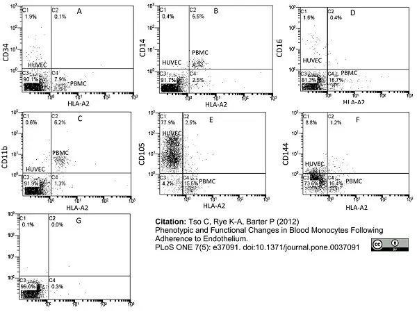
Mouse anti Human CD105 antibody, clone SN6 (MCA1557) used for flow cytometry.
Image caption:
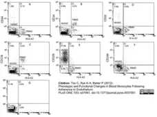
Endothelial adherence of blood monocytes. PBMCs were isolated from HLA-A2+ donors and incubated with HLA-A2− HUVECs (1×10⁶ cells/well) for 2 h, after which the non-adherent PBMCs were removed by washing. The co-cultured cell layers were immediately analysed with dual-colour flow cytometry for HLA-A2 and (A) CD34, (B) CD14, (C) CD11b, (D) CD16, (E) CD105 and (F) CD144 expression. Representative plots from 4–6 individual experiments are shown. (G) Two parameters dot plot showing typical isotype controls.
From: Tso C, Rye K-A, Barter P (2012)
Phenotypic and Functional Changes in Blood Monocytes Following Adherence to Endothelium.
PLoS ONE 7(5): e37091.
This image is from an open access article distributed under the terms of a Creative Commons Attribution License.
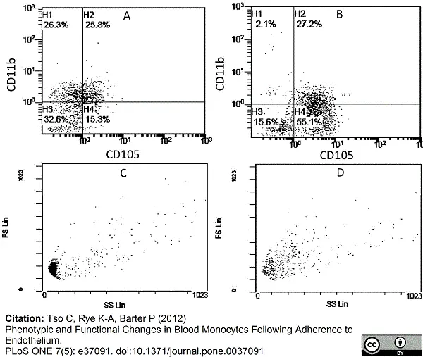
Mouse anti Human CD105 antibody, clone SN6 (MCA1557) used for flow cytometry.
Image caption:
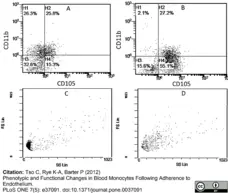
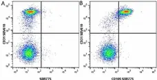
Phenotype change from HLA-A2+/CD11b+/CD105− to HLA-A2+/CD11b−/CD105+ on endothelium-adherent blood monocyte-derived cells with increase in size and granularity during co-culture. HLA-A2+ PBMCs (1×10⁶ cells/well) were incubated for 2 h (Day 0) with HLA-A2− HUVECs, after which the non-adherent cells were removed by washing. The cell layers were analysed by three-colour flow cytometry staining for HLA-A2, CD11b and CD105 on (A) Day 1 and (B) Day 2. These plots were gated for HLA-A2+ cells. Forward scatter/side scatter dot plots gated for HLA-A2+ cells on Day 0 (C) and Day 2 (D) was shown. These are representative of 2 individual experiments.
From: Tso C, Rye K-A, Barter P (2012)
Phenotypic and Functional Changes in Blood Monocytes Following Adherence to Endothelium.
PLoS ONE 7(5): e37091.
This image is from an open access article distributed under the terms of a Creative Commons Attribution License.
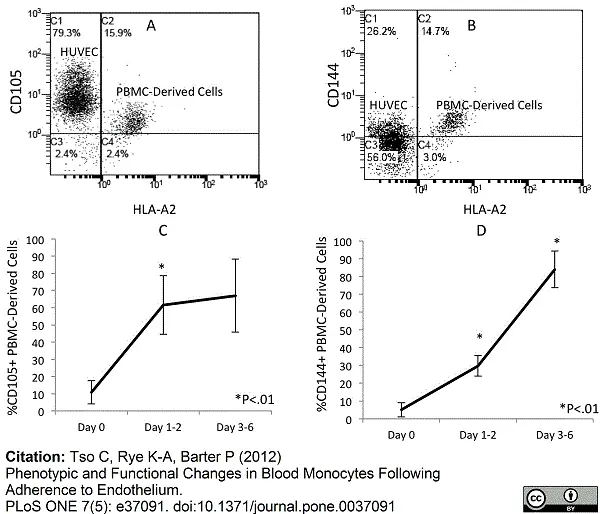
Mouse anti Human CD105 antibody, clone SN6 (MCA1557) used for flow cytometry.
Image caption:
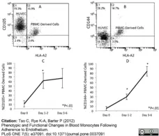
Increased expression of CD105 and CD144 in the endothelium-adherent monocytes during co-culture. HLA-A2+ PBMCs (1×10⁶ cells/well) were incubated for 2 h (Day 0) with HLA-A2− HUVECs, after which the non-adherent cells were removed by washing. The cell layers were maintained in co-culture up to Day 6, then assessed by dual-colour flow cytometry for HLA-A2 and (A) CD105 and (B) CD144 expression on Day 3 of co-culture. Representative plots from 4–7 individual experiments are shown. The increase in CD105 from Day 0 to Day 6 (C) and CD144 expression from Day 0 to Day 6 (D) is also shown.
From: Tso C, Rye K-A, Barter P (2012)
Phenotypic and Functional Changes in Blood Monocytes Following Adherence to Endothelium.
PLoS ONE 7(5): e37091.
This image is from an open access article distributed under the terms of a Creative Commons Attribution License.
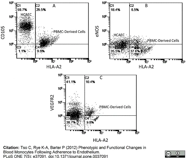
Mouse anti Human CD105 antibody, clone SN6 (MCA1557) used for flow cytometry.
Image caption:
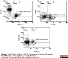
Expression of endothelial antigens in endothelium-adherent monocytes in co-culture with HCAECs. HLA-A2+ PBMCs (1×10⁶ cells/well) were incubated for 2 h (Day 0) with HLA-A2− HCAECs, after which the non-adherent cells were removed by washing. The cell layers were analysed by dual-colour flow cytometry for HLA-A2 and (A) CD105, (B) eNOS and (C) VEGFR2 expression on Day 2 of co-culture.
From: Tso C, Rye K-A, Barter P (2012)
Phenotypic and Functional Changes in Blood Monocytes Following Adherence to Endothelium.
PLoS ONE 7(5): e37091.
This image is from an open access article distributed under the terms of a Creative Commons Attribution License.
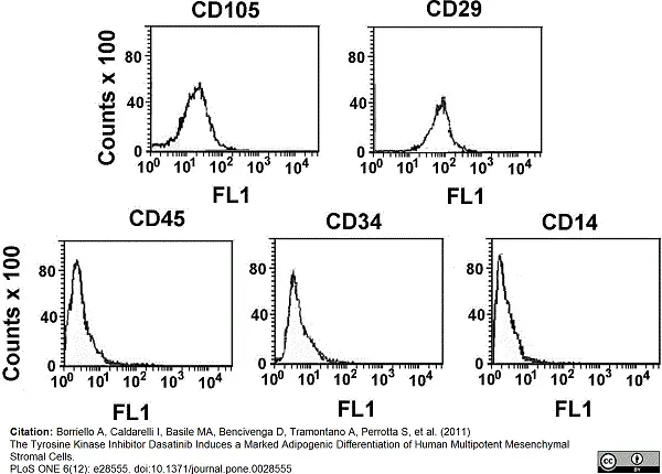
Mouse anti Human CD105 antibody, clone SN6 (MCA1557) used for flow cytometry
Image caption:
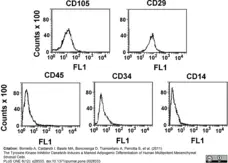
Immunophenotype of mesenchymal stem cells from human bone marrow. MSC cells were prepared as reported in Materials and Methods. MSCs from passage 2 were harvested and labeled with antibodies against CD105 and CD29 (positive MSCs markers) and CD45, CD34 and CD14 (MSCs negative markers) and analyzed by FACS. Histograms represent the staining of cells with the indicated antibody.
From: Borriello A, Caldarelli I, Basile MA, Bencivenga D, Tramontano A, et al.
(2011)
The Tyrosine Kinase Inhibitor Dasatinib Induces a Marked Adipogenic Differentiation of Human Multipotent Mesenchymal Stromal Cells.
PLoS ONE 6(12): e28555.
This image is from an open access article distributed under the terms of a Creative Commons Attribution License.
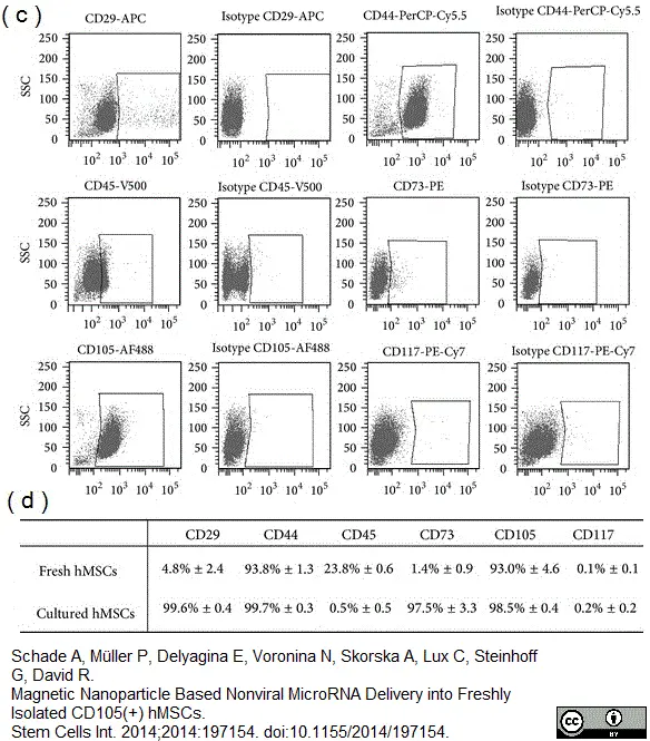
AlexaFlourA488® conjugated Mouse anti Human CD105 antibody, clone SN6 (MCA1557A488) used for flow cytometry on human mesenchymal stem cells.
Image caption:
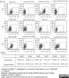
Immunophenotyping of freshly isolated ((c), (d)) and cultured CD105+ hMSCs (d) was evaluated by flow cytometry after staining of specific cell surface markers. Corresponding isotype controls were used as negative controls
From: Anna Schade, Paula Müller, Evgenya Delyagina, et al.
Magnetic Nanoparticle Based Nonviral MicroRNA Delivery into Freshly Isolated CD105+ hMSCs,”
Stem Cells International, vol. 2014, Article ID 197154, 11 pages.
This image is from an open access article distributed under the terms of a Creative Commons Attribution License.
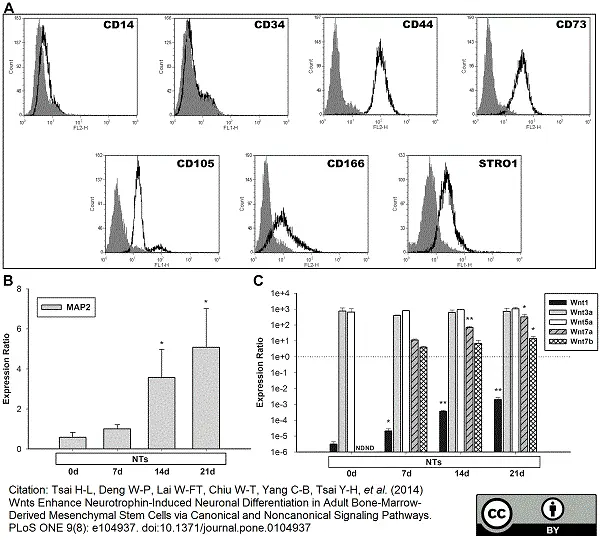
FITC conjugated Mouse anti Human CD105 antibody, clone SN6 (MCA1557F) used for flow cytometry on human bone marrow deived mesenchymal stem cells.
Image caption:
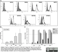
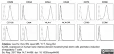
Flow cytometric analysis and Wnt profiles of hMSCs by the induction of NTs. (A) Bone marrow-derived hMSCs were analyzed following four cell passages. hMSCs were positive for CD44, CD73, CD105, CD166, and Stro-1, and negative for CD14 and CD34. The solid curves indicate each type of antibody, and the filled curves indicate mouse IgG as the negative control. (B) mRNA levels of MAP2 were quantified on days 7, 14, and 21 during stimulation with NTs. NTs significantly increased MAP2 levels on days 14 and 21. Untreated hMSCs served as the control. (C) mRNA levels of Wnt1, Wnt3a, Wnt5a, Wnt7a, and Wnt7b were quantified on days 7, 14, and 21 during stimulation with NTs. NTs increased the expression of Wnt1 and induced expressions of Wnt7a and Wnt7b. * p<0.05, ** p<0.01 (i.e., treated vs. control in the Wnt1, Wnt3a, and Wnt5a groups; NTs at 14 and 21 days vs. NTs at 7 days in the Wnt7a and Wnt7b groups). Data are presented as the mean ± SD of one triplicate experiment that was representative of three independent experiments. * p<0.05, ** p<0.01 (i.e., treated vs. control). ND, not determined.
From: Tsai H-L, Deng W-P, Lai W-FT, Chiu W-T, Yang C-B, et al. (2014)
Wnts Enhance Neurotrophin-Induced Neuronal Differentiation in Adult Bone-Marrow-Derived Mesenchymal Stem Cells via Canonical and Noncanonical Signaling Pathways.
PLoS ONE 9(8): e104937.
This image is from an open access article distributed under the terms of a Creative Commons Attribution License.
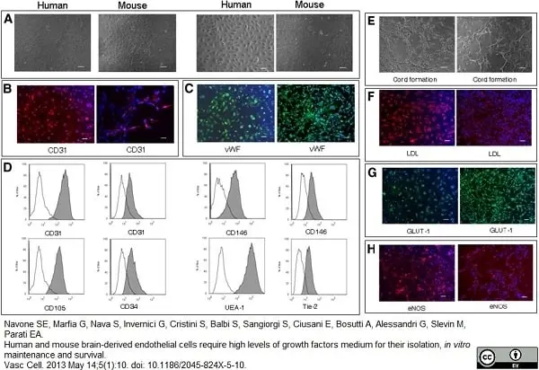
Mouse anti Human CD105 antibody, clone SN6 (MCA1557) used for flow cytometry on brain microvascular endothelial cells
Image caption:
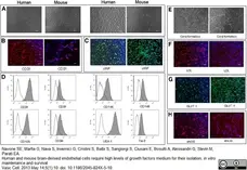
Characterization and functional features of human and murine BMVECs. (A) Phase contrast micrographs of confluent monolayers of human (left image) and murine (right image) BMVECs. BMVECs present the typical “cobblestone appearance”. Scale bar, 100 μm and 200 μm for human BMVECs and murine BMVECs. B) Human (left image) and murine (right image) BMVECs showed a clear cytoplasmic staining for CD31. Scale bar, 50 μm. C) Human (left image) and murine (right image) cells displayed an intense positive immunofluorescence for vWf. Scale bar, 50 μm. D) Flow cytometric analysis of BMVECs. Human BMVECs resulted positive (gray histograms) for CD31 (left graph), CD105, CD146 (left gaph), UEA-1 staining; murine BMVECs resulted positive for CD31 (right graph), CD34, CD146 (right graph) and Tie-2 staining. White histograms represent the isotype controls of each antibody. E) Capillary tube-like structure produced by human (left image) and murine (right image) BMVECs, 7 h after plating onto Matrigel. Scale bar, 100 μm. F) LDL-uptake assay on human (left image) and murine (right image) BMVECs. Scale bar, 50 μm. G) Human (left image) and murine (right image) BMVECs were labelled for GLUT-1. Scale bar, 50 μm. H) Immunofluorescence for eNOS in human (left image) and murine (right image) BMVECs. Scale bar, 50 μm. All nuclei were counterstained with DAPI (blue). One representative of three independent experiments performed in blind is shown for each figure.
From: Navone SE, Marfia G, Nava S, Invernici G, Cristini S, Balbi S, Sangiorgi S, Ciusani E, Bosutti A, Alessandri G, Slevin M, Parati EA.
Human and mouse brain-derived endothelial cells require high levels of growth factors medium for their isolation, in vitro maintenance and survival.
Vasc Cell. 2013 May 14;5(1):10.
This image is from an open access article distributed under the terms of a Creative Commons Attribution License.
FITC conjugated Mouse anti Human CD105 antibody, clone SN6 (MCA1557F) used for the evaluation of endoglin expression on mesenchymal stem cells by flow cytometry.
Image caption:
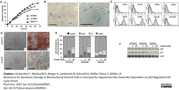
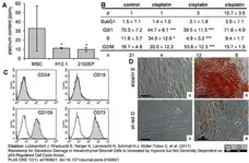
(A) Platinum accumulation in MSC and TGCT cell lines upon 24h treatment with 3 μM cisplatin and analyzed by atomic absorption spectroscopy. Mean ± standard deviation; MSC n = 8, TGCT both n = 3; * p<0.05 vs MSC. (B) Cell cycle populations from analyses as shown in Fig 1C (propidium iodide staining). Data are presented as% of cells distributed to cell cycle phases as mean ± standard deviation; n≥4; * p<0.05, *** p<0.001 vs. control. (C) MSC after subapoptotic damage by cisplatin upon reconstitution of proliferation were analyzed for surface antigen expression by flow cytometry. Data are shown as histograms of fluorescence. Isotype controls (no filling) are overlaid on specific FITC- or PE-conjugated antibodies. Data are representative of at least 4 independent experiments. (D) MSC from (C) were incubated in growth medium (u) or specific osteogenic (o) and adipogenic (a) differentiation media. Cells were stained with alizarin pH4 and oil red for calcium deposition and lipid droplets, respectively. Data are representative of at least 4 independent experiments. Light microscopy, scale bar– 100μm.
From: Lützkendorf J, Wieduwild E, Nerger K, Lambrecht N, Schmoll H-J, Müller-Tidow C, et al. (2017)
Resistance for Genotoxic Damage in Mesenchymal Stromal Cells Is Increased by Hypoxia but Not Generally Dependent on p53-Regulated Cell Cycle Arrest.
PLoS ONE 12(1): e0169921.
This image is from an open access article distributed under the terms of a Creative Commons Attribution License.
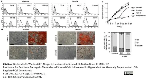
FITC conjugated Mouse anti Human CD105 antibody, clone SN6 (MCA1557F) used for the evaluation of endoglin expression on mesenchymal stem cells by flow cytometry.
Image caption:
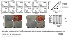
(A) MSC cultured for up to 14 days under physioxia or hypoxia were analyzed for surface antigen expression by flow cytometry. Data are representative of at least 3 independent experiments. (B) MSC from (A) were incubated in growth medium (u) or specific osteogenic (o) and adipogenic (a) differentiation media. Cells were stained with alizarin pH4 and oil red for calcium deposition and lipid droplets, respectively. Data are representative of 3 independent experiments. Light microscopy, scale bar– 100 μm. (C) Growth kinetics of MSC under normoxic, physioxic and hypoxic conditions. Cultivation under physioxia/hypoxia started on day 0. An aliquot of hypoxic cells was reoxygenated to normoxic conditions on d15. Data are representative of 5 independent experiments. (D) Cell cycle analyses of normoxic and hypoxic MSC were performed upon pyronin/7-AAD staining. Data are presented as% of cells in cell cycle phase as mean—standard deviation; n = 5.
From: Lützkendorf J, Wieduwild E, Nerger K, Lambrecht N, Schmoll H-J, Müller-Tidow C, et al. (2017)
Resistance for Genotoxic Damage in Mesenchymal Stromal Cells Is Increased by Hypoxia but Not Generally Dependent on p53-Regulated Cell Cycle Arrest.
PLoS ONE 12(1): e0169921.
This image is from an open access article distributed under the terms of a Creative Commons Attribution License.
FITC conjugated Mouse anti Human CD105 antibody, clone SN6 (MCA1557F) used for the evaluation of endoglin expression on mesenchymal stem cells by flow cytometry.
Image caption:
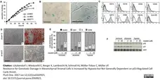
(A) Growth kinetic was performed with MSC with lentiviral p53 knock down (MSCp53kd), MSC with lentiviral control sh-RNA (ctr-MSC) and wildtype MSC (wt-MSC) from the same donor. Lentiviral transduction was performed on day 0. Data are representative of 4 independent experiments. (B) Late passage MSCp53kd were stained for senescence-associated beta-galactosidase activity. Data are representative of 2 independent experiments. Light microscopy, scale bar– 200 μm. (C) MSCp53kd were analyzed for surface antigen expression by flow cytometry. Data are shown as histograms of fluorescence. Isotype controls (no filling) are overlaid on specific FITC- or PE-conjugated antibodies. Data are representative of 4 independent experiments. (D) MSCp53kd were incubated in growth medium (u) or specific osteogenic (o) and adipogenic (a) differentiation media. Cells were stained with alizarin pH4 and oil red for calcium deposition and lipid droplets, respectively. Data are representative of 4 independent experiments. Light microscopy, scale bar– 200 μm. (E) MSCp53kd were treated 72 h with cisplatin under normoxic, physioxic and hypoxic conditions and analyzed for cell cycle distribution. Data are presented as% of cells in cell cycle phase as mean—standard deviation; n = 3. (F) Whole protein lysates from the experiment shown in (E) were analyzed by western blot. Data are representative of 3 independent experiments.
From: Lützkendorf J, Wieduwild E, Nerger K, Lambrecht N, Schmoll H-J, Müller-Tidow C, et al. (2017)
Resistance for Genotoxic Damage in Mesenchymal Stromal Cells Is Increased by Hypoxia but Not Generally Dependent on p53-Regulated Cell Cycle Arrest.
PLoS ONE 12(1): e0169921.
This image is from an open access article distributed under the terms of a Creative Commons Attribution License.
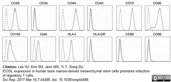
Mouse anti Human CD105 antibody, clone SN6 (MCA1557) used for the identification of mesenchymal stem cells in mixed bone marrow cell populations by flow cytometry.
Image caption:
MSCs used in this study were characterized for marker expression by flow cytometric analysis. Dashed histograms indicate staining with isotype-matched control antibody and solid histograms denote the specific expression of each indicated marker.
From: Lee HJ, Kim SN, Jeon MS, Yi T, Song SU.
ICOSL expression in human bone marrow-derived mesenchymal stem cells promotes induction of regulatory T cells.
Sci Rep. 2017 Mar 14;7:44486.
This image is from an open access article distributed under the terms of a Creative Commons Attribution License.
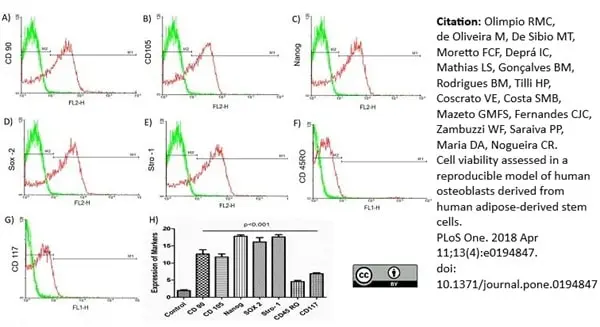
Mouse anti Human CD105 antibody, clone SN6 (MCA1557) used for the evaluation of endoglin expression on human adipose derived stem cells by flow cytometry.
Image caption:
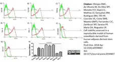
Analysis of hASC markers.
(A) CD90. (B) CD105. (C) Nanog. (D) Sox-2. (E) STRO-1. (F) CD45RO. (G) CD117. (H) Quantification of expression of positive and negative markers. Graphs show the expression of different markers in hASCs. The expression of the markers was assessed using flow cytometry by means of the intensity of the emitted fluorescence (FL1 and FL2). The expression of the positive markers, CD90, CD105, and STRO-1 (surface markers mesenchymal) and Nanog and Sox-2 (pluripotency markers) is compared to the decreased expression of negative markers (CD45RO and CD117). Data are represented as mean ± standard deviation. The statistical analyses were obtained using an ANOVA followed by Dunnett's test with P <0.05 being considered significant.
From: Olimpio RMC, de Oliveira M, De Sibio MT, Moretto FCF, Deprá IC, Mathias LS, et al. (2018)
Cell viability assessed in a reproducible model of human osteoblasts derived from human adipose-derived stem cells.
PLoS ONE 13(4): e0194847.
This image is from an open access article distributed under the terms of a Creative Commons Attribution License.
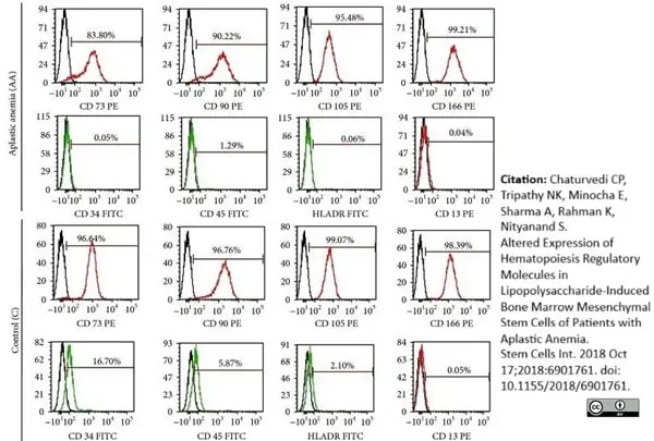
Phycoerythrin conjugated Mouse anti Human CD90 antibody, clone AD2 (MCA1557PE) used to evaluate CD90 expression on bone-marrow derived mesenchymal stem cells by flow cytometry
Image caption:
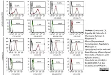
Immunophenotypic and differentiation characterization of BM-MSCs of aplastic anemia (AA) patients. (a) Representative flow cytometric histograms showing immunophenotype of BM-MSCs of AA patients and controls.
From: Chaturvedi CP, Tripathy NK, Minocha E, Sharma A, Rahman K, Nityanand S.
Altered Expression of Hematopoiesis Regulatory Molecules in Lipopolysaccharide-Induced Bone Marrow Mesenchymal Stem Cells of Patients with Aplastic Anemia.
Stem Cells Int. 2018 Oct 17;2018:6901761.
This image is from an open access article distributed under the terms of a Creative Commons Attribution License.
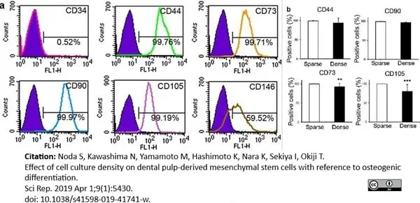
Mouse anti Human CD105 antibody, clone SN6 (MCA1557) used for the evaluation of CD105 expression on dental pulp derived cells by flow cytometry.
Image caption:
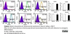
Cell surface markers. (a) Cell surface markers before separation into sparse (sDPSCs) and dense (dDPSCs) groups. A representative case among seven donors is shown. (b) MSC marker expression in sparse (sDPSCs) and dense (dDPSCs) groups. **p = 0.0079 and ***p = 0.0006 (Mann-Whitney U test). The error bar is SD (n = 7). Solid colored histograms represent IgGκ treated cells for control, and open colored histograms represent fluorophore labeled antibody treated cells.
From: Noda S, Kawashima N, Yamamoto M, Hashimoto K, Nara K, Sekiya I, Okiji T.
Effect of cell culture density on dental pulp-derived mesenchymal stem cells with reference to osteogenic differentiation.
Sci Rep. 2019 Apr 1;9(1):5430.
This image is from an open access article distributed under the terms of a Creative Commons Attribution License.

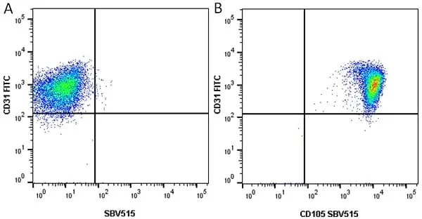
Figure B. FITC conjugated Mouse anti Human CD31 antibody, clone WM59 (MCA1738F) and StarBright Violet 515 conjugated Mouse anti Human CD105 antibody, clone SN6 (MCA1557SBV515). All experiments performed on HUVEC cells gated on live single cells in the presence of 10% human serum.
Data acquired on the ZE5 Cell Analyzer.
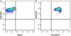
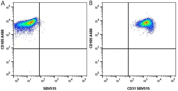
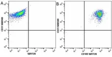
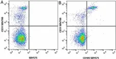
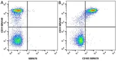
Figure B. Alexa Fluor® 488 conjugated Mouse anti Human CD105 antibody, clone SN6 (MCA1557A488) and StarBright Violet 515 conjugated Mouse anti Human CD31 antibody, clone WM59 (MCA1738SBV515). All experiments performed on HUVEC cells gated on live single cells in the presence of 10% human serum.
Data acquired on the ZE5 Cell Analyzer.

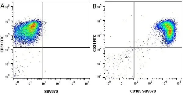
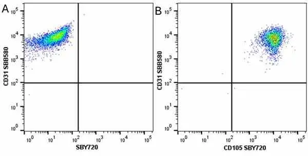
Figure B. FITC conjugated Mouse anti Human CD31 antibody, clone WM59 (MCA1738F). and StarBright Violet 670 conjugated Mouse anti Human CD105 antibody, clone SN6 (MCA1557SBV670). All experiments performed on HUVEC cells gated on live single cells in the presence of 10% human serum.
Data acquired on the ZE5 Cell Analyzer.

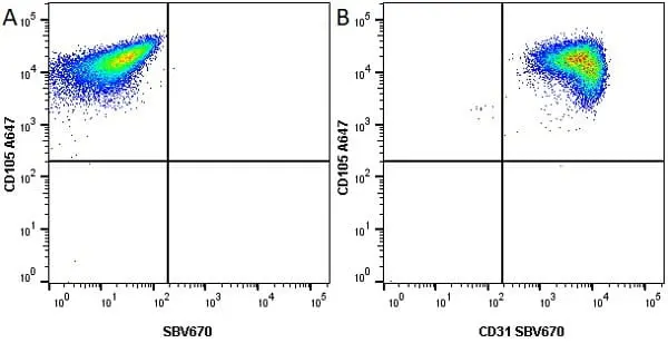
Figure B. Alexa Fluor® 647 conjugated Mouse anti Human CD105 antibody, clone SN6 (MCA1557A647) and StarBright Violet 670 conjugated Mouse anti Human CD31 antibody, clone WM59 (MCA1738SBV670). All experiments performed on HUVEC cells gated on live single cells in the presence of 10% human serum.
Data acquired on the ZE5 Cell Analyzer.
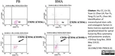
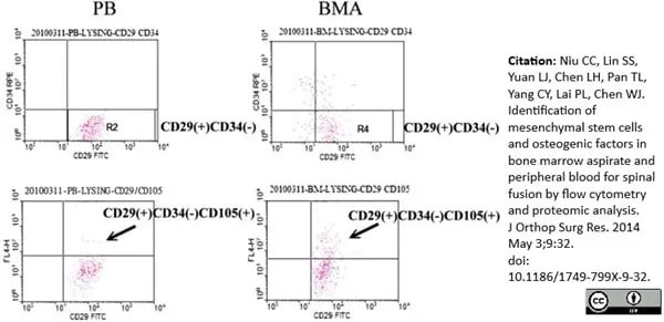
Alexa Fluor® 647 conjugated Mouse anti Human CD105 antibody, clone SN6 (MCA1557A647) used to evaluate levels of CD105 expression on peripheral blood (PB) and bone marrow aspirate (BMA) samples by flow cytometry.
Image caption:
Flow cytometry analysis of MSC-like cells from PB and BMA.
The proportion of CD34−/CD29+/CD105+-nucleated cells in the PB was significantly lower than that in the BMA (t test, p <0.01, n = 4).
From: Niu CC, Lin SS, Yuan LJ, Chen LH, Pan TL, Yang CY, Lai PL, Chen WJ.
Identification of mesenchymal stem cells and osteogenic factors in bone marrow aspirate and peripheral blood for spinal fusion by flow cytometry and proteomic analysis.
J Orthop Surg Res. 2014 May 3;9:32.
doi: 10.1186/1749-799X-9-32.
This image is from an open access article distributed under terms of a Creative Commons Attribution License.
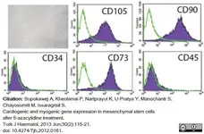
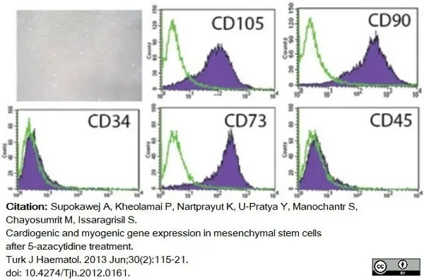
FITC conjugated Mouse anti Human CD105 antibody, clone SN6 (MCA1557F) used to evaluate expression of mesenchymal stem cell markers in vitro by flow cytometry.
Image caption:
In culture, MSCs presented a spindle-shaped and fibroblast-like morphology. Various cell surface markers were analyzed by flow cytometry; positive results were obtained for CD73, CD90, and CD105 (MSC markers), whereas negative results were obtained for CD34 and CD45 (hematopoietic markers).
From: Supokawej A, Kheolamai P, Nartprayut K, U-Pratya Y, Manochantr S, Chayosumrit M, Issaragrisil S.
Cardiogenic and myogenic gene expression in mesenchymal stem cells after 5-azacytidine treatment.
Turk J Haematol. 2013 Jun;30(2):115-21.
doi: 10.4274/Tjh.2012.0161.
This image is from an open access article distributed under terms of a Creative Commons Attribution License.
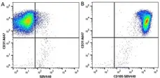
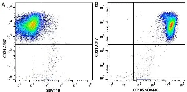
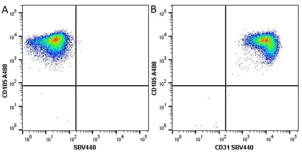
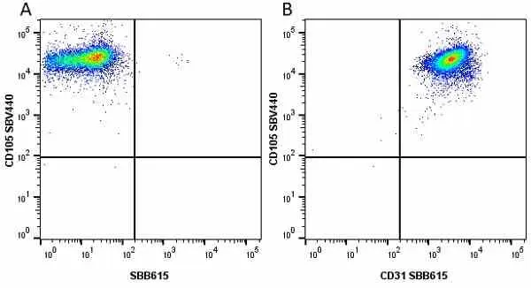
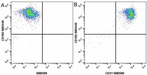
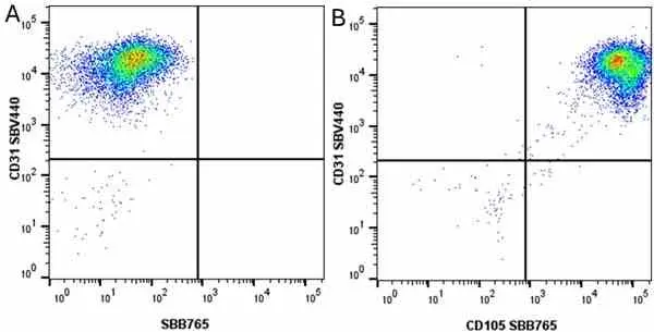
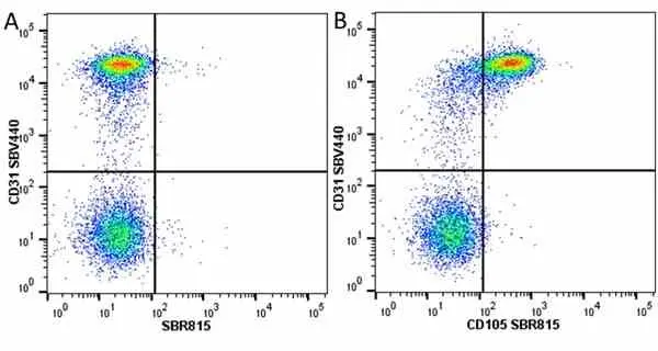
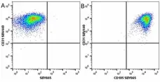
Figure B. Alexa Fluor® 647 conjugated Mouse anti Human CD31 antibody, clone WM59 (MCA1738A647) and StarBright Violet 440 conjugated Mouse anti Human CD105 antibody, clone SN6 (MCA1557SBV440). All experiments performed on HUVEC cells gated on live single cells in the presence of 10% human serum.
Data acquired on the ZE5 Cell Analyzer.
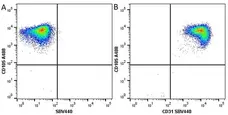
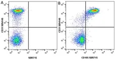
Figure B. Alexa Fluor® 488 conjugated Mouse anti Human CD105 antibody, clone SN6 (MCA1557A488) and StarBright Violet 440 conjugated Mouse anti Human CD31 antibody, clone WM59 (MCA1738SBV440). All experiments performed on HUVEC cells gated on live single cells in the presence of 10% human serum.
Data acquired on the ZE5 Cell Analyzer.

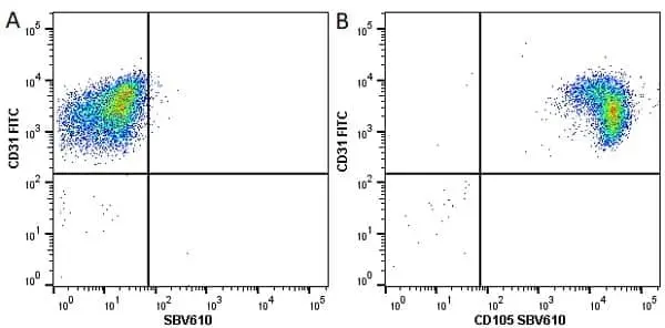
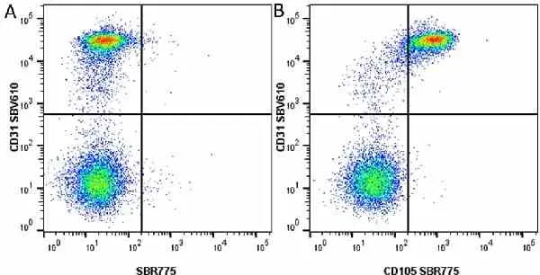
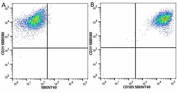
Figure B. FITC conjugated Mouse anti Human CD31 (MCA1738F) and StarBright Violet 610 conjugated Mouse anti Human CD105 antibody, clone SN6 (MCA1557SBV610). All experiments performed on HUVEC cells gated on live single cells in the presence of 10% human serum.
Data acquired on the ZE5 Cell Analyzer.
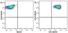
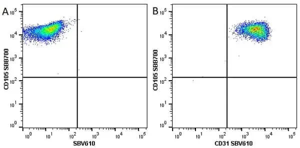
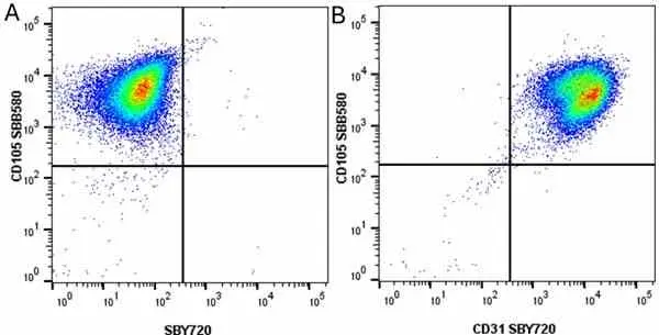
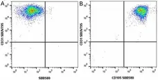
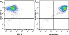
Figure B. StarBright Blue 700 conjugated Mouse anti Human CD105 antibody, clone SN6 (MCA1557SBB700) and StarBright Violet 610 conjugated Mouse anti Human CD31 antibody, clone WM59 (MCA1738SBV610). All experiments performed on HUVEC cells gated on live single cells in the presence of 10% human serum.
Data acquired on the ZE5 Cell Analyzer.
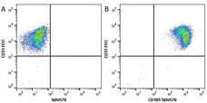
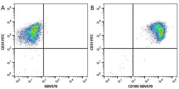
Figure B. FITC conjugated Mouse anti Human CD31 (MCA1738F) and StarBright Violet 570 conjugated Mouse anti Human CD105 (MCA1557SBV570). All experiments performed on HUVEC cells gated on live single cells in the presence of 10% human serum.
Data acquired on the ZE5 Cell Analyzer.
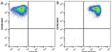
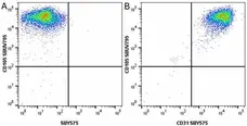
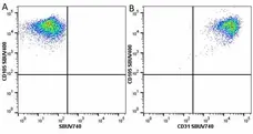
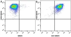
Figure B. Alexa Fluor® 647 conjugated Mouse anti Human CD105 antibody, clone SN6 (MCA1557A647) and StarBright Violet 570 conjugated Mouse anti Human CD31 antibody, clone WM59 (MCA1738SBV570). All experiments performed on HUVEC cells gated on live single cells in the presence of 10% human serum.
Data acquired on the ZE5 Cell Analyzer.
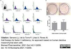
FITC conjugated Mouse anti Human CD105 antibody, clone SN6 (MCA1557F) used to evaluate CD105 expression on dicidual MSC by flow cytometry.
Image caption:
Characterization of decidua mesenchymal stem cells. (A) Plots show the specific antibody staining profile (purple or blue area) versus the isotype Ig control staining (green line). (B) Representative images of crystal violet stained plates of CFU-assays performed at two different cell densities (upper panel) and CFU quantification of DMSC initially plated at 100 (n = 7) or 500 (n = 3) cells/100-mm plate.
From: Serrano LJ, de la Torre P, Liras A, Flores AI.
Cell therapy for factor V deficiency: An approach based on human decidua mesenchymal stem cells.
Biomed Pharmacother. 2021 Oct;142:112059.
doi: 10.1016/j.biopha.2021.112059.
This image is from an open access article distributed under terms of a Creative Commons Attribution License.
FITC conjugated Mouse anti Human CD105 antibody, clone SN6 (MCA1557F) used to label CD105 expressing cells from cynomolgus monkey samples by flow cytometry.
Image caption:
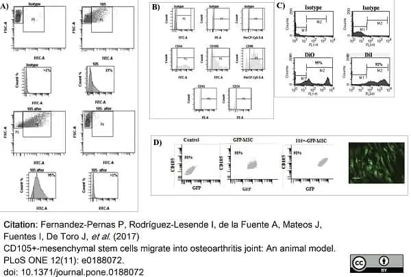
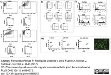
Characterization of CD105+-MSCs used in the cell therapy.
A) Selective enrichment of CD105+ expressing cells. Fluorescence-activated cell sorting (FACS) analysis of the subpopulations of CD105+ pre-sorted (on the top) and post-sorted (on the bottom) from synovial membrane. 105 after was subpopulation of CD105+ post-sorted (on the left bottom) and 105- after was subpopulation of CD105- post-sorted (on the right bottom). B) Characterization by flow cytometry analysis of the CD105+-MSC population using MSCs markers, CD44, CD105, CD90 and haematopoietic markers CD45 and CD34. C) Flow cytometry of subpopulation of CD105+-MSC labelled with oxacarbocyanine (DiO) and octadecyl (C18) indocarbocyanine (DiI) respectively. D) Characterization of GFP-CD105+-MSCs used to validate previous data obtained using DiO- CD105+-MSCs and DiI-CD105+-MSCs in animal model. Fluorescence-activated cell sorting (FACS) analysis of the MSCs populations (on the left), GFP-MSCs (on the middle) and post-sorted for CD105+ (on the right) from synovial membrane. Representative image of GFP-CD105+-MSCs in a plate before injection, bar represents 20 μm.
From: Fernandez-Pernas P, Rodríguez-Lesende I, de la Fuente A, Mateos J, Fuentes I, De Toro J, et al. (2017)
CD105+-mesenchymal stem cells migrate into osteoarthritis joint: An animal model.
PLoS ONE 12(11): e0188072.
doi: 10.1371/journal.pone.0188072
This image is from an open access article distributed under terms of a Creative Commons Attribution License.
FITC conjugated Mouse anti Human CD105 antibody, clone SN6 (MCA1557F) used to label CD105 expressing cells from cynomolgus monkey samples by immunofluorescence.
Image caption:
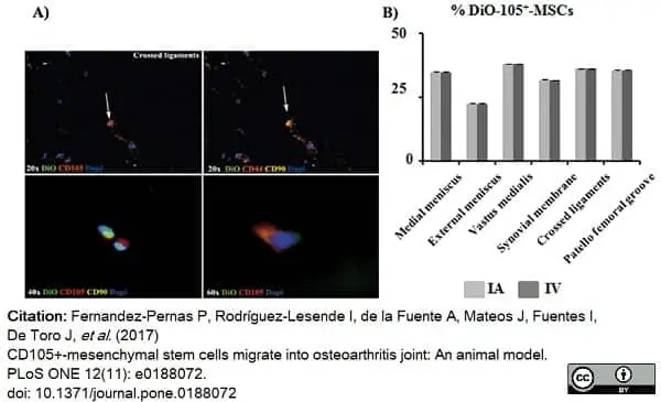
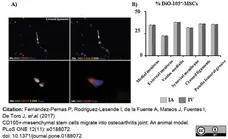
DiO-CD105+-MSCs migration study.
A) Co-localization of DiO-CD105+-MSCs with antibodies against anti-CD105, anti-CD44 and anti-CD90 in crossed ligaments sections of left knee from animals injected with DiO-CD105+-MSCs IV (magnification were 20x, 40x and 60x). Arrows point cells which co-localized for CD44 and CD90 antibodies. B) Histogram showed percentage of DiO positives signal normalized with DAPI signal from animals IA injected in front of animals IV injected. AnalySIS Image Processing was used.
From: Fernandez-Pernas P, Rodríguez-Lesende I, de la Fuente A, Mateos J, Fuentes I, De Toro J, et al. (2017)
CD105+-mesenchymal stem cells migrate into osteoarthritis joint: An animal model.
PLoS ONE 12(11): e0188072.
doi: 10.1371/journal.pone.0188072
This image is from an open access article distributed under terms of a Creative Commons Attribution License.
FITC conjugated Mouse anti Human CD105 antibody, clone SN6 (MCA1557F) used to label CD105 expressing cells from cynomolgus monkey samples by immunofluorescence.
Image caption:
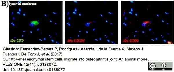
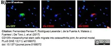
GFP-CD105+-MSCs migration study.
B) Co-localization of GFP-CD105+-MSCs with antibodies against anti-CD105 and anti-CD90 in synovial membrane sections of left knee from animals IV injected with GFP-CD105+-MSCs. All pictures have same magnification 40x.
From: Fernandez-Pernas P, Rodríguez-Lesende I, de la Fuente A, Mateos J, Fuentes I, De Toro J, et al. (2017)
CD105+-mesenchymal stem cells migrate into osteoarthritis joint: An animal model.
PLoS ONE 12(11): e0188072.
doi: 10.1371/journal.pone.0188072
This image is from an open access article distributed under terms of a Creative Commons Attribution License.
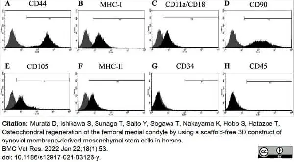
Mouse anti Human CD105 antibody, clone SN6 (MCA1557GA) used to evaluate phenotype of equine synovial membrane derived mesenchymal stem cells by flow cytometry.
Image caption:
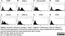
Results of flow cytometry using immunological markers on synovial membrane-derived mesenchymal stem cells. The grey area represents the negative control. The horizontal line in individual histograms indicates the population of the positive cells
From: Murata D, Ishikawa S, Sunaga T, Saito Y, Sogawa T, Nakayama K, Hobo S, Hatazoe T. Osteochondral regeneration of the femoral medial condyle by using a scaffold-free 3D construct of synovial membrane-derived mesenchymal stem cells in horses.
BMC Vet Res. 2022 Jan 22;18(1):53.
doi: 10.1186/s12917-021-03126-y.
This image is from an open access article distributed under terms of a Creative Commons Attribution License.
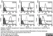
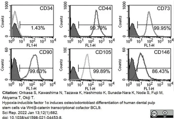
Mouse anti Human CD105 antibody, clone SN6 (MCA1557) used to assess endoglin expression on cultured mesenchymal stem cells by flow cytometry.
Image caption:
hDPSCs express typical mesenchymal stem cell (MSC) markers and exhibit multi-differentiation potential. (a) Typical MSC markers (CD44, CD73, CD90, CD105, CD146) are highly expressed in hDPSCs, and hDPSCs are mostly negative for a hematopoietic marker (CD34). hDPSCs possess neurogenic, adipogenic, chondrogenic, and osteogenic differentiation potential, as determined by the expression of neurogenic markers (GFAP and NF-M)
From: Orikasa S, Kawashima N, Tazawa K, Hashimoto K, Sunada-Nara K, Noda S, Fujii M, Akiyama T, Okiji T.
Hypoxia-inducible factor 1α induces osteo/odontoblast differentiation of human dental pulp stem cells via Wnt/β-catenin transcriptional cofactor BCL9.
Sci Rep. 2022 Jan 13;12(1):682.
doi: 10.1038/s41598-021-04453-8.
This image is from an open access article distributed under terms of a Creative Commons Attribution License.

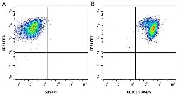
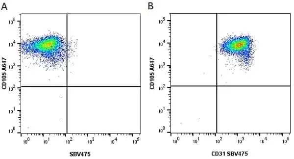
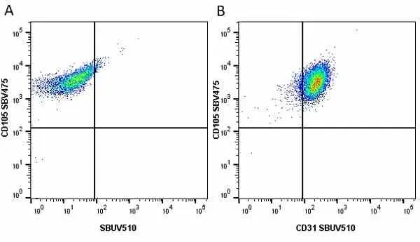
Figure B. FITC conjugated Mouse anti Human CD31 antibody, clone WM59 (MCA1738F) and StarBright Violet 475 conjugated Mouse anti Human CD105 antibody, clone SN6 (MCA1557SBV475). All experiments performed on HUVEC cells gated on live single cells in the presence of 10% human serum.
Data acquired on the ZE5 Cell Analyzer.
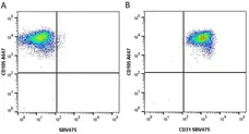
Figure B. Alexa Fluor® 647 conjugated Mouse anti Human CD105 antibody, clone SN6 (MCA1557A647) and StarBright Violet 475 conjugated Mouse anti Human CD31 antibody, clone WM59 (MCA1738SBV475). All experiments performed on HUVEC cells gated on live single cells in the presence of 10% human serum.
Data acquired on the ZE5 Cell Analyzer.
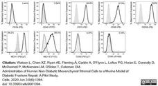
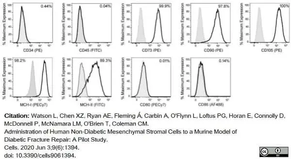
Phycoerythrin conjugated Mouse anti Human CD105 antibody, clone SN6 (MCA1557PE) used to evaluate endoglin expression on human MSCs by flow cytometry.
Image caption:
Characterization of bone marrow cell isolates demonstrated a mesenchymal phenotype with tri-lineage potential. To validate the isolation of mesenchymal stromal cells (MSCs) from the bone marrow, morphologic and phenotypic characterizations were conducted.
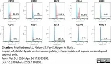
Representative samples, low expression of CD34 (0.44%) and CD45 (0.04%) and high expression of CD73 (99.9%), CD90 (97.8%), and CD105 (100%), indicated the isolation of a population of cells conforming to the ISCT standards. The cells were also positive for MHC-I (98.2%) and MHC-II (89.3%), but negative for co-stimulatory CD80 (0.01%) and CD86 (0.14%). Gray lines indicate the isotype control while black lines indicate an antibody stained sample.
From: Watson L, Chen XZ, Ryan AE, Fleming Á, Carbin A, O'Flynn L, Loftus PG, Horan E, Connolly D, McDonnell P, McNamara LM, O'Brien T, Coleman CM.
Administration of Human Non-Diabetic Mesenchymal Stromal Cells to a Murine Model of Diabetic Fracture Repair: A Pilot Study.
Cells. 2020 Jun 3;9(6):1394.
doi: 10.3390/cells9061394.
This image is from an open access article distributed under terms of a Creative Commons Attribution License.
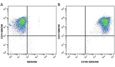
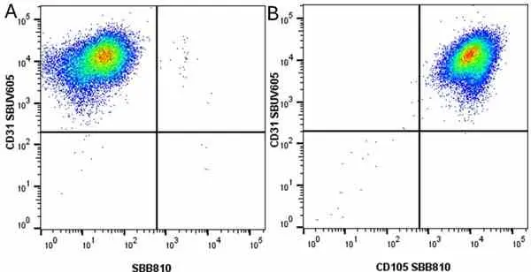
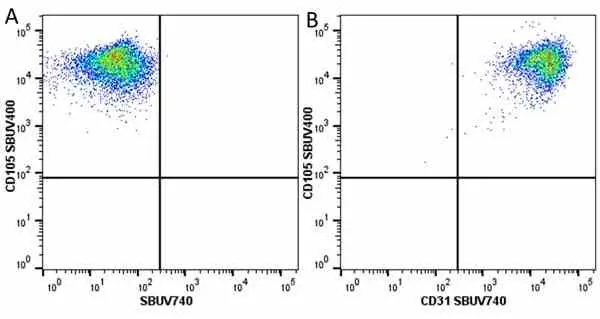
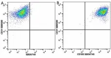
Figure B. StarBright Blue 700 conjugated Mouse anti Human CD31 antibody, clone WM59 (MCA1738SBB700) and StarBright UltraViolet 400 conjugated Mouse anti Human CD105 antibody, clone SN6 (MCA1557SBUV400). All experiments performed on HUVEC cells gated on live single cells in the presence of 10% human serum.
Data acquired on the ZE5 Cell Analyzer.
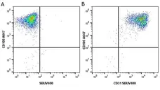
Figure B. Alexa Fluor® 647 conjugated Mouse anti Human CD105 clone SN6 (MCA1557A647) and StarBright UltraViolet 400 conjugated Mouse anti Human CD31 antibody, clone WM59 (MCA1738SBUV400). All experiments performed on HUVEC cells gated on live single cells in the presence of 10% human serum.
Data acquired on the ZE5 Cell Analyzer.

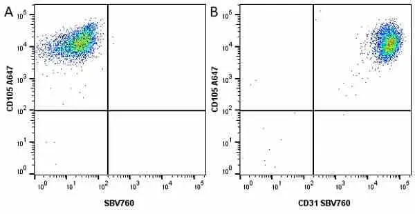
Figure B. Alexa Fluor® 647 conjugated Mouse anti Human CD105 clone SN6 (MCA1557A647) and StarBright Violet 760 conjugated Mouse anti Human CD31 antibody, clone WM59 (MCA1738SBV760). All experiments performed on HUVEC cells gated on live single cells in the presence of 10% human serum.
Data acquired on the ZE5 Cell Analyzer.
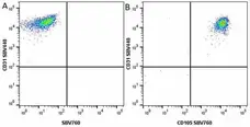
Figure B. StarBright Violet 440 conjugated Mouse anti Human CD31 antibody, clone WM59 (MCA1738SBV440) and StarBright Violet 760 conjugated Mouse anti Human CD105 antibody, clone SN6 (MCA1557SBV760). All experiments performed on HUVEC cells gated on live single cells in the presence of 10% human serum.
Data acquired on the ZE5 Cell Analyzer.
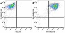
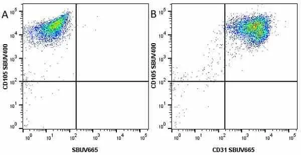
Figure B. StarBright UltraViolet 400 conjugated Mouse anti Human CD105 antibody, clone SN6 (MCA1557SBUV400) and StarBright UltraViolet 665 conjugated Mouse anti Human CD31 antibody, clone WM59 (MCA1738SBUV665). All experiments performed on HUVEC cells gated on live single cells in the presence of 10% human serum.
Data acquired on the ZE5 Cell Analyzer.
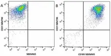
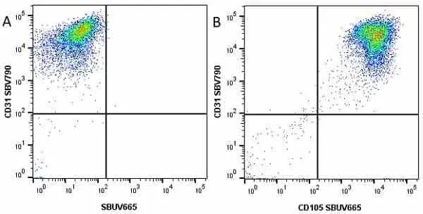
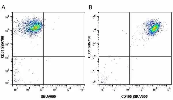
Figure B. StarBright Violet 790 conjugated Mouse anti Human CD31 antibody, clone WM59 (MCA1738SBV790) and StarBright UltraViolet 665 conjugated Mouse anti Human CD105 antibody, clone SN6 (MCA1557SBUV665). All experiments performed on HUVEC cells gated on live single cells in the presence of 10% human serum.
Data acquired on the ZE5 Cell Analyzer.
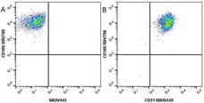
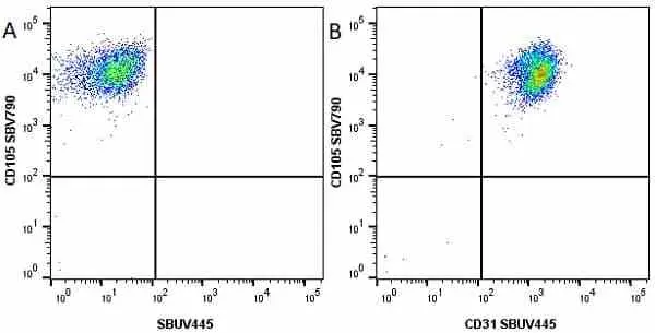
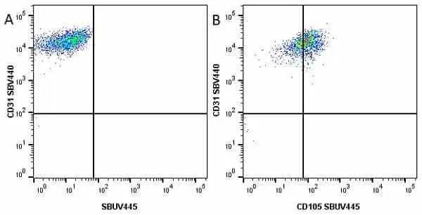
Figure B. Starbright Violet 440 conjugated Mouse anti Human CD31 antibody, clone WM59 (MCA1738SBV440) and StarBright UltraViolet 445 conjugated Mouse anti Human CD105 antibody, clone SN6 (MCA1557SBUV445) . All experiments performed on HUVEC cells gated on live single cells in the presence of 10% human serum.
Data acquired on the ZE5 Cell Analyzer.

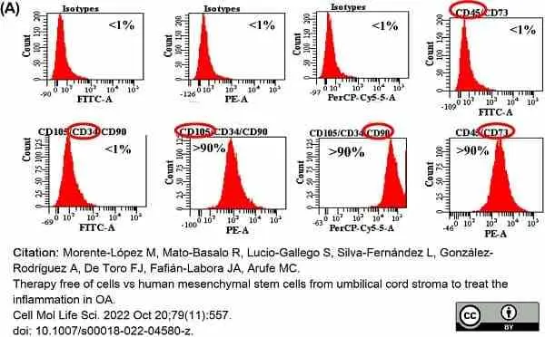
R-Phycoerythrin conjugated Mouse anti Humam CD105 antibody, clone SN6 (MCA1557PE) used to evaluate endoglin expression on human mesenchymal stem cells by flow cytometry.
Image caption:
Characterisation of MSCs and their derived EV from human umbilical cord estroma. A One representative fluorescence-activated cell sorting (FACS) assay is shown. The antibody is indicated at the top of each plot and its linked fluorochrome at the bottom. Positive MSC markers (CD105, CD90 and CD73) and negative haematopoietic markers (CD34 and CD45).
From: Morente-López M, Mato-Basalo R, Lucio-Gallego S, Silva-Fernández L, González-Rodríguez A, De Toro FJ, Fafián-Labora JA, Arufe MC.
Therapy free of cells vs human mesenchymal stem cells from umbilical cord stroma to treat the inflammation in OA.
Cell Mol Life Sci. 2022 Oct 20;79(11):557.
doi: 10.1007/s00018-022-04580-z.
This image is from an open access article distributed under terms of a Creative Commons Attribution License.
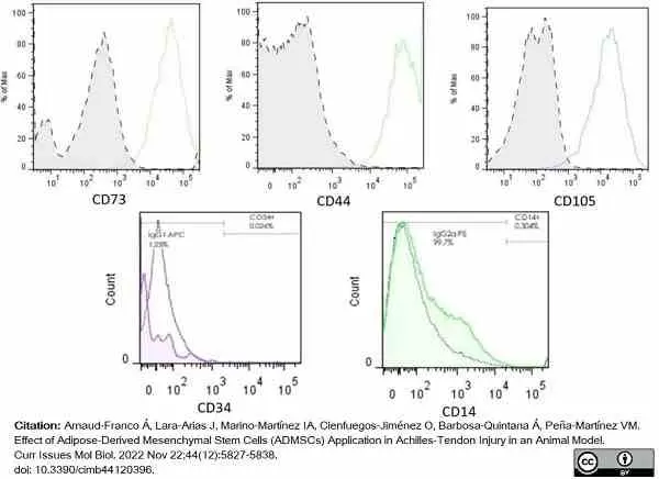
FITC conjugated Mouse anti Human CD105 antibody, clone SN6 (MCA1557F) used for the characterization of rabbit mesenchymal stem cells by flow cytometry.
Image caption:
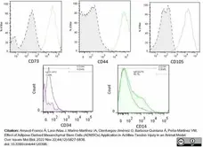
Flow cytometry analysis. Adherent cells that showed MSC morphology were characterized using specific markers for stem cells according to the International Society for Cellular Therapy (ISCT). Negative controls of each antibody’s own isotype were included to rule out nonspecific signals and unlabeled cells (dotted line curves). CD73, CD44, and CD105 stained datasets were found positive after overlay, whereas CD34 and CD14 stained datasets were negative, thus discarding a hematopoietic origin of the sampled cells and suggesting an MSC phenotype.
From: Arnaud-Franco Á, Lara-Arias J, Marino-Martínez IA, Cienfuegos-Jiménez O, Barbosa-Quintana Á, Peña-Martínez VM.
Effect of Adipose-Derived Mesenchymal Stem Cells (ADMSCs) Application in Achilles-Tendon Injury in an Animal Model.
Curr Issues Mol Biol. 2022 Nov 22;44(12):5827-38.
doi: 10.3390/cimb44120396.
This image is from an open access article distributed under terms of a Creative Commons Attribution License.
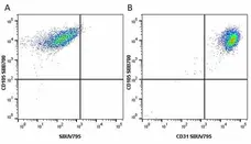
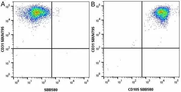
Figure B. StarBright Blue 700 conjugated Mouse anti Human CD105 antibody, clone SN6 (MCA1557SBB700) and StarBright UltraViolet 795 conjugated Mouse anti Human CD31 antibody, clone WM59 (MCA1738SBUV795). All experiments performed on HUVEC cells gated on live single cells in the presence of 10% human serum.
Data acquired on the ZE5 Cell Analyzer.
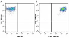
Figure B. StarBright UltraViolet 400 conjugated Mouse anti Human CD31 antibody, clone WM59 (MCA1738SBUV400) and StarBright UltraViolet 795 conjugated Mouse anti human CD105 antibody, clone SN6 (MCA1557SBUV795). All experiments performed on HUVEC cells gated on live single cells in the presence of 10% human serum.
Data acquired on the ZE5 Cell Analyzer.
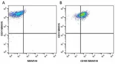
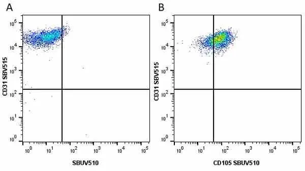
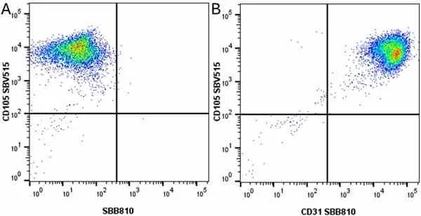
Figure B. StarBright Violet 515 conjugated Mouse anti Human CD31 antibody, clone WM59 (MCA1738SBV515) and StarBright UltraViolet 510 conjugated Mouse anti Human CD105 antibody, clone SN6 (MCA1557SBUV510). All experiments performed on HUVEC cells gated on live single cells in the presence of 10% human serum.
Data acquired on the ZE5 Cell Analyzer.

Figure B. StarBright Violet 475 conjugated Mouse anti Human CD105 antibody, clone SN6 (MCA1557SBV475) and StarBright UltraViolet 510 conjugated Mouse anti Human CD31 antibody, clone WM59 (MCA1738SBUV510). All experiments performed on HUVEC cells gated on live single cells in the presence of 10% human serum.
Data acquired on the ZE5 Cell Analyzer.
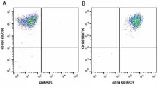
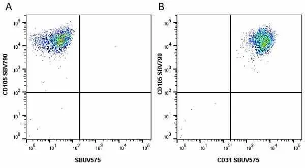
Figure B. StarBright Violet 790 conjugated Mouse anti Human CD105 antibody, clone SN6 (MCA1557SBV790) and StarBright UltraViolet 575 conjugated Mouse anti Human CD31 antibody, clone WM59 (MCA1738SBUV575). All experiments performed on HUVEC cells gated on live single cells in the presence of 10% human serum.
Data acquired on the ZE5 Cell Analyzer.
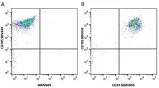
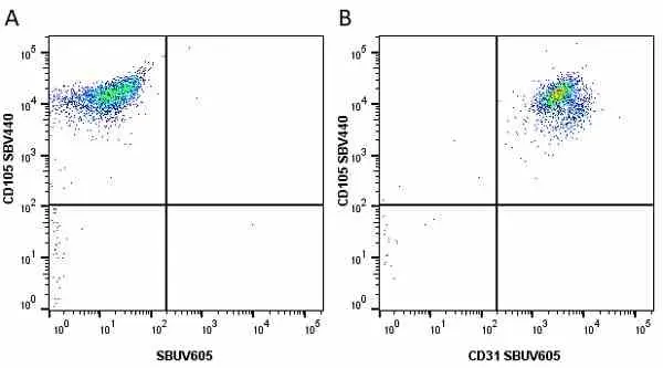
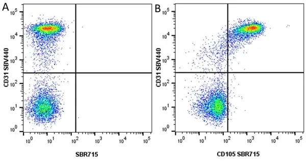
Figure B. StarBright Violet 440 conjugated Mouse anti Human CD105 antibody, clone SN6 (MCA1557SBV440) and StarBright UltraViolet 605 conjugated Mouse anti Human CD31 antibody, clone WM59 (MCA1738SBUV605). All experiments performed on HUVEC cells gated on live single cells in the presence of 10% human serum.
Data acquired on the ZE5 Cell Analyzer.

Data acquired on the ZE5 Cell Analyzer.
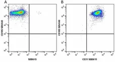
Figure B. StarBright Violet 515 conjugated Mouse anti Human CD31 antibody, clone WM59 (MCA1738SBV515) and StarBright Blue 615 conjugated Mouse anti Human CD105 antibody, clone SN6 (MCA1557SBB615). All experiments performed on HUVEC cells gated on live single cells in the presence of 10% human serum.
Data acquired on the ZE5 Cell Analyzer.

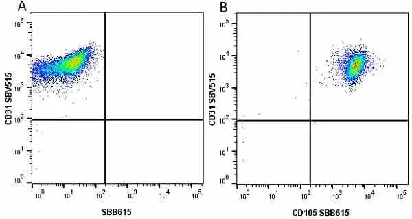
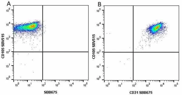
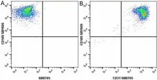
Figure B. StarBright Violet 515 conjugated Mouse anti Human CD105 antibody, clone SN6 (MCA1557SBV515) and StarBright Blue 675 conjugated Mouse anti Human CD31 antibody, clone WM59 (MCA1738SBB675). All experiments performed on HUVEC cells gated on live single cells in the presence of 10% human serum.
Data acquired on the ZE5 Cell Analyzer.
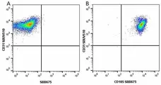
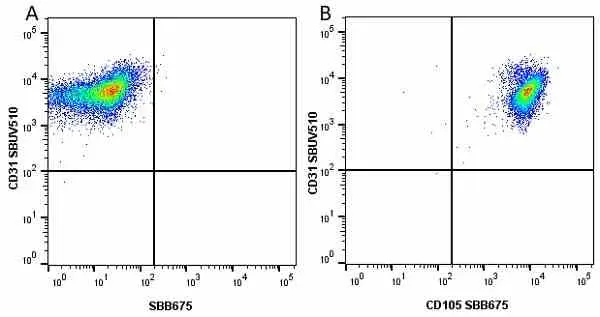
Figure B. StarBright UltraViolet 510 conjugated Mouse anti Human CD31 antibody, clone WM59 (MCA1738SBUV510) and StarBright Blue 675 conjugated Mouse anti Human CD105 clone SN6 (MCA1557SBB675). All experiments performed on HUVEC cells gated on live single cells in the presence of 10% human serum.
Data acquired on the ZE5 Cell Analyzer.

Fitc conjugated Mouse anti Human CD105 antibody, clone SN6 (MCA1557F) used to characterize umbilical cord stroma derived mesenchymal stem cells by flow cytometry.
Image caption:
Characterization of MSCs from umbilical cord stroma and their EV. A One representative fluorescence-activated cell sorting (FACS) assay is shown. Positive MSC markers (CD105, CD90 and CD73) and negative hematopoietic markers (CD34 and CD45).
From: Morente-López M, Mato-Basalo R, Lucio-Gallego S, Gil C, Carrera M, Fafián-Labora JA, Mateos J, Arufe MC.
Effect of miR-21 in mesenchymal stem cells-derived extracellular vesicles behavior.
Stem Cell Res Ther. 2023 Dec 21;14(1):383.
doi: 10.1186/s13287-023-03613-z.
This image is from an open access article distributed under terms of a Creative Commons Attribution License.
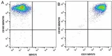
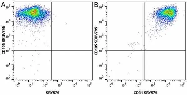
Figure B. StarBright UltraViolet 795 conjugated Mouse anti Human CD105 antibody, clone SN6 (MCA1557SBUV795) and StarBright Yellow 575 conjugated Mouse anti Human CD31 antibody, clone WM59 (MCA1738SBY575). All experiments performed on HUVEC cells gated on live single cells in the presence of 10% human serum.
Data acquired on the ZE5 Cell Analyzer.

Figure B. Alexa Fluor® 647 conjugated Mouse anti Human CD105 antibody, clone SN6 (MCA1557A647) and StarBright Yellow 605 conjugated Mouse anti Human CD31 antibody, clone WM59 (MCA1738SBY605). All experiments performed on HUVEC cells gated on live single cells in the presence of 10% human serum.
Data acquired on the ZE5 Cell Analyzer.
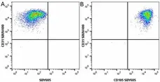
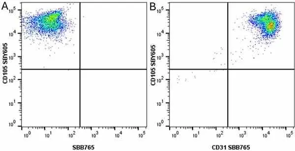
Figure B. StarBright UltraViolet 400 conjugated Mouse anti Human CD31 antibody, clone WM59 (MCA1738SBUV400) and StarBright Yellow 605 conjugated Mouse anti Human CD105 antibody, clone SN6 (MCA1557SBY605). All experiments performed on HUVEC cells gated on live single cells in the presence of 10% human serum.
Data acquired on the ZE5 Cell Analyzer.


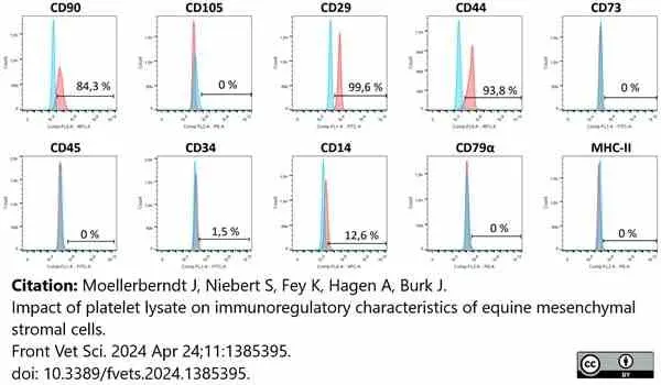
RPE conjugated Mouse anti Human CD105 antibody, clone SN6 (MCA1557PE) used to label equine mesenchymal stem cells and analysed by flow cytometry.
Image caption:
MSC were harvested, stained with fluorescent-labeled monoclonal antibodies
The figure displays representative histograms of surface marker stainings (red) and the corresponding isotype controls (blue).
From: Moellerberndt J, Niebert S, Fey K, Hagen A, Burk J.
Impact of platelet lysate on immunoregulatory characteristics of equine mesenchymal stromal cells.
Front Vet Sci. 2024 Apr 24;11:1385395.
doi: 10.3389/fvets.2024.1385395.
This image is from an open access article distributed under terms of a Creative Commons Attribution License.

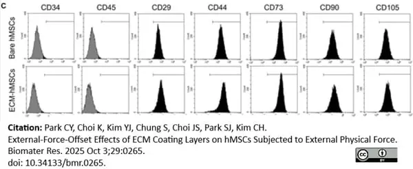
FITC conjugated Mouse anti Human CD105 antibody, clone SN6 (MCA1557F) used for the evaluation of phenotype of cultured mesenchymal stem cells by flow cytometry.
Image caption:
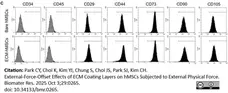
The maintenance of stemness of bare hMSCs and ECM-hMSCs. C) Stemness analysis to confirm stem cell characteristics by flow cytometry analysis.
From: Park CY, Choi K, Kim YJ, Chung S, Choi JS, Park SJ, Kim CH.
External-Force-Offset Effects of ECM Coating Layers on hMSCs Subjected to External Physical Force.
Biomater Res. 2025 Oct 3;29:0265.
doi: 10.34133/bmr.0265.
This image is from an open access article distributed under terms of a Creative Commons Attribution License.
Filter by Application:
F IF Reset| Mouse anti Human CD105 antibody, clone SN6 recognizes human endoglin, also known as CD105. CD105 is a glycoprotein homodimer of ~95 kDa subunits expressed by endothelial cells, activated monocytes and some leukemia cells. |

Our CD105 (SN6) Antibody has been referenced in >73 publications* *Based on June 2020 data from CiteAb's antibody search engine. |
- Target Species
- Human
- Species Cross-Reactivity
-
Target Species Cross Reactivity Horse Cynomolgus monkey Rhesus Monkey Primate Expected from Sequence - N.B. Antibody reactivity and working conditions may vary between species.
- Product Form
- Purified IgG - liquid
- Preparation
- MCA1557T: Purified IgG prepared by affinity chromatography on Protein G from tissue culture supernatant
- MCA1557: Purified IgG prepared by affinity chromatography on Protein A from tissue culture supernatant
- Buffer Solution
- Phosphate buffered saline
- Preservative Stabilisers
- 0.09% sodium azide (NaN3)
- Carrier Free
- Yes
- Immunogen
- Partially purified cell membrane antigens from fresh leukemia cells
- Approx. Protein Concentrations
- IgG concentration 1.0 mg/ml
- Fusion Partners
- Spleen cells from immunized BALB/c mice were fused with cells of the mouse P3/NS1/1-Ag4-1 myeloma cell line
- Regulatory
- For research purposes only
- Guarantee
- 12 months from date of despatch
This product is shipped at ambient temperature. It is recommended to aliquot and store at -20°C on receipt. When thawed, aliquot the sample as needed. Keep aliquots at 2-8°C for short term use (up to 4 weeks) and store the remaining aliquots at -20°C.
Avoid repeated freezing and thawing as this may denature the antibody. Storage in frost-free freezers is not recommended.
Avoid repeated freezing and thawing as this may denature the antibody. Storage in frost-free freezers is not recommended.
This product has been reported to work in the following applications. This information is derived from testing within our laboratories, peer-reviewed publications or personal communications from the originators. Please refer to references indicated for further information. For general protocol recommendations, please visit the antibody protocols page.
| Application Name | Verified | Min Dilution | Max Dilution |
|---|---|---|---|
| Flow Cytometry |  |
1/10 | 1/50 |
| Immunohistology - Frozen 1 | |
||
| Immunohistology - Paraffin | |||
| Immunoprecipitation | |
||
| Western Blotting | |
- 1The epitope recognised by this antibody is reported to be sensitive to formaldehyde fixation and tissue processing. Bio-Rad recommends the use of acetone fixation for frozen sections.
Where this product has not been tested for use in a particular technique this does not necessarily exclude its use in such procedures. Suggested working dilutions are given as a guide only. It is recommended that the user titrates the product for use in their own system using appropriate negative/positive controls.
- Flow Cytometry
- Use 10μl of the suggested working dilution to label 106 cells in 100μl
| Description | Product Code | Applications | Pack Size | List Price | Your Price | Quantity | |
|---|---|---|---|---|---|---|---|
| Mouse IgG1 Negative Control | MCA928 | F | 100 Tests |

|
Log in | ||
| List Price | Your Price | ||||||
|
|
Log in | ||||||
| Description | Mouse IgG1 Negative Control | ||||||
References for CD105 antibody
-
Haruta, Y. & Seon, B.K. (1986) Distinct human leukemia-associated cell surface glycoprotein GP160 defined by monoclonal antibody SN6.
Proc Natl Acad Sci USA 83 (20): 7898-902. -
Pierelli, L. et al. (2000) Modulation of bcl-2 and p27 in human primitive proliferating hematopoietic progenitors by autocrine TGF-beta1 is a cell cycle-independent effect and influences their hematopoietic potential.
Blood 95: 3001-9. -
Nagano, M. et al. (2007) Identification of functional endothelial progenitor cells suitable for the treatment of ischemic tissue using human umbilical cord blood.
Blood 110 (1): 151-60. -
Lozanoska-Ochser, B. et al. (2008) Expression of CD86 on human islet endothelial cells facilitates T cell adhesion and migration.
J Immunol. 181: 6109-16. -
Benetti, A. et al. (2008) Transforming growth factor-beta1 and CD105 promote the migration of hepatocellular carcinoma-derived endothelium.
Cancer Res. 68: 8626-34. -
Diaz-Romero, J. et al. (2008) Immunophenotypic changes of human articular chondrocytes during monolayer culture reflect bona fide dedifferentiation rather than amplification of progenitor cells.
J Cell Physiol. 214: 75-83. -
Sallustio, F. et al. (2010) TLR2 plays a role in the activation of human resident renal stem/progenitor cells.
FASEB J. 24: 514-25. -
Arufe, M.C. et al. (2010) Chondrogenic potential of subpopulations of cells expressing mesenchymal stem cell markers derived from human synovial membranes.
J Cell Biochem. 111: 834-45.
View The Latest Product References
-
Agha-Hosseini, F. et al. (2010) In vitro isolation of stem cells derived from human dental pulp.
Clin Transplant. 24: E23-8. -
Ferro, F. et al. (2010) Biochemical and biophysical analyses of tissue-engineered bone obtained from three-dimensional culture of a subset of bone marrow mesenchymal stem cells.
Tissue Eng Part A 16: 3657-67. -
Jin, H.J. et al. (2010) GD2 expression is closely associated with neuronal differentiation of human umbilical cord blood-derived mesenchymal stem cells.
Cell Mol Life Sci. 67 (11): 1845-58. -
Hauser, P.V. et al. (2010) Stem cells derived from human amniotic fluid contribute to acute kidney injury recovery.
Am J Pathol. 177: 2011-21. -
Braun, J. et al. (2010) Evaluation of the osteogenic and chondrogenic differentiation capacities of equine adipose tissue-derived mesenchymal stem cells.
Am J Vet Res. 71 (10): 1228-36. -
Balmayor, E.R. et al. (2011) Synthesis and functionalization of superparamagnetic poly-ε-caprolactone microparticles for the selective isolation of subpopulations of human adipose-derived stem cells.
J R Soc Interface 8: 896-908. -
Ciccocioppo, R. et al. (2011) Autologous bone marrow-derived mesenchymal stromal cells in the treatment of fistulising Crohn's disease.
Gut 60: 788-98. -
Cox, G. et al. (2011) The use of the reamer-irrigator-aspirator to harvest mesenchymal stem cells.
J Bone Joint Surg Br. 93: 517-24. -
De Schauwer, C. et al. (2012) In search for cross-reactivity to immunophenotype equine mesenchymal stromal cells by multicolor flow cytometry.
Cytometry A 81: 312-23. -
Tso, C. et al. (2012) Phenotypic and functional changes in blood monocytes following adherence to endothelium.
PLoS One 7: e37091. -
Supokawej, A. et al. (2013) Cardiogenic and myogenic gene expression in mesenchymal stem cells after 5-azacytidine treatment.
Turk J Haematol. 30 (2): 115-21. -
Mehrkens, A. et al. (2013) Non-adherent mesenchymal progenitors from adipose tissue stromal vascular fraction.
Tissue Eng Part A 20: 1081-8. -
Kang, S.D. et al. (2013) Isolation of Functional Human Endothelial Cells from Small Volumes of Umbilical Cord Blood.
Ann Biomed Eng. 41: 2181-92. -
Cho, H.J. et al. (2013) Generation of human secondary cardiospheres as a potent cell processing strategy for cell-based cardiac repair.
Biomaterials 34: 651-61. -
Hu, N. et al. (2013) Long-term outcome of the repair of 50 mm long median nerve defects in rhesus monkeys with marrow mesenchymal stem cells-containing, chitosan-based tissue engineered nerve grafts.
Biomaterials 34: 100-11. -
Niu, C.C. et al. (2014) Identification of mesenchymal stem cells and osteogenic factors in bone marrow aspirate and peripheral blood for spinal fusion by flow cytometry and proteomic analysis.
J Orthop Surg Res. 9: 32. -
Williamson, K.A. et al. (2015) Restricted differentiation potential of progenitor cell populations obtained from the equine superficial digital flexor tendon (SDFT).
J Orthop Res. 33 (6): 849-58. -
Yi, T. et al. (2015) Manufacture of Clinical-Grade Human Clonal Mesenchymal Stem Cell Products from Single Colony Forming Unit-Derived Colonies Based on the Subfractionation Culturing Method.
Tissue Eng Part C Methods. 21 (12): 1251-62. -
Mumaw, J.L. et al. (2015) Feline mesenchymal stem cells and supernatant inhibit reactive oxygen species production in cultured feline neutrophils.
Res Vet Sci. 103: 60-9. -
Zhang, J. et al. (2016) Bone mesenchymal stem cells differentiate into myofibroblasts in the tumor microenvironment.
Oncol Lett. 12 (1): 644-50. -
Morsing, M. et al. (2016) Evidence of two distinct functionally specialized fibroblast lineages in breast stroma.
Breast Cancer Res. 18 (1): 108. -
Boccardo, S. et al. (2016) Engineered mesenchymal cell-based patches as controlled VEGF delivery systems to induce extrinsic angiogenesis.
Acta Biomater. 42: 127-35. -
Fernandez-Pernas, P. et al. (2017) CD105+-mesenchymal stem cells migrate into osteoarthritis joint: An animal model.
PLoS One. 12 (11): e0188072. -
Lee, H.J. et al. (2017) ICOSL expression in human bone marrow-derived mesenchymal stem cells promotes induction of regulatory T cells.
Sci Rep. 7: 44486. -
Bertolo, A. et al. (2017) Oxidative status predicts quality in human mesenchymal stem cells.
Stem Cell Res Ther. 8 (1): 3. -
Lützkendorf, J. et al. (2017) Resistance for Genotoxic Damage in Mesenchymal Stromal Cells Is Increased by Hypoxia but Not Generally Dependent on p53-Regulated Cell Cycle Arrest.
PLoS One. 12 (1): e0169921. -
GarikipatiV, N.S. et al. (2018) Isolation and characterization of mesenchymal stem cells from human fetus heart.
PLoS One. 13 (2): e0192244. -
Olimpio, R.M.C. et al. (2018) Cell viability assessed in a reproducible model of human osteoblasts derived from human adipose-derived stem cells.
PLoS One. 13 (4): e0194847. -
Lotfi, R. et al. (2018) ATP promotes immunosuppressive capacities of mesenchymal stromal cells by enhancing the expression of indoleamine dioxygenase.
Immun Inflamm Dis. 6 (4): 448-55. -
May, J.E. et al. (2018) Chemotherapy-induced genotoxic damage to bone marrow cells: long-term implications.
Mutagenesis. 33 (3): 241-251. -
Santos,V.H.D. et al. (2019) Evaluation of alginate hydrogel encapsulated mesenchymal stem cell migration in horses.
Res Vet Sci. 124: 38-45. -
Rey, F. et al. (2019) Adipose-Derived Stem Cells from Fat Tissue of Breast Cancer Microenvironment Present Altered Adipogenic Differentiation Capabilities.
Stem Cells Int. 2019: 1480314. -
Kim, S.H. et al. (2019) Forkhead box O1 (FOXO1) controls the migratory response of Toll-like receptor (TLR3)-stimulated human mesenchymal stromal cells.
J Biol Chem. 294 (21): 8424-37. -
Cargnoni, A. et al. (2020) Amniotic MSCs reduce pulmonary fibrosis by hampering lung B-cell recruitment, retention, and maturation.
Stem Cells Transl Med. 9 (9): 1023-35. -
Tripathy, N.K. et al. (2018) Cardiomyogenic Heterogeneity of Clonal Subpopulations of Human Bone Marrow Mesenchymal Stem Cells.
J Stem Cells Regen Med. 14 (1): 27-33. -
Karpyuk, V. et al. (2019) Innovation-based Approach in Reconstruction of Reduced Jaw Alveolar Ridge Bone using Cell Regeneration Technologies
Archiv Euromedica 9 (2): 147-55. -
Di Paola, A. et al. (2021) Eltrombopag in paediatric immune thrombocytopenia: Iron metabolism modulation in mesenchymal stromal cells.
Br J Haematol. 97 (1): 110-119. -
Watson, L. et al. (2020) Administration of Human Non-Diabetic Mesenchymal Stromal Cells to a Murine Model of Diabetic Fracture Repair: A Pilot Study.
Cells. 9 (6): 1394. -
Noda, S. et al. (2019) Effect of cell culture density on dental pulp-derived mesenchymal stem cells with reference to osteogenic differentiation.
Sci Rep. 9 (1): 5430. -
Kim, M. et al. (2020) A Small-Sized Population of Human Umbilical Cord Blood-Derived Mesenchymal Stem Cells Shows High Stemness Properties and Therapeutic Benefit.
Stem Cells Int. 2020: 5924983. -
Lotfi, R. et al. (2020) Validation of Microbiological Testing of Cellular Medicinal Products Containing Antibiotics.
Transfus Med Hemother. 47 (2): 144-51. -
Piñeiro-Ramil, M. et al. (2020) Immortalizing Mesenchymal Stromal Cells from Aged Donors While Keeping Their Essential Features.
Stem Cells Int. 2020: 5726947. -
Piñeiro-Ramil, M. et al. (2021) Generation of Mesenchymal Cell Lines Derived from Aged Donors.
Int J Mol Sci. 22 (19): 10667. -
Serrano, L.J. et al. (2021) Cell therapy for factor V deficiency: An approach based on human decidua mesenchymal stem cells.
Biomed Pharmacother. 142: 112059. -
Manini, I. et al. (2020) Heterogeneity Matters: Different Regions of Glioblastoma Are Characterized by Distinctive Tumor-Supporting Pathways.
Cancers (Basel). 12 (10): 2960. -
Murata, D. et al. (2022) Osteochondral regeneration of the femoral medial condyle by using a scaffold-free 3D construct of synovial membrane-derived mesenchymal stem cells in horses.
BMC Vet Res. 18 (1): 53. -
Connolly, D.M. et al. (2023) Early Human Pathophysiological Responses to Exertional Hypobaric Decompression Stress.
Aerosp Med Hum Perform. 94 (10): 738-49. -
Jakl, V. et al. (2023) Effect of Expansion Media on Functional Characteristics of Bone Marrow-Derived Mesenchymal Stromal Cells.
Cells. 12 (16): 2105. -
Orikasa, S. et al. (2022) Hypoxia-inducible factor 1α induces osteo/odontoblast differentiation of human dental pulp stem cells via Wnt/β-catenin transcriptional cofactor BCL9.
Sci Rep. 12 (1): 682. -
Tiraihi, T. et al. (2023) A Sequential Culturing System for Generating Epithelial-Like Stem Cells from Human Mesenchymal Stem Cells Derived from Adipose Tissue
Cell and Tissue Biology. 17 (6): 639-52. -
Freitag, N. et al. (2022) Eutopic endometrial immune profile of infertility-patients with and without endometriosis.
J Reprod Immunol. 150: 103489. -
Morente-López, M. et al. (2022) Therapy free of cells vs human mesenchymal stem cells from umbilical cord stroma to treat the inflammation in OA.
Cell Mol Life Sci. 79 (11): 557. -
Creamer, D.G. et al. (2022) Influence of exposure to microbial ligands, immunosuppressive drugs and chronic kidney disease on endogenous immunomodulatory gene expression in feline adipose-derived mesenchymal stem cells.
J Feline Med Surg. 24 (6): e43-e56. -
Arnaud-Franco, Á. et al. (2022) Effect of Adipose-Derived Mesenchymal Stem Cells (ADMSCs) Application in Achilles-Tendon Injury in an Animal Model.
Curr Issues Mol Biol. 44 (12): 5827-38. -
Morente-López, M. et al. (2023) Effect of miR-21 in mesenchymal stem cells-derived extracellular vesicles behavior.
Stem Cell Res Ther. 14 (1): 383. -
Lo, H.Y. et al. (2021) High Induction of IL-6 Secretion From hUCMSCs Optimize the Potential of hUCMSCs and TCZ as Therapy for COVID-19-Related ARDS.
Cell Transplant. 30: 9636897211054481. -
de Barcelos, M.S. et al. (2024) Extracellular vesicles derived from bovine adipose-derived mesenchymal stromal cells enhance in vitro embryo production from lesioned ovaries
Cytotherapy. 20 May [Epub ahead of print]. -
Moellerberndt, J. et al. (2024) Impact of platelet lysate on immunoregulatory characteristics of equine mesenchymal stromal cells.
Front Vet Sci. 11: 1385395. -
Piñeiro-Ramil, M. et al. (2023) Generation of human immortalized chondrocytes from osteoarthritic and healthy cartilage : a new tool for cartilage pathophysiology studies.
Bone Joint Res. 12 (1): 46-57. -
Tafuri, W.L. et al. (2022) Skin fibrosis associated with keloid, scleroderma and Jorge Lobo's disease (lacaziosis): An immuno-histochemical study.
Int J Exp Pathol. 103 (6): 234-44. -
Giesen, M. et al. (2025) Rap1 Guanosine Triphosphate Hydrolase (GTPase) Regulates Shear Stress-Mediated Adhesion of Mesenchymal Stromal Cells.
Biology (Basel). 14 (1): 96. -
Abreu, C.A. et al. (2025) Early ultrastructural damage in retina and optic nerve following intraocular pressure elevation.
Vision Res. 227: 108544. -
Parker, J.L. et al. (2025) Impact of IL-6 and TGF-β1 on equine mesenchymal stromal cell homing gene expression.
Res Vet Sci. 197: 105969. -
Park, C.Y. et al. (2025) External-Force-Offset Effects of ECM Coating Layers on hMSCs Subjected to External Physical Force.
Biomater Res. 29: 0265. -
Moreno, S. et al. (2025) Generation and characterization of an ovine cell line derived from peripheral blood and its potential use in the study of livestock and zoonotic viral infections.
Virol J. 22 (1): 392. -
Seo, M.G. et al. (2026) Application of infrapatellar fat pad-derived stem cells after surgical reconstruction of a ruptured cranial cruciate ligament in a dog.
Vet Res Commun. 50 (3): 235.
Further Reading
-
Carrade, D.D. et al. (2012) Comparative Analysis of the Immunomodulatory Properties of Equine Adult-Derived Mesenchymal Stem Cells.
Cell Med. 4: 1-11. -
Burk, J. et al. (2013) Equine cellular therapy--from stall to bench to bedside?
Cytometry A 83 (1): 103-13.
- Synonyms
- Endoglin
- RRID
- AB_321986
- UniProt
- P17813
- Entrez Gene
- ENG
- GO Terms
- GO:0001300 chronological cell aging
- GO:0001569 patterning of blood vessels
- GO:0001937 negative regulation of endothelial cell proliferation
- GO:0001947 heart looping
- GO:0003084 positive regulation of systemic arterial blood pressure
- GO:0007155 cell adhesion
- GO:0004888 transmembrane receptor activity
- GO:0005024 transforming growth factor beta receptor activity
- GO:0005072 transforming growth factor beta receptor, cytoplasmic mediator activity
- View More GO Terms
- GO:0005114 type II transforming growth factor beta receptor binding
- GO:0005534 galactose binding
- GO:0005539 glycosaminoglycan binding
- GO:0005615 extracellular space
- GO:0005624 membrane fraction
- GO:0007179 transforming growth factor beta receptor signaling pathway
- GO:0050431 transforming growth factor beta binding
- GO:0009897 external side of plasma membrane
- GO:0009986 cell surface
- GO:0010552 positive regulation of gene-specific transcription from RNA polymerase II promoter
- GO:0010553 negative regulation of gene-specific transcription from RNA polymerase II promoter
- GO:0010862 positive regulation of pathway-restricted SMAD protein phosphorylation
- GO:0017015 regulation of transforming growth factor beta receptor signaling pathway
- GO:0022009 central nervous system vasculogenesis
- GO:0022617 extracellular matrix disassembly
- GO:0030155 regulation of cell adhesion
- GO:0030509 BMP signaling pathway
- GO:0030512 negative regulation of transforming growth factor beta receptor signaling pathway
- GO:0030513 positive regulation of BMP signaling pathway
- GO:0031953 negative regulation of protein autophosphorylation
- GO:0034713 type I transforming growth factor beta receptor binding
- GO:0042060 wound healing
- GO:0042127 regulation of cell proliferation
- GO:0042803 protein homodimerization activity
- GO:0045449 regulation of transcription
- GO:0048185 activin binding
- GO:0048745 smooth muscle tissue development
- GO:0048844 artery morphogenesis
- GO:0048845 venous blood vessel morphogenesis
- GO:0051001 negative regulation of nitric-oxide synthase activity
- GO:0060326 cell chemotaxis
- GO:0060394 negative regulation of pathway-restricted SMAD protein phosphorylation
- GO:0070022 transforming growth factor beta receptor complex
- GO:0070483 detection of hypoxia
MCA1557T
MCA1557
If you cannot find the batch/lot you are looking for please contact our technical support team for assistance.
View more products with CD105 specificity
Please Note: All Products are "FOR RESEARCH PURPOSES ONLY"
View all Anti-Human ProductsAlways be the first to know.
When we launch new products and resources to help you achieve more in the lab.
Yes, sign me up