CD4 antibody | CA13.1E4
Mouse anti Dog CD4
- Product Type
- Monoclonal Antibody
- Clone
- CA13.1E4
- Isotype
- IgG1
- Specificity
- CD4

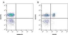
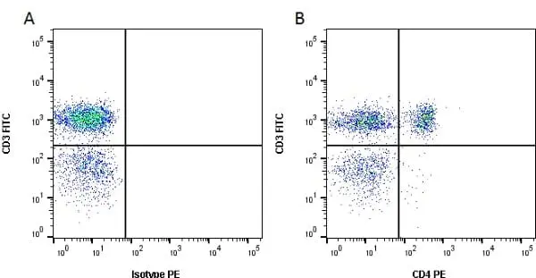
Figure B. FITC conjugated Mouse anti Dog CD3 antibody, clone CA17.2A12 (MCA1774F) and unconjugated Mouse anti Dog CD4 antibody, clone CA13.1E4 (MCA1998S) detected with RPE conjugated Rabbit anti Mouse IgG1 antibody (STAR12A). All experiments performed on red cell lysed canine blood gated on lymphoid cells in the presence of 10% dog serum.
Data acquired on the ZE5 Cell Analyzer.

Mouse anti Dog CD4 antibody, clone CA13.1E4 (MCA1998S) used to label canine CD4 expressing cells by immunofluorescence.
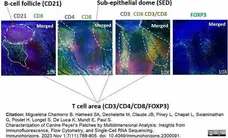
Image caption:
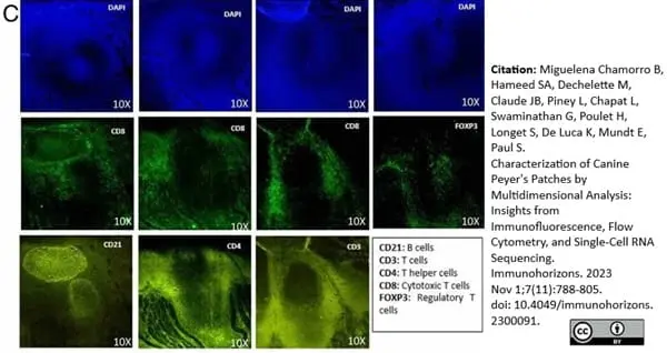
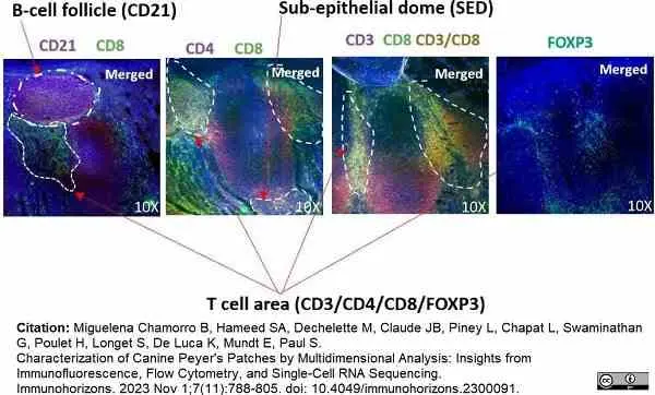
Structural organization of canine Peyer’s patches, according to immunofluorescence data.(C) Immunofluorescence staining of PPs. PPs were stained with DAPI in combination with Abs against CD8 and one of the following markers: CD21, CD4, or FOXP3. Epifluorescence microscopy is shown at original magnification ×10, visualizing specific structures, including the subepithelial dome (SED), T cell areas, and B cell follicles.
From: Miguelena Chamorro B, Hameed SA, Dechelette M, Claude JB, Piney L, Chapat L, Swaminathan G, Poulet H, Longet S, De Luca K, Mundt E, Paul S.
Characterization of Canine Peyer's Patches by Multidimensional Analysis: Insights from Immunofluorescence, Flow Cytometry, and Single-Cell RNA Sequencing.
Immunohorizons. 2023 Nov 1;7(11):788-805.
doi: 10.4049/immunohorizons.2300091.
This image is from an open access article distributed under terms of a Creative Commons Attribution License.
Mouse anti Dog CD4 antibody, clone CA13.1E4 (MCA1998S) used to label canine CD4 expressing cells by immunofluorescence.
Image caption:
Structural organization of canine Peyer’s patches, according to immunofluorescence data.(C) Immunofluorescence staining of PPs. PPs were stained with DAPI in combination with Abs against CD8 and one of the following markers: CD21, CD4, or FOXP3. Epifluorescence microscopy is shown at original magnification ×10, visualizing specific structures, including the subepithelial dome (SED), T cell areas, and B cell follicles.
From: Miguelena Chamorro B, Hameed SA, Dechelette M, Claude JB, Piney L, Chapat L, Swaminathan G, Poulet H, Longet S, De Luca K, Mundt E, Paul S.
Characterization of Canine Peyer's Patches by Multidimensional Analysis: Insights from Immunofluorescence, Flow Cytometry, and Single-Cell RNA Sequencing.
Immunohorizons. 2023 Nov 1;7(11):788-805.
doi: 10.4049/immunohorizons.2300091.
This image is from an open access article distributed under terms of a Creative Commons Attribution License.
Filter by Application:
F IF Reset| Mouse anti Dog CD4 antibody (CA13.1E4), a monoclonal antibody specific for canine CD4. CA13.1E4 was clustered at the first Canine Leukocyte Antigen Workshop (Claw) [Cobbold et al. 1992 ] and identifies, by immunoprecipitation a ~60 kDa monomeric protein under both reducing and non-reducing conditions. Canine CD4 is a surface glycoprotein expressed by non CD8 expressing T lymphocytes, with similar developmental and functional characteristics to T helper cells described in other mammalian species. Mouse anti Dog CD4 also recognizes a population of CD8 positive cells in the canine thymus (Moore et al. 1992). CD4 expression has also been reported on a population of CD3 positive peripheral blood T lymphocytes which are double positive for CD4 and CD8 (Bismarck et al. 2012 ). Similar reports have been made for cynomolgus monkey (Nam et al. 2000), pig (Saalmuller et al. 1987 , Pescowitz et al. 1990) as well as rat, chicken and human (reviewed Zuckermann 1999 ). Uniquely amongst mammalian species clone CA13.1E4 also recognizes CD4 expressed on canine neutrophils at a similar density to that expressed on canine T helper cells. The functional significance of this is not understood and remains enigmatic in view of the roles described for CD4 in canine and other mammalian species (Moore et al. 1992). Clone CA13.1E4 also demonstrates immunohistological staining of splenic marginal zone macrophages (Moore et al. 1992). Other macrophage populations, such as splenic red pulp macrophages and Langerhans cells do not stain with clone CA13.1E4. Canine CD4 positive T cells can be further characterized according to their simultaneous expression of CD45RA as recognized by Rat anti Canine anti CD45RA clone CA4.1D3 in a manner analogous to that seen with human T cells (Moore et al. 1992). Clone CA13.1E4 has been used amongst a large panel of anti-canine monoclonal antibodies for the study of various lymphoproliferative diseases. T lymphocytes in large granular lymphocyte (LGL) lymphocytosis are negative for CD4 (McDonough and Moore 2000 ). Dogs with chronic myelogenous leukemia demonstrate CD4 positive staining on neutrophils (Tarrant et al. 2001). CD4 expression varies considerably between various canine leukemias. |
- Target Species
- Dog
- Product Form
- Tissue culture supernatant - liquid
- Preservative Stabilisers
- < 0.1% sodium azide (NaN3)
- Immunogen
- Canine Thymocytes
- Fusion Partners
- Spleen cells from immunised Balb/c mice were fused with cells of the P3X63-Ag.653 mouse myeloma cell line.
- Regulatory
- For research purposes only
- Guarantee
- 12 months from date of despatch
This product is shipped at ambient temperature. It is recommended to aliquot and store at -20°C on receipt. When thawed, aliquot the sample as needed. Keep aliquots at 2-8°C for short term use (up to 4 weeks) and store the remaining aliquots at -20°C.
Avoid repeated freezing and thawing as this may denature the antibody. Storage in frost-free freezers is not recommended.
Avoid repeated freezing and thawing as this may denature the antibody. Storage in frost-free freezers is not recommended.
This product has been reported to work in the following applications. This information is derived from testing within our laboratories, peer-reviewed publications or personal communications from the originators. Please refer to references indicated for further information. For general protocol recommendations, please visit the antibody protocols page.
| Application Name | Verified | Min Dilution | Max Dilution |
|---|---|---|---|
| Flow Cytometry |  |
Neat | |
| Immunohistology - Frozen | |
||
| Immunoprecipitation | |
Where this product has not been tested for use in a particular technique this does not necessarily exclude its use in such procedures. Suggested working dilutions are given as a guide only. It is recommended that the user titrates the product for use in their own system using appropriate negative/positive controls.
- Flow Cytometry
- Use 25μl of the suggested working dilution to label 106 cells or 100μl whole blood
- Histology Positive Control Tissue
- Canine spleen or lymph node
| Description | Product Code | Applications | Pack Size | List Price | Your Price | Quantity | |
|---|---|---|---|---|---|---|---|
| Mouse IgG1 Negative Control | MCA928 | F | 100 Tests |

|
Log in | ||
| List Price | Your Price | ||||||
|
|
Log in | ||||||
| Description | Mouse IgG1 Negative Control | ||||||
References for CD4 antibody
-
Moore, P.F. et al. (1992) Monoclonal antibodies specific for canine CD4 and CD8 define functional T-lymphocyte subsets and high-density expression of CD4 by canine neutrophils.
Tissue Antigens 40(2): 75-85. -
Cobbold, S. & Metcalfe, S. (1994) Monoclonal antibodies that define canine homologues of human CD antigens: summary of the First International Canine Leukocyte Antigen Workshop (CLAW).
Tissue Antigens. 43 (3): 137-54. -
Veenhof, E.Z. et al. (2011) Characterisation of T cell phenotypes, cytokines and transcription factors in the skin of dogs with cutaneous adverse food reactions.
Vet J. 187 (3): 320-4. -
Izci C et al. (2015) Clinical and light microscopic studies of the conjunctival tissues of dogs with bilateral keratoconjunctivitis sicca before and after treatment with topical 2% cyclosporine.
Biotech Histochem. 90 (3): 223-30. -
Ricklin, M.E. et al. (2010) Characterization of canine dendritic cells in healthy, atopic, and non-allergic inflamed skin.
J Clin Immunol. 30 (6): 845-54. -
Wijewardana, V. et al. (2013) Production of canine soluble CD40 ligand to induce maturation of monocyte derived dendritic cells for cancer immunotherapy.
Vet Immunol Immunopathol. 156 (1-2): 121-7. -
Kamiie, J. et al. (2014) Quantitative analysis of CD3ε in a cloned canine lymphoma cell line by selected reaction monitoring assay.
Biosci Biotechnol Biochem. 78 (2): 271-5. -
Heinrich, F. et al. (2015) Immunophenotyping of immune cell populations in the raccoon (Procyon lotor).
Vet Immunol Immunopathol. 168 (3-4): 140-6.
View The Latest Product References
-
Yuasa, K. et al. (2007) Injection of a recombinant AAV serotype 2 into canine skeletal muscles evokes strong immune responses against transgene products.
Gene Ther. 14 (17): 1249-60. -
Lin, S. et al. (2015) IMMUNE CHARACTERIZATION OF PERIPHERAL BLOOD MONONUCLEAR CELLS OF THE DOGS RESTORED FROM INOCULATION OF CANINE TRANSMISSIBLE VENEREAL TUMOR CELLS
Taiwan Veterinary Journal. 40 (04): 181-90. -
Constantinoiu, C.C. et al. (2015) Mucosal tolerance of the hookworm Ancylostoma caninum in the gut of naturally infected wild dogs.
Parasite Immunol. 37 (10): 510-520. -
Lin, C.S. et al. (2018) Activating natural killer (NK) cytotoxicity of canine CD5-CD21- cells requires low surface CD5 density NK cells.
Iran J Vet Res. 19 (2): 87-95. -
Bertolo, P.H.L. et al. (2022) Influence of serum progesterone levels on the inflammatory response of female dogs with visceral leishmaniosis.
Vet Parasitol. 302: 109658. -
Troupel, T. et al. (2022) Generalised idiopathic polymyositis mimicking masticatory myositis in a dog
Veterinary Record Case Reports. 10 (4) e452. -
Anthonyraj, S. et al. (2024) Chicory root powder included as a prebiotic in different cereal-based diets for dogs: Influences on gut health, metabolic and immunological status
Bioactive Carbohydrates and Dietary Fibre. : 100414. -
Miguelena Chamorro, B. et al. (2023) Characterization of Canine Peyer's Patches by Multidimensional Analysis: Insights from Immunofluorescence, Flow Cytometry, and Single-Cell RNA Sequencing.
Immunohorizons. 7 (11): 788-805. -
Mushati, K.A. et al. (2026) Modulation of uterine responses in dogs: The divergent effects of semen and embryonal signals on steroidogenic receptors and selected immune system-related factors.
Theriogenology. 250: 117644.
- RRID
- AB_2077610
- UniProt
- P33705
- Entrez Gene
- CD4
- GO Terms
- GO:0007155 cell adhesion
- GO:0016021 integral to membrane
- GO:0006955 immune response
- GO:0045058 T cell selection
View more products with CD4 specificity
Please Note: All Products are "FOR RESEARCH PURPOSES ONLY"
View all Anti-Dog ProductsAlways be the first to know.
When we launch new products and resources to help you achieve more in the lab.
Yes, sign me up