CD34 antibody | 1H6
Mouse anti Dog CD34
- Product Type
- Monoclonal Antibody
- Clone
- 1H6
- Isotype
- IgG1
- Specificity
- CD34

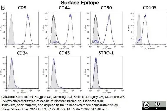
Mouse anti Canine CD34 antibody, clone 1H6 (MCA2411GA) used as part of a panel of antibodies to demonstrate the phenotype of canine mesenchymal stem cells by flow cytometry together with mesenchymal markers CD9, CD44, CD90 and CD105. Negative results for lineage cell markers CD34, CD45 and STRO-1 are shown.
Image caption:
b. Representative histograms demonstrating positive and negative staining of marrow cMSCs from a single donor
From: Bearden RN, Huggins SS, Cummings KJ, Smith R, Gregory CA, Saunders WB.
In-vitro characterization of canine multipotent stromal cells isolated from synovium, bone marrow, and adipose tissue: a donor-matched comparative study.
Stem Cell Res Ther. 2017 Oct 3;8(1):218.
This image is from an open access article distributed under terms of a Creative Commons Attribution License.
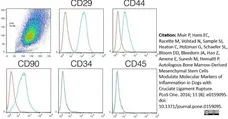
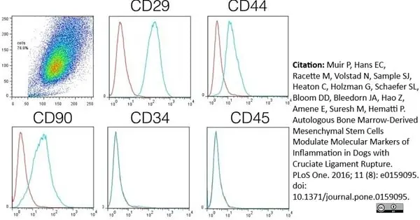
Mouse anti Canine CD34 antibody, clone 1H6 (MCA2411GA) used for the evaluation of CD34 expression on canine bone marrow-derived mesenchymal stem cells by flow cytometry.
Image caption:
The phenotype of cultured bone marrow-derived mesenchymal stem cells (BM-MSCs) from each dog was confirmed before autologous injection.
Expression of CD29, CD34, CD44, CD45, and CD90 by cultured cells was evaluated by flow cytometric analysis using anti-CD29, anti-CD34, anti-CD44, anti-CD45, and anti-CD90 antibodies, after selection of the cell population of interest. Control isotype antibody is plotted in red in each histogram plot. Cultured cells were confirmed as BM-MSCs by their appearance in culture and by the CD29+CD44+CD90+CD34-CD45- phenotype.
From: Muir P, Hans EC, Racette M, Volstad N, Sample SJ, Heaton C, et al. (2016)
Autologous Bone Marrow-Derived Mesenchymal Stem Cells Modulate Molecular Markers of Inflammation in Dogs with Cruciate Ligament Rupture.
PLoS ONE 11(8): e0159095.
This image is from an open access article distributed under the terms of the Creative Commons Attribution License.
Filter by Application:
F Reset| Mouse anti dog CD34 antibody, clone 1H6 recognizes the canine homologue of CD34, a glycosylated type 1 transmembrane protein of approximately 110 kDa (McSweeney et al. 1998) expressed on the cell suface of endothelial cells and haematopoietic stem cells. Mouse anti dog CD34 antibody, clone 1H6 is a key marker of canine hematopoietic progenitor cells and is reported for use in CD34+ enrichment assays, (Goerner et al. 2001) and (Horn et al. 2004). |
- Target Species
- Dog
- Product Form
- Purified IgG - liquid
- Preparation
- Purified IgG prepared by affinity chromatography on Protein A from tissue culture supernatant
- Buffer Solution
- Phosphate buffered saline
- Preservative Stabilisers
- 0.09% Sodium Azide (NaN3)
- Carrier Free
- Yes
- Immunogen
- Canine CD34 fusion protein.
- Approx. Protein Concentrations
- IgG concentration 1.0 mg/ml
- Fusion Partners
- Spleen cells from immunized BALB/c mice were fused with cells of the mouse NS-1/FOX-NY myeloma cell line.
- Regulatory
- For research purposes only
- Guarantee
- 12 months from date of despatch
This product is shipped at ambient temperature. It is recommended to aliquot and store at -20°C on receipt. When thawed, aliquot the sample as needed. Keep aliquots at 2-8°C for short term use (up to 4 weeks) and store the remaining aliquots at -20°C.
Avoid repeated freezing and thawing as this may denature the antibody. Storage in frost-free freezers is not recommended.
Avoid repeated freezing and thawing as this may denature the antibody. Storage in frost-free freezers is not recommended.
This product has been reported to work in the following applications. This information is derived from testing within our laboratories, peer-reviewed publications or personal communications from the originators. Please refer to references indicated for further information. For general protocol recommendations, please visit the antibody protocols page.
| Application Name | Verified | Min Dilution | Max Dilution |
|---|---|---|---|
| Flow Cytometry |  |
1/50 | 1/100 |
| Western Blotting | |
Where this antibody has not been tested for use in a particular technique this does not necessarily exclude its use in such procedures. Suggested working dilutions are given as a guide only. It is recommended that the user titrates the antibody for use in their own system using appropriate negative/positive controls.
- Flow Cytometry
- Use 10ul of the suggested working dilution to label 1x10⁶ cells in 100ul.
- Western Blotting
- MCA2411GA detects a band of approximately 110kDa.
| Description | Product Code | Applications | Pack Size | List Price | Your Price | Quantity | |
|---|---|---|---|---|---|---|---|
| Mouse IgG1 Negative Control | MCA928 | F | 100 Tests |

|
Log in | ||
| List Price | Your Price | ||||||
|
|
Log in | ||||||
| Description | Mouse IgG1 Negative Control | ||||||
Source Reference
-
McSweeney, P.A. et al. (1998) Characterization of monoclonal antibodies that recognize canine CD34.
Blood. 91 (6): 1977-86.
References for CD34 antibody
-
Goerner, M. et al. (1999) The use of granulocyte colony-stimulating factor during retroviral transduction on fibronectin fragment CH-296 enhances gene transfer into hematopoietic repopulating cells in dogs.
Blood. 94 (7): 2287-92. -
Bhattacharya, V. et al. (2000) Enhanced endothelialization and microvessel formation in polyester grafts seeded with CD34(+) bone marrow cells.
Blood. 95 (2): 581-5. -
Goerner, M. et al. (2001) Sustained multilineage gene persistence and expression in dogs transplanted with CD34(+) marrow cells transduced by RD114-pseudotype oncoretrovirus vectors.
Blood. 98 (7): 2065-70. -
Georges, G. et al. (2001) Engraftment of DLA-haploidentical marrow with ex vivo expanded, retrovirally transduced cytotoxic T lymphocytes.
Blood. 98:3447-55. -
Horn, P.A. et al. (2004) Efficient lentiviral gene transfer to canine repopulating cells using an overnight transduction protocol.
Blood. 103 (10): 3710-6. -
Avallone, G. et al. (2007) The spectrum of canine cutaneous perivascular wall tumors: morphologic, phenotypic and clinical characterization.
Vet Pathol. 44 (5): 607-20. -
Palmieri, C. et al. (2013) Use of electron microscopy to classify canine perivascular wall tumors.
Vet Pathol. 50 (2): 226-33. -
Bearden, R.N. et al. (2017) In-vitro characterization of canine multipotent stromal cells isolated from synovium, bone marrow, and adipose tissue: a donor-matched comparative study.
Stem Cell Res Ther. 8 (1): 218.
View The Latest Product References
-
Trindade, A.B. et al. (2017) Mesenchymal-like stem cells in canine ovary show high differentiation potential.
Cell Prolif. Oct 08 [Epub ahead of print]. -
Lee, S.H. et al. (2016) Impact of local injection of brain-derived neurotrophic factor-expressing mesenchymal stromal cells (MSCs) combined with intravenous MSC delivery in a canine model of chronic spinal cord injury.
Cytotherapy. Oct 28 [Epub ahead of print]. -
Muir, P. et al. (2016) Autologous Bone Marrow-Derived Mesenchymal Stem Cells Modulate Molecular Markers of Inflammation in Dogs with Cruciate Ligament Rupture.
PLoS One. 11 (8): e0159095. -
Rajawat, Y.S. et al. (2021) In Vivo Gene Therapy for Canine SCID-X1 Using Cocal-Pseudotyped Lentiviral Vector.
Hum Gene Ther. 32 (1-2): 113-27. -
Grudzien, M. et al. (2021) A newly established canine NK-type cell line and its cytotoxic properties.
Vet Comp Oncol. 19 (3): 567-77. -
Tongu, E.A.O. et al. (2021) Allogenic mesenchymal stem cell-conditioned medium does not affect sperm parameters and mitigates early endometrial inflammatory responses in mares.
Theriogenology. 169: 1-8. -
Jaensch, S. et al. (2022) Clinicopathologic and immunophenotypic features in dogs with presumptive large granular lymphocyte leukaemia
Australian Veterinary Journal. [Epub ahead of print]. -
Salari Sedigh, H. et al. (2023) In vitro investigation of canine periodontal ligament-derived mesenchymal stem cells: A possibility of promising tool for periodontal regeneration.
J Oral Biol Craniofac Res. 13 (3): 403-11. -
Papa, P.M. et al. (2023) Intratesticular transplantation of allogenic mesenchymal stem cells mitigates testicular destruction after induced heat stress in Miniature-horse stallions.
J Equine Vet Sci. 132: 104961. -
Rezaei, M. et al. (2019) Transplantation of Bone Marrow-Derived Mesenchymal Stem Cells, Platelet-Rich Plasma, and Fibrin Glue for Periodontal Regeneration.
Int J Periodontics Restorative Dent. 39 (1): e32-e45. -
Yang, V.K. et al. (2021) Intravenous administration of allogeneic Wharton jelly-derived mesenchymal stem cells for treatment of dogs with congestive heart failure secondary to myxomatous mitral valve disease.
Am J Vet Res. 82 (6): 487-93. -
Crain, S.K. et al. (2019) Extracellular Vesicles from Wharton's Jelly Mesenchymal Stem Cells Suppress CD4 Expressing T Cells Through Transforming Growth Factor Beta and Adenosine Signaling in a Canine Model.
Stem Cells Dev. 28 (3): 212-26. -
Sheng, R. et al. (2023) Prognostic significance of CD25 expression in dogs with a noninvasive diagnosis of B-cell lymphoma treated with CHOP chemotherapy.
Vet Comp Oncol. 21 (1): 28-35. -
Millanta, F. et al. (2020) Cytologic grading of canine and feline spindle-cell sarcomas of soft tissues and its correlation with histologic grading.
Top Companion Anim Med. 41: 100458. -
Rogato, F. et al. (2024) Leukemia cutis as a prominent clinical sign in a dog with acute myeloid leukemia.
Vet Clin Pathol. 53 (4): 448-57. -
Blockeel, A. et al. (2025) CD94 as a novel marker for immunophenotyping of leukemia and lymphoma in dogs.
Front Vet Sci. 12: 1716800.
Further Reading
-
McSweeney, P. et al. (1996) Canine CD34: cloning of the cDNA and evaluation of an antiserum to recombinant protein.
Blood. 88:1992-2003.
- UniProt
- Q28270
- Entrez Gene
- CD34
- GO Terms
- GO:0016021 integral to membrane
- GO:0030246 carbohydrate binding
- GO:0016337 cell-cell adhesion
- GO:0050900 leukocyte migration
MCA2411GA
If you cannot find the batch/lot you are looking for please contact our technical support team for assistance.
View more products with CD34 specificity
Please Note: All Products are "FOR RESEARCH PURPOSES ONLY"
View all Anti-Dog ProductsAlways be the first to know.
When we launch new products and resources to help you achieve more in the lab.
Yes, sign me up