Fluorescently Labeled Streptavidin for Flow Cytometry
Overview
Highly specific streptavidin-biotin binding is utilized in many applications, for example, HRP labeled streptavidin (STAR5B) and alkaline phosphatase labeled streptavidin (STAR6B) are commonly used in ELISA, western blotting, and immunohistochemistry. We have handy protocols describing the use of streptavidin in immunostaining (frozen tissue sections and paraffin sections) and ELISA (direct and sandwich).
Fluorescently labeled streptavidin is also ideal for flow cytometry to visualize primary antibodies conjugated to biotin, also known as biotinylated primary antibodies. They are compatible with multiplexing panels, making them a great option when the ideal directly labeled fluorescent antibody is not available. We will describe their many benefits for use in flow cytometry, and how to use them successfully in your experiments.
The Biology of Streptavidin
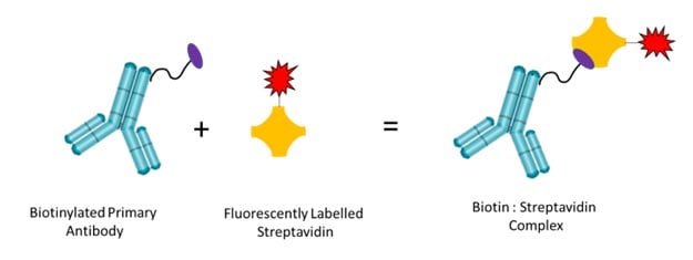
Streptavidin is isolated from Streptomyces avidinii and has four binding sites with strong affinity to biotin, a naturally occurring vitamin. The relatively small size of biotin means that when bound to an antibody it does not hinder the antibody binding site allowing it to still bind to its target antigen.

Fig. 1. Binding of biotinylated antibodies to fluorescently labeled streptavidin. Primary antibodies are biotinylated and bind fluorescently labeled streptavidin with high affinity.
Benefits when using Biotinylated Antibodies with Fluorescently Labeled Streptavidin in Flow Cytometry
There are many features and benefits when using the biotin:streptavidin system, as shown in Table 1, that not only aid multiplex panel design, but also help to achieve good quality flow data.
Table 1. Features and benefits of using biotin: streptavidin in flow cytometry.
Feature |
Benefit |
|---|---|
|
Low background staining |
Streptavidin has a high affinity to biotin, with negligible binding to non-biotinylated macromolecules, and therefore gives very low background. |
|
Stable |
The strong biotin-streptavidin bond can withstand extremes in pH and temperature therefore their interaction remains very stable during assays. |
|
Signal amplification |
Biotinylated antibodies may be conjugated to more than one biotin molecule, each biotin will bind a labeled streptavidin resulting in signal amplification. This makes it ideal to detect low expressing antigens. |
|
Expands antibody choice |
When there is no directly conjugated antibody available or not one in your preferred format using the biotin:streptavidin system can provide the solution. |
|
Suitable for use with all isotypes |
In flow cytometry secondary antibodies need to be carefully chosen to match the isotype of your primary antibody, while also ensuring there is no unwanted binding to other primary antibodies in your experiment. As the biotin:streptavidin system is isotype independent, it overcomes any problems of isotype compatibility. |
|
Large range of biotinylated antibodies |
Many antibodies validated for flow cytometry are available biotinylated. |
|
Simple to conjugate purified antibodies to biotin |
If an antibody of interest is not available biotinylated, it’s easy to conjugate a purified version of the antibody. For example, Biotin (Type 1) Conjugation Kit from Bio-Rad is simple to use, adding biotin to an antibody in minutes. |
|
Large choice of fluorophores |
Bio-Rad’s labeled streptavidin conjugates are available in a range of fluorophores across all the common laser lines, including new StarBright™ Dyes. This gives a wide choice of fluorophores to easily incorporate into new or existing flow panels. |
How to Use Fluorescently Labeled Streptavidin in Flow Cytometry

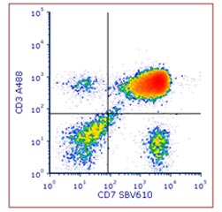
Fig. 2. Staining of human peripheral blood with anti-human CD7 biotin: StarBright Violet 610 streptavidin and Anti-CD3 Alexa Fluor 488. Red cell lysed human blood was stained with Alexa Fluor 488 conjugated Mouse Anti Human CD3 (MCA463A488) and biotin conjugated Mouse Anti Human CD7 (MCA1197B) detected with streptavidin StarBright Violet 610 (STAR210SBV610). Cells were acquired on the ZE5 Cell Analyzer and gated on live single cell lymphocytes.
1. Indirect staining — common two-step method
The most common method to use the biotin:streptavidin system is a two-step staining protocol. This method is similar to using a labeled secondary antibody to detect a primary antibody.
Step 1:
- Incubate cells with a biotinylated primary antibody
- Wash to remove excess antibody
Step 2:
- Incubate cells with a fluorescently labeled streptavidin
- Wash to remove excess streptavidin and acquire samples on a flow cytometer
An example of indirect staining can be seen in Figure 2, where human blood was stained with biotinylated CD7 (MCA1197B) and detected with StarBright Violet 610 labeled streptavidin (STAR210SBV610).
2. Direct staining - premix one-step method
An alternative method is to mix a biotinylated antibody with a fluorescently labeled streptavidin. Cells can be directly stained with this premix complex using a one-step method.
Step 1:
- Incubate cells with a premixed biotinylated primary antibody: fluorescently labeled streptavidin complex
- Wash to remove excess antibody and streptavidin, and acquire samples on a flow cytometer
This method requires optimization of the ratio of biotinylated antibody to fluorescently labeled streptavidin. As shown in Figure 3, Anti-Human CD19 biotin and StarBright UltraViolet 795 labeled streptavidin (STAR210SBUV795) were premixed at three different molar ratios prior to staining. For this pair the 1:3 ratio gave the best staining, having the greatest separation of the positive and negative population and a higher stain index.

Fig. 3. Staining of human PBMCs with biotinylated Anti CD19 premixed with streptavidin StarBright UltraViolet 795. Anti-CD19 biotin antibodies were mixed with streptavidin StarBright UltraViolet 795 (STAR210SBUV795) at different molar ratios for 30 minutes at room temperature prior to staining human PBMCs. Cells were acquired on the ZE5 Cell Analyzer and gated on single cell lymphocytes.
Optimization of the ratio only needs to be done once for each biotinylated antibody: labeled streptavidin pair. If a pairing will be used more than twice this method will save time as staining is reduced by 60-80 minutes over an indirect staining protocol. Also, as the premix is stable for at least seven days a master premix can be made and used over the course of at least a week.
It is important to note that there may be rare cases where this method doesn’t work well. If too many biotins are attached to an antibody or biotin residues are too close to the antigen binding site, there will be steric hinderance resulting in loss of binding. In these rare cases an indirect method will be required to achieve bright staining.
A direct staining protocol will be required when more than one biotinylated antibody is incorporated into a multiplexing panel. Each biotinylated antibody needs to be premixed with a different fluorescent labeled streptavidin. In Figure 4 you can see an example of using three different premixed biotinylated antibody:streptavidin complexes together. The optimal ratio of each pairing was determined and titrated prior to use.

Fig. 4. Staining of human PBMCs with three premixed biotinylated antibody: fluorescently labeled streptavidin complexes. PBMCs were stained with CD2B: STAR210SBV670. CD8B: STAR210SBUV400 and CD19B :STAR210SBV790. Cells were acquired on the ZE5 Cell Analyzer and gated on single cell lymphocytes.
Biotinylated Antibodies
Bio-Rad has a range of biotinylated antibodies validated for use in flow cytometry.
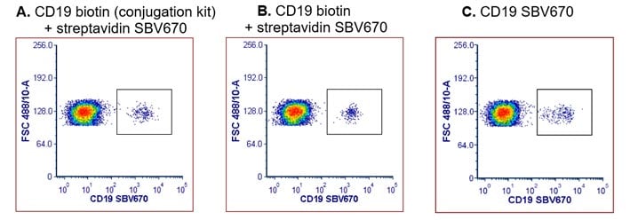
However, if an antibody of interest is not available biotinylated it is easy to conjugate the purified version using a simple kit, such as Biotin (Type 1) Conjugation Kit. As shown in Figure 5, an antibody conjugated using this kit gave similar staining to a commercial biotinylated antibody and a directly conjugated antibody.

Fig. 5. CD19 staining of human peripheral blood using a biotinylated CD19 antibody detected with StarBright Violet 670 streptavidin or a directly conjugated CD19 SBV670 antibody. Red cell lysed human blood was stained using either A. Anti CD19 (MCA1940GA) biotinylated using a conjugation kit (LNK261B) and detected with streptavidin StarBright Violet 670 (STAR210SBV670). B. A commercially available biotinylated Anti CD19 Antibody (MCA1940B) and detected with streptavidin StarBright Violet 670 (STAR210SBV670). C. Anti CD19 directly conjugated to StarBright Violet 670 (MCA1940SBV670). Cells were acquired on the ZE5 Cell Analyzer and gated on live single cell lymphocytes.
Top Tips When Using Biotinylated Antibodies with Fluorescently Labeled Streptavidin in Flow Cytometry
The following tips should be considered when designing flow experiments.
- Perform a titration. It is best practice to titrate your antibody to determine the best concentration of antibody and streptavidin to use. Start with a fixed amount of the biotinylated antibody, using the recommended amount from the supplier, and titrate the fluorescently labeled streptavidin. If there is high non-specific staining next titrate the primary antibody using the fixed amount of the fluorescently labeled streptavidin that has been determined.
- Include a blocking step. Although streptavidin gives negligible nonspecific staining it is recommended to still perform an antibody blocking step that is in most staining protocols. This is due to the presence of primary antibodies that may bind non-specifically to Fc receptors.
- Optimize premixed antibody: streptavidin ratios. The optimal molar ratio of antibody to streptavidin needs to be determined when using premixed complexes. Recommended ratios for initial testing are 3:1, 1:1, and 1:3 of antibody to streptavidin, as these cover the ratios that commonly give the highest stain index.
Streptavidin Formats
Bio-Rad offers a wide range of fluorescently labeled streptavidin including our new StarBright Dyes as well as traditional dyes, shown in Table 2 below.
Table 2. Fluorescently labeled streptavidin available from Bio-Rad.
Fluorophore |
Optimal excitation laser |
Excitation Max, nm |
Emission Max, nm |
Product code |
|---|---|---|---|---|
|
StarBright UltraViolet 400 |
355 |
347 |
394 |
|
|
StarBright UltraViolet 445 |
355 |
347 |
440 |
|
|
StarBright UltraViolet 510 |
355 |
340 |
513 |
|
|
StarBright UltraViolet 575 |
355 |
340 |
569 |
|
|
StarBright UltraViolet 605 |
355 |
340 |
609 |
|
|
StarBright UltraViolet 665 |
355 |
340 |
684 |
|
|
StarBright UltraViolet 740 |
355 |
344 |
742 |
|
|
StarBright UltraViolet 795 |
355 |
340 |
792 |
|
|
StarBright Violet 440 |
405 |
385 |
438 |
|
|
StarBright Violet 475 |
405 |
405 |
479 |
|
|
StarBright Violet 515 |
405 |
401 |
516 |
|
|
StarBright Violet 570 |
405 |
402 |
570 |
|
|
StarBright Violet 610 |
405 |
402 |
607 |
|
|
StarBright Violet 670 |
405 |
400 |
667 |
|
|
StarBright Violet 760 |
405 |
403 |
754 |
|
|
StarBright Violet 790 |
405 |
401 |
782 |
|
|
DyLight 405 |
405 |
400 |
420 |
|
|
StarBright Blue 580 |
488 |
475 |
582 |
|
|
StarBright Blue 615 |
488 |
475 |
612 |
|
|
StarBright Blue 675 |
488 |
476 |
675 |
|
|
StarBright Blue 700 |
488 |
470 |
700 |
|
|
StarBright Blue 765 |
488 |
476 |
764 |
|
|
StarBright Blue 810 |
488 |
477 |
802 |
|
|
FITC |
488 |
490 |
525 |
|
|
DyLight 488 |
488 |
493 |
518 |
|
|
StarBright Yellow 575 |
561 |
548 |
579 |
|
|
StarBright Yellow 605 |
561 |
572 |
606 |
|
|
StarBright Yellow 665 |
561 |
554 |
670 |
Coming soon |
|
StarBright Yellow 720 |
561 |
548 |
719 |
|
|
StarBright Yellow 800 |
561 |
549 |
788 |
|
|
RPE |
561 |
496 |
578 |
|
|
DyLight 550 |
561 |
562 |
576 |
|
|
DyLight 650 |
561 |
564 |
673 |
|
|
StarBright Red 670 |
640 |
653 |
666 |
Coming soon |
|
StarBright Red 715 |
640 |
638 |
712 |
Coming soon |
|
StarBright Red 775 |
640 |
653 |
778 |
Coming soon |
|
StarBright Red 815 |
640 |
654 |
811 |
Coming soon |
|
APC |
640 |
650 |
661 |


