Myelin Proteolipid Protein antibody | plpc1
Mouse anti Myelin Proteolipid Protein
- Product Type
- Monoclonal Antibody
- Clone
- plpc1
- Isotype
- IgG2a
- Specificity
- Myelin Proteolipid Protein


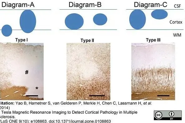
Mouse anti myelin proteolipid protein antibody, clone plpc1 (MCA839G) used for the detection of proteolipid protein on formalin fixed, paraffin embedded brain tissue.
Image caption:
NL scheme and PLP staining. (A) Type-I NL with demyelination of the whole width of the cortex (#) and adjacent WM (*). (B) Type-II intracortical lesion evolving around a vessel. (C) Type-III subpial NL. Demyelination spreads from the pial surface until cortical layer 3. Scale bars represent 500 μm.
From:
Yao B, Hametner S, van Gelderen P, Merkle H, Chen C, et al. (2014)
7 Tesla Magnetic Resonance Imaging to Detect Cortical Pathology in Multiple Sclerosis.
PLoS ONE 9(10): e108863.
This image is from an open access article distributed under the terms of a Creative Commons Attribution License.
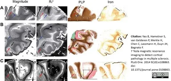
Mouse anti Bovine myelin proteolipid protein antibody, clone plpc1 (MCA839G) used for the detection of proteolipid protein on formalin fixed, paraffin embedded brain tissue.
Image caption:
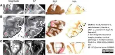
NLs on MRI and tissue sections stained for PLP and iron. Examples Examples of NLs from tissue MS1 (A, B and C) identified on MRI magnitude (TE = 25.2 ms) and R2* images as well as color-coded PLP-staining and iron staining. Red arrows point towards NLs identified by MRI and confirmed to correspond to area of demyelination by the color-coded PLP staining. Blue arrows point towards NLs identified by the color-coded PLP staining and only retrospectively identified by MRI. Black arrows and black box point towards NLs identified by the color-coded PLP staining and not by MRI even upon a second retrospective image inspection. In the black box we include a large area of demyelination which goes entirely undetected by MRI. In the color-coded PLP staining of figure: green = complete WM demyelination, red = complete GM demyelination, yellow = areas of variably reduced myelin density.
From:
Yao B, Hametner S, van Gelderen P, Merkle H, Chen C, et al. (2014)
7 Tesla Magnetic Resonance Imaging to Detect Cortical Pathology in Multiple Sclerosis.
PLoS ONE 9(10): e108863.
This image is from an open access article distributed under the terms of a Creative Commons Attribution License.
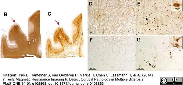
Mouse anti myelin proteolipid protein antibody, clone plpc1 (MCA839G) used for the detection of proteolipid protein on formalin fixed, paraffin embedded brain tissue.
Image caption:
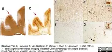
Iron loss in NLs. PLP myelin staining (B) and iron staining (C) on two consecutive slides of tissue MS2 disclose iron loss together with myelin loss in a NL (red arrows). Higher magnification (D, E, F, G) of PLP (D, F) and iron stainings (E, G). In NAGM (D, E), iron is present in oligodendrocytes (black arrow and inset in E) and myelin sheaths. In a NL (F, G), iron is removed from the demyelinated cortical parenchyma and present in perivascular macrophages and microglia (black arrow and inset in G).
From:
Yao B, Hametner S, van Gelderen P, Merkle H, Chen C, et al. (2014)
7 Tesla Magnetic Resonance Imaging to Detect Cortical Pathology in Multiple Sclerosis.
PLoS ONE 9(10): e108863.
doi: 10.1371/journal.pone.0108863
This image is from an open access article distributed under the terms of a Creative Commons Attribution License.
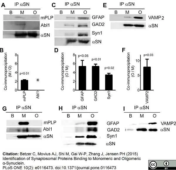
Mouse anti myelin proteolipid protein antibody, clone plpc1 (MCA839G) used for detection of myelin proteolipid protein in human and porcine brain lysates by western blotting
Image caption:
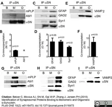
Validation of monomer and oligomer preference of αSN interacting proteins. Proteins pulled down by monomer αSN (M), oligomer αSN (O), and buffer control (B) from porcine (A-F) and human (G-I) brain extracts were analyzed by immunoblotting using antibodies against antigens selected among the monomer and oligomer binding proteins. Monomer binding antigens were myelin proteolipid protein (mPLP) and Abl interactor 1 (Abl1) and oligomer binding proteins were glial fibrillary acidic protein (GFAP), glutamate decarboxylase 2 (GAD2), and synapsin 1 (Syn1). VAMP-2 was tested because it has been reported to bind &apha;SN, although it was not detected in our proteomic analysis. One representative of three experiments is presented for porcine αSN binding proteins (A, C, E), and the quantification of the three experiments is presented in panels B, D, F. The quantification of bands was performed after subtracting the non-specific signal in the buffer control from the specific bands in monomer and oligomer samples. Bars represent mean ratio between monomer and oligomer ± S.D. of the three replicates. The values for binding to monomer and oligomer were compared by Student's t-test and the resulting p-values are listed above the bars. * Indicates that the band intensity from oligomer did not differ significantly from background making quantifications impracticable. In order to ensure that the interaction were not due to species differences between human and porcine proteins we conducted validations in human brain extracts. One representative of two experiments is presented for each validated protein. The validation for both porcine and human of mPLP, Abl1, Syn1 and VAMP-2 was conducted in the LP2 fraction enriched in synaptic vesicle and the validation to GFAP and GAD2 in the LS1 fraction of synaptosomal lysate.
From: Betzer C, Movius AJ, Shi M, Gai W-P, Zhang J, et al. (2015)
Identification of Synaptosomal Proteins Binding to Monomeric and Oligomeric α-Synuclein.
PLoS ONE 10(2): e0116473.
doi: 10.1371/journal.pone.0116473
This image is from an open access article distributed under the terms of a Creative Commons Attribution License.
Mouse anti Bovine myelin proteolipid protein antibody, clone plpc1 (MCA839G) used for detection of proteolipid protein in formalin fixed, paraffin embedded multiple sclerosis brain tissue.
Image caption:
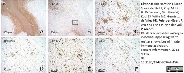
Microglia associated with preactive lesions express NADPH oxidase-2 subunits. Clusters of HLA-DR immunopositive microglia (B and C, HLA-DR) in NAWM (PLP, A) express various NADPH oxidase-2 subunits, including gp91phox (D), p22phox (E) and p47phox (F) in consecutive sections. Figure1C-F represents a magnification of the outlined square in Figure1B. Original magnifications A, B: 4×, C-F: 40×.
From: van Horssen J, Singh S, van der Pol S, Kipp M, Lim JL, Peferoen L, Gerritsen W, Kooi EJ, Witte ME, Geurts JJ, de Vries HE, Peferoen-Baert R, van den Elsen PJ, van der Valk P, Amor S.
Clusters of activated microglia in normal-appearing white matter show signs of innate immune activation.
J Neuroinflammation. 2012 Jul 2;9:156.
This image is from an open access article distributed under the terms of a Creative Commons Attribution License.
Mouse anti Bovine myelin proteolipid protein antibody, clone plpc1 (MCA839G) used for detection of proteolipid protein in formalin fixed, paraffin embedded multiple sclerosis brain tissue
Image caption:
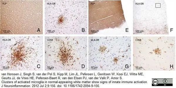
Preactive lesions are composed of clusters of HLA-DR-positive microglia. In NAWM, i.e., in the absence of apparent myelin loss (A, E: proteolipid protein) preactive lesions are defined as circumscribed nodules of activated microglia expressing HLA-DR (B, C) and CD68 (D). Preactive lesions are predominantly observed in blocks containing active lesions (E: proteolipid protein; F, G: HLA-DR). Figure1G represents a magnification of the outlined square in (F). In some cases microglial nodules are surrounded by a halo devoid of microglia (H: HLA-DR). Original magnifications: A, B: 20×; C, D: 40×; E, F: 4×; G, H: 40×.
From: van Horssen J, Singh S, van der Pol S, Kipp M, Lim JL, Peferoen L, Gerritsen W, Kooi EJ, Witte ME, Geurts JJ, de Vries HE, Peferoen-Baert R, van den Elsen PJ, van der Valk P, Amor S.
Clusters of activated microglia in normal-appearing white matter show signs of innate immune activation.
J Neuroinflammation. 2012 Jul 2;9:156.
This image is from an open access article distributed under the terms of a Creative Commons Attribution License.
Mouse anti myelin proteolipid protein antibody, clone plpc1 (MCA839G) used for detection of myelin proteolipid protein in brain lysates by western blotting
Image caption:
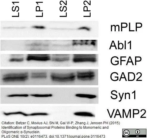
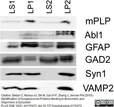
Presence of &apha;SN binding proteins chosen for validation in synaptosomal fractions. 30μg of each of the fractions synaptosomal membranes LP1, synaptosomal lysate (LS1), synaptic vesicles (LP2) and synaptosomal cytosol (LS2) were immunoblotted, and analyzed for the presence of myelin Proteolipid protein (mPLP), Abl interactor 1 (Abl1), Glial fibrillary acidic protein (GFAP), Glutamic acid decarboxylase 2 (GAD2), Synapsin 1 (Syn1), and Vesicle associated membrane protein 2 (VAMP2).
From: Betzer C, Movius AJ, Shi M, Gai W-P, Zhang J, et al. (2015)
Identification of Synaptosomal Proteins Binding to Monomeric and Oligomeric α-Synuclein.
PLoS ONE 10(2): e0116473.
doi: 10.1371/journal.pone.0116473.
This image is from an open access article distributed under the terms of a Creative Commons Attribution License.

Mouse anti Bovine myelin proteolipid protein, clone plpc1 (MCA839G) used for the evaluation of PLP expression in human brain by immunohistochemistry on formalin fixed, paraffin embedded biopsy sections.
Image caption:
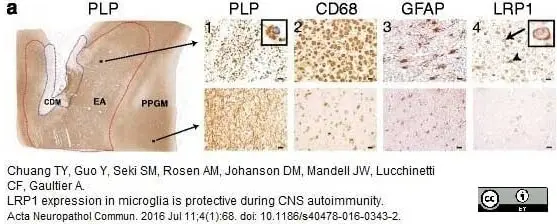
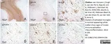
LRP1 expression is increased in MS lesions. a Immunohistochemistry on consecutive sections from an early active (EA) MS lesion (upper row) shows: (1) myelin (PLP) laden macrophages consistent with ongoing demyelinating activity, (2) abundant macrophage infiltration (CD68), (3) hypertrophic reactive astrocytes indicating gliosis (GFAP), and (4) LRP1 immunoreactivity present on both astrocytes (arrowhead) and macrophages (arrow and inset). In contrast, the periplaque gray matter (PPGM, lower row) shows: (1) normal appearing myelin (PLP), (2) limited microglial reactivity (CD68), (3) astrocytes with regular size and morphology (GFAP), and (4) limited LRP1 immunoreactivity. (Scale bars = 20 μm). b Luxol Fast Blue histology (LFB) and immunofluorescence for CD68 and LRP1 shows that myeloid cells express LRP1 within the lesion (Scale bar = 100 μm)
From: Chuang TY, Guo Y, Seki SM, Rosen AM, Johanson DM, Mandell JW, Lucchinetti CF, Gaultier A.
LRP1 expression in microglia is protective during CNS autoimmunity.
Acta Neuropathol Commun. 2016 Jul 11;4(1):68.
This image is from an open access article distributed under the terms of a Creative Commons Attribution License.
Mouse anti Bovine myelin proteolipid protein antibody, clone plpc1 (MCA839G) used for the evaluation of PLP expression in human brain by immuohistochemistry on formalin fixed, paraffin embedded tissue sections.
Image caption:
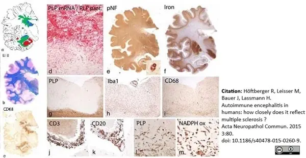
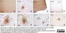
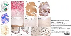
Basic Pathology of HAE: a: Topographical distribution of demyelinated lesions in the brain shows the prominent periventricular demyelination with peri-venous extensions (Dawson Fingers) and demyelinated plaques in the cortex and the deep grey matter nuclei; green: white matter lesions, red: cortical and hippocampal lesions, blue: lesions in thalamus and basal ganglia; the blue dots in the meninges show the location of prominent meningeal inflammatory infiltrates; b: Sections stained with luxol fast blue depicts the demyelinated lesions in the white matter; c: Immunocytochemistry for CD68 shows the accumulation of macrophages at the edge of active white matter lesions; d: Double staining for proteolipid (PLP) protein (red) and mRNA (black) reveals loss of oligodendrocytes within the lesion and the presence of numerous macrophages with PLP degradation products at the lesion edge; e: In sections stained for phosphorylated neurofilament only a mild to moderate reduction of axonal density is seen in the lesions; the insert shows a neuron in the substantia nigra with an α-synuclein reactive Lewy body. f: The section stained for iron shows prominent iron accumulation in the deep grey matter nuclei and at the cortico/subcortical border; some increased iron reactivity is seen within the periventricular demyelinated lesions: g: Subpial cortical lesion in the insular cortex (Fig. 1a) with selective loss of myelin in the cortex; h, i: The subpial lesions shows an actively demyelinating edge with high density of activated microglia (Iba-1, Fig. 1h), expressing the phagocytosis associated marker CD68 (Fig. 1i). j, k: In the meninges, covering the active lesion, inflammatory infiltrates are seen, composed or perivascular T-cells (CD3, Fig. 1j) and B-cells (CD20, Fig. 1k); l: The active lesion edge of the cortical lesions contains numerous macrophages with PLP reactive myelin degradation products; m: Activated microglia and macrophages at the lesions edge express NADPH oxidase.
From: Höftberger R, Leisser M, Bauer J, Lassmann H.
Autoimmune encephalitis in humans: how closely does it reflect multiple sclerosis?
Acta Neuropathol Commun. 2015 Dec 4;3:80.
This image is from an open access article distributed under the terms of a Creative Commons Attribution License.
Mouse anti Bovine myelin proteolipid protein antibody, clone plpc1 (MCA839G) used for the demonstration of plp expression in brain tissue from multiple sclerosis affected specimens.
Image caption:
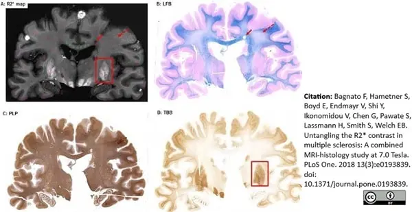
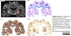
Side-by-side R2* (A), LFB (B), PLP (C) and TBB (iron, D) staining. One can appreciate examples of ROIs showing WM-L (solid red arrow on PLP and R2* maps), DWMI (dashed red arrow on PLP and R2* maps), and areas of increased iron accumulation in the dGM (red rectangle on TBB and R2* maps).
From: Bagnato F, Hametner S, Boyd E, Endmayr V, Shi Y, Ikonomidou V, et al. (2018)
Untangling the R2* contrast in multiple sclerosis: A combined MRI-histology study at 7.0 Tesla.
PLoS ONE 13(3): e0193839.
This image is from an open access article distributed under the terms of a Creative Commons Attribution License.
Mouse anti Bovine myelin proteolipid protein antibody, clone plpc1 (MCA839G) used for the visualization of myelin in a model of experimental autoimmune encephalomyelitis in rhesus monkey brain by immunohistochemistry on formalin fixed paraffin embedded tissue sections.
Image caption:
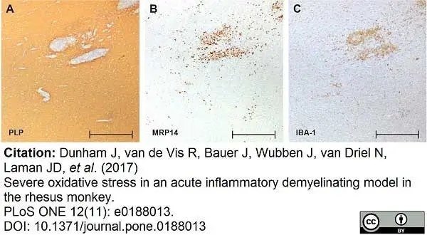
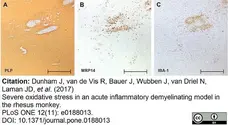
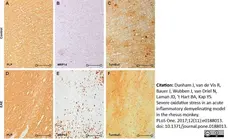
Expression of oxidative stress pathway markers.
Expression of key markers of the oxidative stress pathway was analyzed by immunohistochemistry. Shown is a representative brain area with EAE lesions stained for PLP (A), MRP14 (B) and Iba-1 (C). Image scale bars are 500 μm.
From: Dunham J, van de Vis R, Bauer J, Wubben J, van Driel N, Laman JD, et al. (2017)
Severe oxidative stress in an acute inflammatory demyelinating model in the rhesus monkey.
PLoS ONE 12(11): e0188013.
This image is from an open access article distributed under the terms of a Creative Commons Attribution License.
Mouse anti myelin proteolipid protein antibody, clone plpc1 (MCA839G) used for the visualization of myelin in a model of experimental autoimmune encephalomyelitis in rhesus monkey brain by immunohistochemistry on formalin fixed paraffin embedded tissue sections.
Image caption:
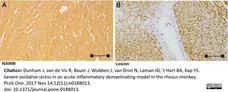
Characterization of rhesus monkey EAE brain pathology.
Tissue was stained for myelin (PLP)The image scale bar is 100 μm.
From: Dunham J, van de Vis R, Bauer J, Wubben J, van Driel N, Laman JD, et al. (2017)
Severe oxidative stress in an acute inflammatory demyelinating model in the rhesus monkey.
PLoS ONE 12(11): e0188013.
This image is from an open access article distributed under the terms of a Creative Commons Attribution License.
Mouse anti myelin proteolipid protein antibody, clone plpc1 (MCA839G) used for the visualization of myelin in a model of experimental autoimmune encephalomyelitis in rhesus monkey brain by immunohistochemistry on formalin fixed paraffin embedded tissue sections.
Image caption:
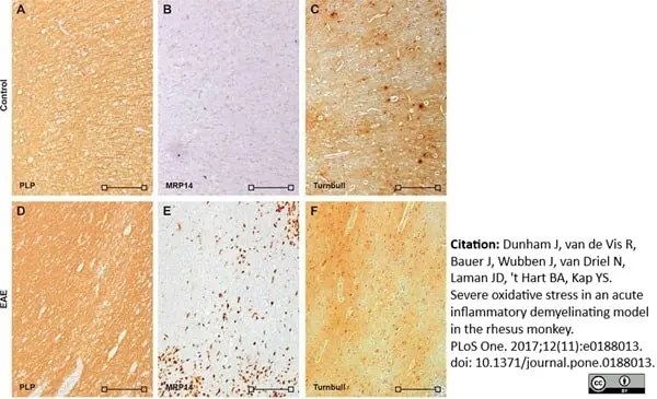
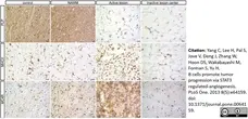
Iron accumulates in rhesus brain.
To determine tissue specific iron, Turnbull staining was performed on healthy control and EAE brain tissue from rhesus monkeys. Shown are overview images of PLP (A,D), MRP14 (B,E), and Turnbull (C,F) of a control brain (A-C) and an EAE brain (D-F). In the control brain, iron accumulation was strongest in the GM (A,C). In the EAE brain no distinct pattern of iron staining emerged. Image scale bars are 200 μm (open square).
From: Dunham J, van de Vis R, Bauer J, Wubben J, van Driel N, Laman JD, et al. (2017)
Severe oxidative stress in an acute inflammatory demyelinating model in the rhesus monkey.
PLoS ONE 12(11): e0188013.
This image is from an open access article distributed under the terms of a Creative Commons Attribution License.

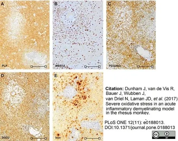
Mouse anti myelin proteolipid protein antibody, clone plpc1 (MCA839G) used for the visualization of myelin in a model of experimental autoimmune encephalomyelitis in rhesus monkey brain by immunohistochemistry on formalin fixed paraffin embedded tissue sections.
Image caption:
Iron and oxidative stress.
Shown are adjacent stains of PLP (A), MRP14 (B), p22phox (C), SOD2 (D) and Iron (E) of an EAE lesion. The image scale bar is 100 μm.
From: Dunham J, van de Vis R, Bauer J, Wubben J, van Driel N, Laman JD, et al. (2017)
Severe oxidative stress in an acute inflammatory demyelinating model in the rhesus monkey.
PLoS ONE 12(11): e0188013.
This image is from an open access article distributed under the terms of a Creative Commons Attribution License.
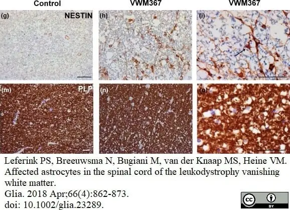
Mouse anti Human myelin proteolipid protein, clone plpc1 (MCA839G) used for the demonstration of plp expressing cells in the spinal cord of a Vanishing White Matter disease patient by immunohistochemistry on formalin fixed, paraffin embedded tissue sections.
Image caption:
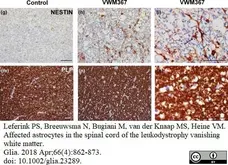
The glial cells in the spinal cord white matter of a VWM patient are affected. Stains on a cross‐section of lateral white matter of the thoracic spinal cord of (g and m) a healthy control, and (h, and o) VWM patient VWM367. Stains are performed for (g–I) nestin; (m–o) PLP. Arrows indicate examples of (I) a nestin‐expressing cell. Scale bar 75 μm.
From: Leferink PS, Breeuwsma N, Bugiani M, van der Knaap MS, Heine VM.
Affected astrocytes in the spinal cord of the leukodystrophy vanishing white matter.
Glia. 2018 Apr;66(4):862-73.
doi: 10.1002/glia.23289.
This image is from an open access article distributed under the terms of a Creative Commons Attribution License.
Mouse anti Bovine myelin proteolipid protein antibody, clone plpc1 (MCA839G) used to demonstrate myelin in mouse brain section by immunohistochemistry on vibratome sections.
Image caption:
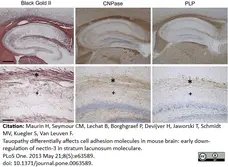
Histology and IHC of Myelin. Histological staining using Black Gold II, and IHC for CNPase and PLP of hippocampus (upper panels) and higher magnifications of TA (*) and PP (+) in respectively CA1 SLM and molecular layer of the DG (lower panels). Scale bars: upper panels:400 µm; lower panels: 100 μm.
From: Maurin H, Seymour CM, Lechat B, Borghgraef P, Devijver H, Jaworski T, et al. (2013)
Tauopathy Differentially Affects Cell Adhesion Molecules in Mouse Brain: Early Down-Regulation of Nectin-3 in Stratum Lacunosum Moleculare.
PLoS ONE 8(5): e63589.
This image is from an open access article distributed under terms of a Creative Commons Attribution License.
Mouse anti Human myelin proteolipid protein antibody, clone plpc1 (MCA839G) used to stain myelin in human brain tissue by immunohistochemistry on human white matter tissue sections.
Image caption:
Protein expression of MC4R in MS lesions. Immunostainings of representative control white matter, NAWM, an active lesion, and inactive lesion center. Active MS lesions are characterized by loss of proteolipid protein (PLP) and presence of MHCII positive leukocytes, while inactive lesions are characterized by a demyelinated core, with little presence of MHCII+ cells. MC4R is expressed in control and NAWM and increased immunoreactivity is observed in active lesions (open arrowheads, scale-bar = 25 μm).
From:Kamermans A, Verhoeven T, van Het Hof B, Koning JJ, Borghuis L, Witte M, van Horssen J, de Vries HE, Rijnsburger M.
Setmelanotide, a Novel, Selective Melanocortin Receptor-4 Agonist Exerts Anti-inflammatory Actions in Astrocytes and Promotes an Anti-inflammatory Macrophage Phenotype.
Front Immunol. 2019 Oct 4;10:2312.
doi: 10.3389/fimmu.2019.02312.
This image is from an open access article distributed under terms of a Creative Commons Attribution License.
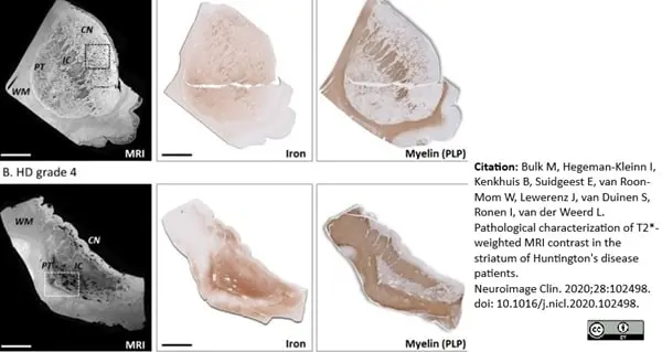
Mouse anti Bovine myelin proteolipid protein antibody, clone plpc1 (MCA839G) used to demonstrate myelin in brain of Huntington's disease patients by immunohistochemistry on formalin fixed paraffin embedded tissue sections.
Image caption:
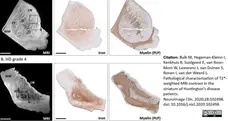
Macroscopic colocalization of MRI and histological staining for iron and immunohistological visualization of myelin. (A) Control subject showed macroscopic colocalization of the small focal hypointensities on MRI with small regions of increased staining intensity in both the iron and myelin staining. (B) HD patients exhibited larger focal hypointensities within the caudate nucleus and putamen. Histology showed high iron, but low myelin staining intensity within these regions. The hypointensities as observed on MRI frequently colocalized with vessels and enlarged perivascular spaces.CN = caudate nucleus; PT = putamen; IC = internal capsule; WM = white matter. Scale bar = 1 cm.
From: Bulk, M. et al.
Pathological characterization of T2*-weighted MRI contrast in the striatum of Huntington’s disease patients.
NeuroImage: Clinical 28, 2020, 102498.
doi: 10.1016/j.nicl.2020.102498
This image is from an open access article distributed under terms of a Creative Commons Attribution License.

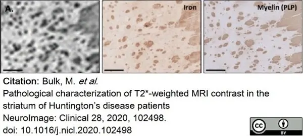
Mouse anti myelin proteolipid protein antibody, clone plpc1 (MCA839G) used to demonstrate myelin in brain of Huntington's disease patients by immunohistochemistry on formalin fixed paraffin embedded tissue sections.
Image caption:
Macroscopic structures affecting the MRI contrast in control subjects and HD patients. (A) Small focal hypointensities on MRI originated from myelinated fiber bundles traversing the caudate nucleus and putamen, which were also characterized by high iron concentrations.
From: Bulk, M. et al.
Pathological characterization of T2*-weighted MRI contrast in the striatum of Huntington’s disease patients.
NeuroImage: Clinical 28, 2020, 102498.
doi: 10.1016/j.nicl.2020.102498
This image is from an open access article distributed under terms of a Creative Commons Attribution License.
Mouse anti Bovine myelin proteolipid protein antibody, clone plpc1 (MCA839G) used to demonstrate myelin staining in human post mortem brain tissue by immunohistochemistry on cryostat sections.
Image caption:
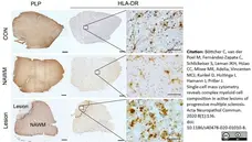
Representative brain sections of control white matter (CON, upper image; scale bar = 1 mm) and normal appearing white matter (NAWM) as well as lesion-enriched white matter (Lesion) of donors with MS pathology (lower image; scale bar = 1 mm). NAWM tissue shows ramified microglia and intact myelin, based on HLA-DR and PLP staining, respectively. In active lesion, demyelinated center and active microglia/macrophages could be detected throughout the whole lesion. The majority of HLADR+ cells in lesion MS are foamy microglia/macrophages. Scale bar of high resolution pictures is 50 μm.
From: Böttcher C, van der Poel M, Fernández-Zapata C, Schlickeiser S, Leman JKH, Hsiao CC, Mizee MR, Adelia, Vincenten MCJ, Kunkel D, Huitinga I, Hamann J, Priller J.
Single-cell mass cytometry reveals complex myeloid cell composition in active lesions of progressive multiple sclerosis.
Acta Neuropathol Commun. 2020 Aug 18;8(1):136.
doi: 10.1186/s40478-020-01010-8.
This image is from an open access article distributed under terms of a Creative Commons Attribution License.
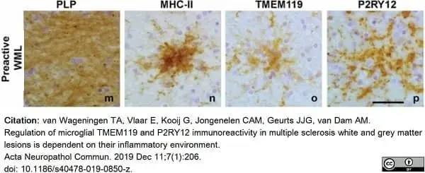
Mouse anti myelin proteolipid protein antibody, clone plpc1 (MCA839G) used to label myelin in human brain by immunohistochemistry on formalin fixed, paraffin embedded tissue sections.
Image caption:
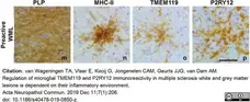
Representative images of PLP, MHC-II, TMEM119 and P2RY12 (j, k, l) immunoreactivity in in pre-active lesions (m, n, o, p). Scalebars (m-p) = 50 μm
From: van Wageningen TA, Vlaar E, Kooij G, Jongenelen CAM, Geurts JJG, van Dam AM.
Regulation of microglial TMEM119 and P2RY12 immunoreactivity in multiple sclerosis white and grey matter lesions is dependent on their inflammatory environment.
Acta Neuropathol Commun. 2019 Dec 11;7(1):206.
doi: 10.1186/s40478-019-0850-z.
This image is from an open access article distributed under terms of a Creative Commons Attribution License.
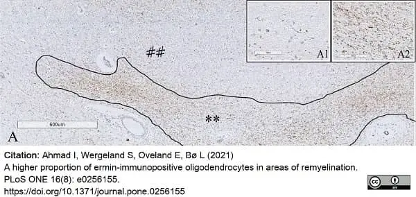
Mouse anti myelin proteolipid protein antibody, clone plpc1 (MCA839G) used to stain PLP in mouse brains by immunohistochemistry on formalin fixed, paraffin embedded tissue sections
Image caption:
Ermin positive cell density in MS white matter lesions.
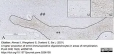
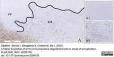
Myelin proteolipid protein (PLP) stained tissue section (** represents remyelinated white matter area and demyelinated white matter area is represented by ##); insertion A1 shows a demyelinated area with no remyelination, and insertion (A2) shows a partly remyelinated area
From: Ahmad I, Wergeland S, Oveland E, Bø L (2021)
A higher proportion of ermin-immunopositive oligodendrocytes in areas of remyelination.
PLoS ONE 16(8): e0256155.
doi: 10.1371/journal.pone.0256155
This image is from an open access article distributed under terms of a Creative Commons Attribution License.
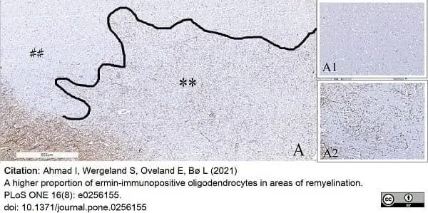
Mouse anti Bovine myelin proteolipid protein antibody, clone plpc1 (MCA839G) used to stain PLP in mouse brains by immunohistochemistry on formalin fixed, paraffin embedded tissue sections
Image caption:
Ermin positive cell density in MS gray matter lesions.
Myelin proteolipid protein (PLP) stained tissue section (** represents remyelinated area, demyelinated area is represented by ##); the insertion A1 shows an area of complete demyelination in gray matter. The insertion A2 shows partial remyelination.
From: Ahmad I, Wergeland S, Oveland E, Bø L (2021)
A higher proportion of ermin-immunopositive oligodendrocytes in areas of remyelination.
PLoS ONE 16(8): e0256155.
doi: 10.1371/journal.pone.0256155
This image is from an open access article distributed under terms of a Creative Commons Attribution License.
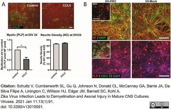
Mouse anti Bovine myelin proteolipid protein antibody, clone plpc1 (MCA839G) used to label plp expressing cells in human CNS cultures by immunofluorescence.
Image caption:
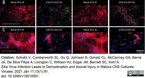
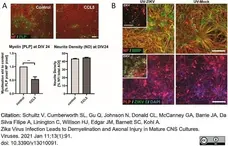
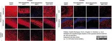
Effect of human CCL5 and UV treatment in CNS cultures. (A) CCL5 treatment (100 ng/mL) of rat CNS cultures from DIV 16 until DIV 24. Representative images of a control culture and a human CCL5 treated culture at DIV 24 are shown. Myelin and axons are shown by the immunofluorescence labeling of PLP (green signal) and NF (red signal). Graphs show the quantification of myelin and axons on DIV 24 per field of view of a captured image by CellProfiler software. Values for CCL5-treated cultures were normalized to control. Scale bar = 100μm; n = 7; Mean ± SEM; paired t-test; ** p <0.01. (B) Supernatant of mock- or ZIKV-infected immature mouse CNS cultures was collected at 6 dpi and UV-treated to inactivate viral particles. UV-treated supernatant was mixed 3:1 with fresh medium. Analysis by IF at 6 dpt onto immature cultures. Representative images of myelin staining (MBP, green signal; PLP, red signal) and axon (NF, red signal) staining at 6 dpt (DIV 24) are shown (n = 2). Inset images (upper panels) depict ZIKV (ZIKV E, green signal) infected CNS cells and pathology in cultures used for supernatant collection (6 dpi, DIV 24), with myelin (MBP, green signal; PLP, red signal) and neurofilament (NF, red) shown. Scale bar = 100μm.
From: Schultz V, Cumberworth SL, Gu Q, Johnson N, Donald CL, McCanney GA, Barrie JA, Da Silva Filipe A, Linington C, Willison HJ, Edgar JM, Barnett SC, Kohl A. Zika Virus Infection Leads to Demyelination and Axonal Injury in Mature CNS Cultures. Viruses. 2021 Jan 11;13(1):91. doi: 10.3390/v13010091.
This image is from an open access article distributed under terms of a Creative Commons Attribution License.
Mouse anti myelin proteolipid protein antibody, clone plpc1 (MCA839G) used to label plp expressing cells in human CNS cultures by immunofluorescence.
Image caption:
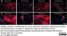
ZIKV infection causes damage to mature, myelinated CNS cultures.
(B,C) IF analysis of myelin (PLP, red signal) and axons (β-Tubulin III, red signal). ZIKV infection was assessed with an antibody recognising ZIKV E protein (green signal). A total of four biological replicates (n = 4) were analyzed.
From: Schultz V, Cumberworth SL, Gu Q, Johnson N, Donald CL, McCanney GA, Barrie JA, Da Silva Filipe A, Linington C, Willison HJ, Edgar JM, Barnett SC, Kohl A.
Zika Virus Infection Leads to Demyelination and Axonal Injury in Mature CNS Cultures.
Viruses. 2021 Jan 11;13(1):91.
doi: 10.3390/v13010091.
This image is from an open access article distributed under terms of a Creative Commons Attribution License.
Mouse anti myelin proteolipid protein antibody, clone plpc1 (MCA839G) used to label myelin in human brain by immunohistochemistry on formalin fixed, paraffin embedded tissue sections.
Image caption:
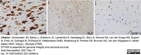
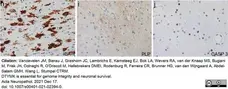
h–j The more peripheral hemispheric white matter shows massive activation of microglia with ameboid morphology (h, IBA-1), lack of myelin (i, proteolipid protein stain for myelin) and loss of oligodendrocytes by apoptosis (j, CASP3 stain for apoptotic cells).
From: Vanoevelen JM, Bierau J, Grashorn JC, Lambrichs E, Kamsteeg EJ, Bok LA, Wevers RA, van der Knaap MS, Bugiani M, Frisk JH, Colnaghi R, O'Driscoll M, Hellebrekers DMEI, Rodenburg R, Ferreira CR, Brunner HG, van den Wijngaard A, Abdel-Salam GMH, Wang L, Stumpel CTRM.
DTYMK is essential for genome integrity and neuronal survival.
Acta Neuropathol. 2021 143(2):245-262..
doi: 10.1007/s00401-021-02394-0.
This image is from an open access article distributed under terms of a Creative Commons Attribution License.
Mouse anti myelin proteolipid protein antibody, clone plpc1 (MCA839G) used to stain myelin in normal appearing white matter (NAWM) and white matter lesions (WML) from human brain by immunohistochemistry of formalin fixed, paraffin embedded tissue sections.
Image caption:
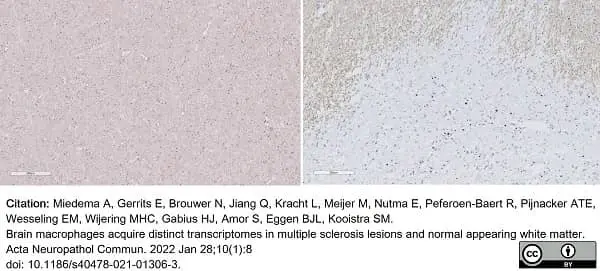
Representative images of DAB staining for PLP that was used for sample scoring.
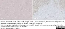
From: Miedema A, Gerrits E, Brouwer N, Jiang Q, Kracht L, Meijer M, Nutma E, Peferoen-Baert R, Pijnacker ATE, Wesseling EM, Wijering MHC, Gabius HJ, Amor S, Eggen BJL, Kooistra SM.
Brain macrophages acquire distinct transcriptomes in multiple sclerosis lesions and normal appearing white matter.
Acta Neuropathol Commun. 2022 Jan 28;10(1):8.
doi: 10.1186/s40478-021-01306-3.
This image is from an open access article distributed under terms of a Creative Commons Attribution License.
Mouse anti myelin proteolipid protein antibody, clone plpc1 (MCA839G) used to label myelin in mouse brain by immunofluorescence of formalin fixed paraffin embedded sections.
Image caption:
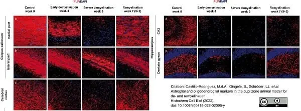
The course of de- and remyelination in representative brain regions of C57BL/6 mice exposed to cuprizone. Double immunofluorescence staining with antibodies against the myelin marker proteolipid protein (PLP) was performed to visualize the course of de- and remyelination in the midline (a) and lateral parts of corpus callosum (b), cerebral cortex (c) (represented layers V–VI), and hippocampal areas CA3 (d) and dentate gyrus (e) and in C57BL/6 mice exposed to cuprizone (nuclei were counterstained with DAPI). Representative pictures in the first column show the corresponding regions with intact myelin of untreated animals (week 0). Cuprizone feeding provoked a progressive loss of myelin (middle columns), with nearly complete demyelination at week 5 in all studied brain regions except for the lateral part of the corpus callosum, where only partial demyelination occurred (third column). Finally, re-expression of PLP could be observed 2 weeks after cuprizone withdrawal during remyelination (last column). Scale bars: 20μm (a, c, d, e); 50μm (b)
From: Castillo-Rodriguez, M.d.A., Gingele, S., Schröder, LJ. et al.
Astroglial and oligodendroglial markers in the cuprizone animal model for de- and remyelination.
Histochem Cell Biol (2022) 158(1):15-38. .
doi: 10.1007/s00418-022-02096-y
This image is from an open access article distributed under terms of a Creative Commons Attribution License.

Mouse anti myelin proteolipid protein antibody, clone plpc1 (MCA839G) used to identify plp expression in human mesial precentral gyrus by immunohistochemistry on formalin fixed, paraffin embedded archival tissue sections.
Image caption:
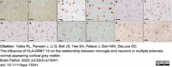
Representative immunohistochemistry. Control cases left column. MS cases right column. PLP (A, B), NeuN (C, D), CD68 (E, F), TMEM119 (G, H), Iba1 (I, J), GFAP (K, L). Scale bar 50μm.
From: Yates RL, Pansieri J, Li Q, Bell JS, Yee SA, Palace J, Esiri MM, DeLuca GC.
The influence of HLA-DRB1*15 on the relationship between microglia and neurons in multiple sclerosis normal appearing cortical grey matter.
Brain Pathol. 2022 Jul;32 (4): e13041.
doi: 10.1111/bpa.13041.
This image is from an open access article distributed under terms of a Creative Commons Attribution License.

Mouse anti myelin proteolipid protein antibody, clone plpc1 (MCA839G) used to evaluate PLP expression levels in human neural samples by western blotting.
Image caption:
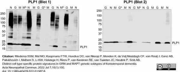
Validation of the distinct involvement of proteins in FTD subtypes using immunoblotting. (B) Analysis of PLP1 in temporal cortical tissues. Annotated immunoblots used for the analysis of PLP1 antibody signals (chemi channel) are shown. Average protein signals are quantified and shown in dot plots. Differences in protein expression levels were analysed per group comparison using a Student’s t-test. PLP1 shows a lower expression in both FTD-GRN (0.44x) and FTD-MAPT (0.39x) compared to NDC, with a strong trend for FTD-MAPT (p = 0.0595). All five independent samples show comparable expression levels to those of the original cohort. Numbers represent apparent molecular weights in kDa. Letters represent sample annotations. Protein signal values are corrected for gel loading differences and are normalized to NDC samples. G; FTD-GRN sample, GN; FTD-GRN sample from the independent cohort, M; FTD-MAPT sample, MN; FTD-MAPT sample from the independent cohort, N; non-demented control sample.
From: Miedema SSM, Mol MO, Koopmans FTW, Hondius DC, van Nierop P, Menden K, de Veij Mestdagh CF, van Rooij J, Ganz AB, Paliukhovich I, Melhem S, Li KW, Holstege H, Rizzu P, van Kesteren RE, van Swieten JC, Heutink P, Smit AB.
Distinct cell type-specific protein signatures in GRN and MAPT genetic subtypes of frontotemporal dementia.
Acta Neuropathol Commun. 2022 Jul 7;10(1):100.
doi: 10.1186/s40478-022-01387-8.
This image is from an open access article distributed under terms of a Creative Commons Attribution License.
Mouse anti myelin proteolipid protein antibody, clone plpc1 (MCA839) used to label myelin by immunohistochemistry on cryosections of human brain.
Image caption:
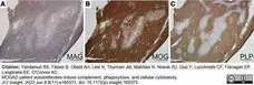
Neuropathology in frontal lobe biopsy of patient with MOGAD with paired serum effector functions. Right frontal lobe biopsy was undertaken in a symptomatic patient with MOGAD based on MRI findings. (A–C) Histology was performed and indicated active demyelinating lesions with loss of (A) MAG, (B) MOG, and (C) PLP.
From:Yandamuri SS, Filipek B, Obaid AH, Lele N, Thurman JM, Makhani N, Nowak RJ, Guo Y, Lucchinetti CF, Flanagan EP, Longbrake EE, O'Connor KC.
MOGAD patient autoantibodies induce complement, phagocytosis, and cellular cytotoxicity.
JCI Insight. 2023 Jun 8;8(11):e165373.
doi: 10.1172/jci.insight.165373.
This image is from an open access article distributed under terms of a Creative Commons Attribution License.
Mouse anti myelin proteolipid protein antibody, clone plpc1 (MCA839G) used to identify myelin expressing regions in human thoracic spinal cord by immunohistochemistry on formalin fixed, paraffin embedded tissue sections.
Image caption:
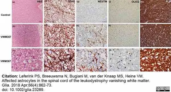
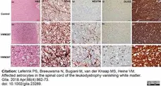
The glial cells in the spinal cord white matter of a VWM patient are affected. Stains on a cross‐section of lateral white matter of the thoracic spinal cord of (a, d, g, j, and m) a healthy control, and (b, c, e, f, h, i, k, l, n, and o) VWM patient VWM367. Stains are performed for (a–c) H&E; (d–f) GFAP; (g–i) nestin; (j–l) Olig2; (m–o) PLP. Arrows indicate examples of (f) a dysmorphic astrocyte; (i) a nestin‐expressing cell; (o) an Olig2‐positive nucleus. Scale bar 75 μm
From: Leferink PS, Breeuwsma N, Bugiani M, van der Knaap MS, Heine VM.
Affected astrocytes in the spinal cord of the leukodystrophy vanishing white matter.
Glia. 2018 Apr;66(4):862-873.
doi: 10.1002/glia.23289.
This image is from an open access article distributed under terms of a Creative Commons Attribution License.

Mouse anti myelin proteolipid protein antibody, clone plpc1 (MCA839G) used to lable nerve sheathss in human brain by immunofluorescence.
Image caption:
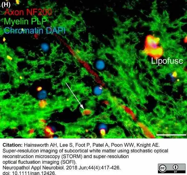
Conventional epifluorescence image of human subcortical white matter, double labelled for axonal neurofilaments (red) and the myelin sheath protein PLP (green). Nuclear chromatin is counterstained with DAPI (blue). Note a myelinated axonal profile (arrow). Autofluorescent bundles of lipofuscin (‘Lipofusc’) are relatively sparse in this field of view. Scale bar:20 μm (H).
From:Hainsworth AH, Lee S, Foot P, Patel A, Poon WW, Knight AE.
Super-resolution imaging of subcortical white matter using stochastic optical reconstruction microscopy (STORM) and super-resolution optical fluctuation imaging (SOFI).
Neuropathol Appl Neurobiol. 2018 Jun;44(4):417-426.
doi: 10.1111/nan.12426.
This image is from an open access article distributed under terms of a Creative Commons Attribution License.
Mouse anti myelin proteolipid protein antibody, clone plpc1 (MCA839G) used to stain myelin in mouse brain by immunohistochemistry on formalin fixed, paraffin embedded tissue sections.
Image caption:
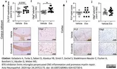
Evobrutinib induces myelin debris clearing in the cuprizone mouse model. All brain sections were immunohistochemically stained and the corpus callosum (CC) as well as a defined area of the cortex were analyzed. e, i Myelinated areas were assessed by e, i anti-myelin proteolipid protein (PLP) staining and are shown as percentage of myelinated CC in relation to the total CC e) or scoring of the cortex area (i), scale bar: 200 μM. Immunostaining of (f, j) oligodendrocyte transcription factor 2 (Olig2), number of cells/mm2 per group, scale bar: 100 μM. b–l Data are shown as mean ± standard error of the mean (SEM). Data are normalized to vehicle and pooled from at least two independent experiments (n = 12–16). Asterisks indicate significant difference calculated using the unpaired two-tailed t-test (*P ≤0.05, **P ≤0.01, ***P ≤0.001)
From: Geladaris A, Torke S, Saberi D, Alankus YB, Streit F, Zechel S, Stadelmann-Nessler C, Fischer A, Boschert U, Häusler D, Weber MS.
BTK inhibition limits microglia-perpetuated CNS inflammation and promotes myelin repair.
Acta Neuropathol. 2024 Apr 24;147(1):75.
doi: 10.1007/s00401-024-02730-0.
This image is from an open access article distributed under terms of a Creative Commons Attribution License.
Mouse anti myelin proteolipid protein antibody, clone plpc1 (MCA839G) used to stain myelin in mouse brain by immunohistochemistry on formalin fixed, paraffin embedded tissue sections.
Image caption:
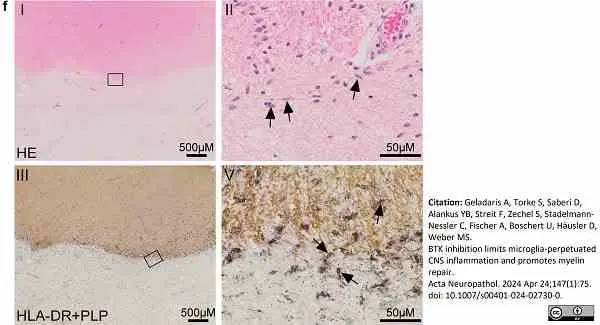
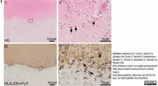
f) Brain biopsy of chronically active (smouldering) MS lesion. I+II: HE stained section with sharply demarcated lesion (bottom of I) displaying a lesion rim consisting of activated microglial cells with elongated nuclei (II: magnification of area depicted in I, arrows point at microglial cells). Note the almost complete absence of foamy macrophages as well as perivascular lymphocytic cuffs. III+V: Double immunolabeling against proteolipid protein (PLP) visualizing myelin loss at the lesion center (bottom of III) and against HLA immunopositive microglial cells (V: magnification of area depicted in III, arrows point at microglial cells). Note the abundance of microglial cells at the lesion rim.
From: Geladaris A, Torke S, Saberi D, Alankus YB, Streit F, Zechel S, Stadelmann-Nessler C, Fischer A, Boschert U, Häusler D, Weber MS.
BTK inhibition limits microglia-perpetuated CNS inflammation and promotes myelin repair.
Acta Neuropathol. 2024 Apr 24;147(1):75.
doi: 10.1007/s00401-024-02730-0.
This image is from an open access article distributed under terms of a Creative Commons Attribution License.
Mouse anti myelin proteolipid protein antibody, clone plpc1 (MCA839G) used to label PLP expressing cells in mouse brain by immunohistochemistry on formalin fixed, paraffin embedded sections.
Image caption:
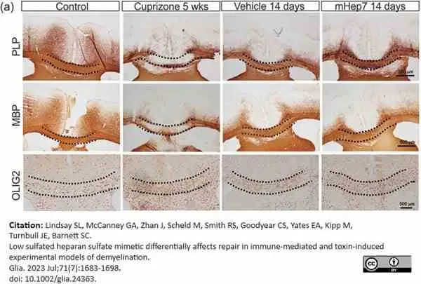
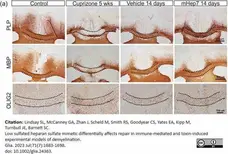
Effect of LS‐mHep7 on PLP, MBP, OLIG2 expression in the acute cuprizone model of demyelination. (a) Representative images of anti‐PLP, anti‐MBP, and anti‐OLIG2 staining within anatomical brain region 215 of a control animal, or after being fed cuprizone diet (0.25%) for 5 weeks (Cup 5 weeks) or after treatment with PBS vehicle or LS‐mHep7 40 mg/kg via subcutaneous injections (s.c.) for 14 days post cuprizone diet removal. Dashed black line demarcates the corpus callosum (CC) region.
From: Lindsay SL, McCanney GA, Zhan J, Scheld M, Smith RS, Goodyear CS, Yates EA, Kipp M, Turnbull JE, Barnett SC.
Low sulfated heparan sulfate mimetic differentially affects repair in immune-mediated and toxin-induced experimental models of demyelination.
Glia. 2023 Jul;71(7):1683-98.
doi: 10.1002/glia.24363.
This image is from an open access article distributed under terms of a Creative Commons Attribution License.
Mouse anti myelin proteolipid protein antibody, clone plpc1 (MCA839G) used to stain cervical spinal cord tissue sections for myelin proteolipid protein by immunohistochemistry of formalin fixed, paraffin embedded sections.
Image caption:
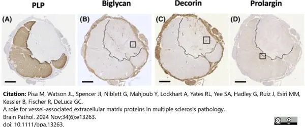
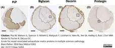
Immunoreactivity of biglycan, decorin, and prolargin along the neuraxis. Cervical spinal cord (A–D) from a prototypical MS case immunolabelled for myelin (PLP) (A), biglycan (B), decorin (C), and prolargin (D).
From: Pisa M, Watson JL, Spencer JI, Niblett G, Mahjoub Y, Lockhart A, Yates RL, Yee SA, Hadley G, Ruiz J, Esiri MM, Kessler B, Fischer R, DeLuca GC.
A role for vessel-associated extracellular matrix proteins in multiple sclerosis pathology.
Brain Pathol. 2024 Nov;34(6):e13263.
doi: 10.1111/bpa.13263.
This image is from an open access article distributed under terms of a Creative Commons Attribution License.
Mouse anti myelin proteolipid protein antibody, clone plpc1 (MCA839G) detection of myelin in MS brain sections by immunofluorescence
Image caption:
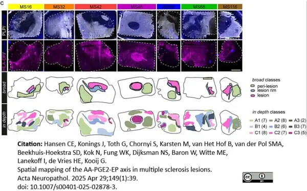
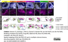
Immunohistochemical depiction of PLP and HLA-DR in MS tissues (color-coded) and annotated ROIs for broad and in-depth tissue classification; scale bar: 5 mm.
From: Hansen CE, Konings J, Toth G, Chornyi S, Karsten M, van Het Hof B, van der Pol SMA, Beekhuis-Hoekstra SD, Kok N, Fung WK, Dijksman NS, Baron W, Witte ME, Lanekoff I, de Vries HE, Kooij G.
Spatial mapping of the AA-PGE2-EP axis in multiple sclerosis lesions.
Acta Neuropathol. 2025 Apr 29;149(1):39.
doi: 10.1007/s00401-025-02878-3.
This image is from an open access article distributed under terms of a Creative Commons Attribution License.
Mouse anti myelin proteolipid protein antibody, clone plpc1 (MCA839G) used to identify proteolipid protein cepression in brain sections deom multiple sclerosis patients by immunohistochemistry of formalin fixed, paraffin embedded tissue sections.
Image caption:
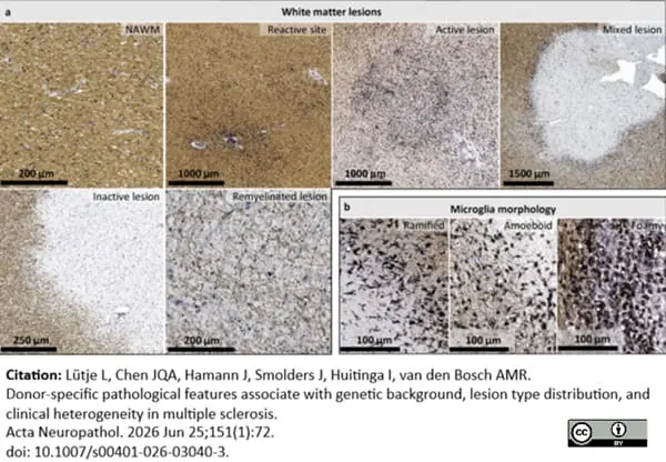
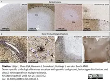
Representative images of common features in MS pathology and donor-specific pathological features. All images are stained for HLA (black) and PLP (brown). a Normal-appearing white matter and white matter lesions characterized in MS tissue of the NBB. b Microglia/macrophages expressing HLA with distinct morphologies at the edge of mixed active/inactive lesions. c Grey matter with and without lesions. d Donor-specific pathological features include perivascular cuffs, nodules of ≥ 4 HLA+ microglia/macrophages surrounded by intact myelin, mixed lesions with broad rim and high remyelination efficiency. GM grey matter; NAGM normal-appearing grey matter; NAWM normal-appearing white matter; RL remyelinated lesion; WM white matter
From: Lütje L, Chen JQA, Hamann J, Smolders J, Huitinga I, van den Bosch AMR.
Donor-specific pathological features associate with genetic background, lesion type distribution, and clinical heterogeneity in multiple sclerosis.
Acta Neuropathol. 2026 Jun 25;151(1):72.
This image is from an open access article distributed under terms of a Creative Commons Attribution License.
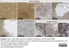
Mouse anti myelin proteolipid protein antibody, clone plpc1 (MCA839G) used to identify proteolipid protein expression in brain sections from multiple sclerosis patients by immunohistochemistry of formalin fixed, paraffin embedded tissue sections.
Image caption:
Representative images of common features in MS pathology and donor-specific pathological features. All images are stained for HLA (black) and PLP (brown). a Normal-appearing white matter and white matter lesions characterized in MS tissue of the NBB. b Microglia/macrophages expressing HLA with distinct morphologies at the edge of mixed active/inactive lesions.
From: Lütje L, Chen JQA, Hamann J, Smolders J, Huitinga I, van den Bosch AMR.
Donor-specific pathological features associate with genetic background, lesion type distribution, and clinical heterogeneity in multiple sclerosis.
Acta Neuropathol. 2026 Jun 25;151(1):72.
doi: 10.1007/s00401-026-03040-3.
This image is from an open access article distributed under terms of a Creative Commons Attribution License.
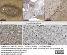
Mouse anti myelin proteolipid protein antibody, clone plpc1 (MCA839G) used to identify proteolipid protein expression in brain sections from multiple sclerosis patients by immunohistochemistry of formalin fixed, paraffin embedded tissue sections.
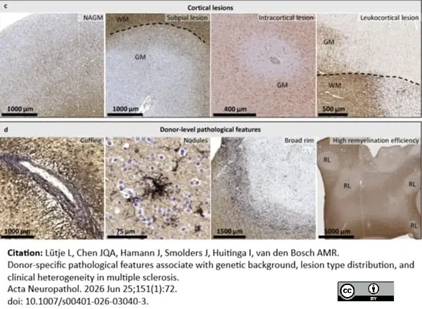
Image caption:
Representative images of common features in MS pathology and donor-specific pathological features. All images are stained for HLA (black) and PLP (brown). c Grey matter with and without lesions. d Donor-specific pathological features include perivascular cuffs, nodules of ≥ 4 HLA+ microglia/macrophages surrounded by intact myelin, mixed lesions with broad rim and high remyelination efficiency. GM grey matter; NAGM normal-appearing grey matter; NAWM normal-appearing white matter; RL remyelinated lesion; WM white matter.
From: Lütje L, Chen JQA, Hamann J, Smolders J, Huitinga I, van den Bosch AMR.
Donor-specific pathological features associate with genetic background, lesion type distribution, and clinical heterogeneity in multiple sclerosis.
Acta Neuropathol. 2026 Jun 25;151(1):72.
doi: 10.1007/s00401-026-03040-3.
This image is from an open access article distributed under terms of a Creative Commons Attribution License.
Filter by Application:
P WB C IF Reset| Mouse anti myelin proteolipid protein antibody, clone plpc1 recognizes myelin proteolipid protein (PLP) in many mammalian species (Stoffel et al. 1985). Clone plpc1 also recognizes the alternative PLP splice variant lacking part of the cytoplasmic domain (amino acids 117-151), known as DM20 (Simons et al. 1987) . PLP encodes the major protein components of compact CNS myelin and mutations in the PLP gene can lead to severe dysmyelinating disease (Hudson et al. 1989). Mouse anti myelin proteolipid protein, clone plpc1 has proved a useful immunohistochemical tool for the study of central nervous system injury in patients with multiple sclerosis (Seewan et al. 2011, Huizinga et al. 2011) |
- Target Species
- Bovine
- Species Cross-Reactivity
-
Target Species Cross Reactivity Human Tenerife lizard (Gallotia galloti) - N.B. Antibody reactivity and working conditions may vary between species.
- Product Form
- Purified IgG - liquid
- Preparation
- Purified IgG prepared by affinity chromatography on Protein G from tissue culture supernatant
- Buffer Solution
- Phosphate buffered saline
- Preservative Stabilisers
- <0.1% Sodium Azide (NaN3)
- Immunogen
- Synthetic peptide GRGTKF corresponding to C terminal region of myelin proteolipid protein.
- Approx. Protein Concentrations
- IgG concentration 1 mg/ml
- Fusion Partners
- Spleen cells from immunized BALB/c mice were fused with cells of the mouse SP2/0 myeloma cell line.
- Regulatory
- For research purposes only
- Guarantee
- 12 months from date of despatch
This product is shipped at ambient temperature. It is recommended to aliquot and store at -20°C on receipt. When thawed, aliquot the sample as needed. Keep aliquots at 2-8°C for short term use (up to 4 weeks) and store the remaining aliquots at -20°C.
Avoid repeated freezing and thawing as this may denature the antibody. Storage in frost-free freezers is not recommended.
Avoid repeated freezing and thawing as this may denature the antibody. Storage in frost-free freezers is not recommended.
This product has been reported to work in the following applications. This information is derived from testing within our laboratories, peer-reviewed publications or personal communications from the originators. Please refer to references indicated for further information. For general protocol recommendations, please visit the antibody protocols page.
| Application Name | Verified | Min Dilution | Max Dilution |
|---|---|---|---|
| Flow Cytometry |  |
||
| Immunofluorescence | |
||
| Immunohistology - Frozen | |
||
| Immunohistology - Paraffin | |
||
| Western Blotting | |
Where this antibody has not been tested for use in a particular technique this does not necessarily exclude its use in such procedures. Suggested working dilutions are given as a guide only. It is recommended that the user titrates the antibody for use in their own system using appropriate negative/positive controls.
| Description | Product Code | Applications | Pack Size | List Price | Your Price | Quantity | |
|---|---|---|---|---|---|---|---|
| Mouse IgG2a Negative Control | MCA929 | F | 100 Tests |

|
Log in | ||
| List Price | Your Price | ||||||
|
|
Log in | ||||||
| Description | Mouse IgG2a Negative Control | ||||||
References for Myelin Proteolipid Protein antibody
-
Boon, L. et al. (2001) Prevention of experimental autoimmune encephalomyelitis in the common marmoset (Callithrix jacchus) using a chimeric antagonist monoclonal antibody against human CD40 is associated with altered B cell responses.
J Immunol. 167: 2942-9. -
Jaśkiewicz, E. et al. (2005) Expression of recombinant forms of human 21.5 kDa myelin basic protein and proteolipid protein in CHO cells.
Acta. Biochim. Pol. 52: 863-6. -
Pomeroy, I.M. et al. (2005) Demyelinated neocortical lesions in marmoset autoimmune encephalomyelitis mimic those in multiple sclerosis.
Brain. 128: 2713-21. -
Jatana, M. et al. (2006) Combination of systemic hypothermia and N-acetylcysteine attenuates hypoxic-ischemic brain injury in neonatal rats.
Pediatr Res. 59 (5): 684-9. -
Santos, E. et al. (2006) Peculiar and typical oligodendrocytes are involved in an uneven myelination pattern during the ontogeny of the lizard visual pathway.
J Neurobiol. 66 (10): 1115-24. -
Gilmore, C.P. et al. (2006) Spinal cord gray matter demyelination in multiple sclerosis-a novel pattern of residual plaque morphology.
Brain Pathol. 16: 202-8. -
van Horssen, J. et al. (2006) NAD(P)H:quinone oxidoreductase 1 expression in multiple sclerosis lesions.
Free Radic Biol Med. 41: 311-7. -
Roemer, S.F. et al. (2007) Pattern-specific loss of aquaporin-4 immunoreactivity distinguishes neuromyelitis optica from multiple sclerosis.
Brain. 130: 1194-205.
View The Latest Product References
-
Geurts, J.J. et al. (2007) Extensive hippocampal demyelination in multiple sclerosis.
J Neuropathol Exp Neurol. 66: 819-27. -
Moharregh-Khiabani, D. et al. (2010) Effects of fumaric acids on cuprizone induced central nervous system de- and remyelination in the mouse.
PLoS One. 5:e11769. -
van Horssen, J. et al. (2010) Nrf2 and DJ1 are consistently upregulated in inflammatory multiple sclerosis lesions.
Free Radic Biol Med. 49: 1283-9. -
Popescu, B.F. et al. (2010) Absence of cortical demyelination in neuromyelitis optica.
Neurology. 75: 2103-9. -
Coulpier, F. et al. (2010) CNS/PNS boundary transgression by central glia in the absence of Schwann cells or Krox20/Egr2 function.
J Neurosci. 30: 5958-67. -
Bramow, S. et al. (2010) Demyelination versus remyelination in progressive multiple sclerosis.
Brain.133: 2983-98. -
Kooij, G. et al. (2010) Adenosine triphosphate-binding cassette transporters mediate chemokine (C-C motif) ligand 2 secretion from reactive astrocytes: relevance to multiple sclerosis pathogenesis.
Brain. 134: 555-70. -
Kooi, E.J. et al. (2011) Cholinergic imbalance in the multiple sclerosis hippocampus.
Acta Neuropathol. 122: 313-22. -
Haider, L. et al. (2011) Oxidative damage in multiple sclerosis lesions.
Brain. 134: 1914-24. -
Baeten, K. et al. (2011) Tracking of myelin-reactive T cells in experimental autoimmune encephalomyelitis (EAE) animals using small particles of iron oxide and MRI.
NMR Biomed. 23: 601-9. -
Bagnato, F. et al. (2011) Tracking iron in multiple sclerosis: a combined imaging and histopathological study at 7 Tesla.
Brain. 134: 3602-15. -
Hinson, S.R. et al. (2012) Molecular outcomes of neuromyelitis optica (NMO)-IgG binding to aquaporin-4 in astrocytes.
Proc Natl Acad Sci U S A. 109: 1245-50. -
Kooi, E.J. et al. (2012) Heterogeneity of cortical lesions in multiple sclerosis: clinical and pathologic implications.
Neurology. 79 (13): 1369-76. -
van Horssen, J. et al. (2012) Clusters of activated microglia in normal-appearing white matter show signs of innate immune activation.
J Neuroinflammation. 9: 156. -
Seewann, A. et al. (2012) Postmortem verification of MS cortical lesion detection with 3D DIR.
Neurology. 78: 302-8. -
Skripuletz, T. et al. (2013) Astrocytes regulate myelin clearance through recruitment of microglia during cuprizone-induced demyelination.
Brain. 136 (Pt 1): 147-67. -
Skripuletz, T. et al. (2015) Pivotal role of choline metabolites in remyelination.
Brain. 138 (Pt 2): 398-413. -
Fjær, S. et al. (2015) Magnetization transfer ratio does not correlate to myelin content in the brain in the MOG-EAE mouse model.
Neurochem Int. 83-84: 28-40. -
Klok, M.D. et al. (2015) Interferon-α and the calcifying microangiopathy in Aicardi-Goutières syndrome.
Ann Clin Transl Neurol. 2 (7): 774-9. -
Clarner, T. et al. (2015) CXCL10 triggers early microglial activation in the cuprizone model.
J Immunol. 194 (7): 3400-13. -
Alme, M.N. et al. (2015) Fingolimod does not enhance cerebellar remyelination in the cuprizone model.
J Neuroimmunol. 285: 180-6. -
Betzer, C. et al. (2015) Identification of Synaptosomal Proteins Binding to Monomeric and Oligomeric α-Synuclein.
PLoS One. 10: e0116473. -
Jonkman, L.E. et al. (2016) Ultra-high field MTR and qR2* differentiates subpial cortical lesions from normal-appearing gray matter in multiple sclerosis.
Mult Scler. 22 (10): 1306-14. -
Kilsdonk, I.D. et al. (2016) Increased cortical grey matter lesion detection in multiple sclerosis with 7 T MRI: a post-mortem verification study.
Brain. 139 (Pt 5): 1472-81. -
Dooves, S. et al. (2016) Astrocytes are central in the pathomechanisms of vanishing white matter.
J Clin Invest. 126 (4): 1512-24. -
Russi, A.E. et al. (2016) Meningeal mast cell-T cell crosstalk regulates T cell encephalitogenicity.
J Autoimmun. 73: 100-10. -
Nakajima, M. et al. (2016) Auraptene induces oligodendrocyte lineage precursor cells in a cuprizone-induced animal model of demyelination.
Brain Res. 1639: 28-37. -
van Horssen, J. et al. (2016) Human endogenous retrovirus W in brain lesions: Rationale for targeted therapy in multiple sclerosis.
Mult Scler Relat Disord. 8: 11-8. -
Chuang, T.Y. et al. (2016) LRP1 expression in microglia is protective during CNS autoimmunity.
Acta Neuropathol Commun. 4 (1): 68. -
Barateiro, A. et al. (2016) S100B as a Potential Biomarker and Therapeutic Target in Multiple Sclerosis.
Mol Neurobiol. 53 (6): 3976-91. -
Janssen, K. et al. (2016) Absence of CCL2 and CCL3 Ameliorates Central Nervous System Grey Matter But Not White Matter Demyelination in the Presence of an Intact Blood-Brain Barrier.
Mol Neurobiol. 53 (3): 1551-64. -
Dunham, J. et al. (2017) Severe oxidative stress in an acute inflammatory demyelinating model in the rhesus monkey.
PLoS One. 12 (11): e0188013. -
Bihler, K. et al. (2017) Formyl Peptide Receptor 1-Mediated Glial Cell Activation in a Mouse Model of Cuprizone-Induced Demyelination.
J Mol Neurosci. 62 (2): 232-43. -
Tobin, W.O. et al. (2017) Clinical-radiological-pathological spectrum of central nervous system-idiopathic inflammatory demyelinating disease in the elderly.
Mult Scler. 23 (9): 1204-13. -
Michailidou, I. et al. (2017) Complement C3 on microglial clusters in multiple sclerosis occur in chronic but not acute disease: Implication for disease pathogenesis.
Glia. 65 (2): 264-77. -
Maccarrone, G. et al. (2017) MALDI imaging mass spectrometry analysis-A new approach for protein mapping in multiple sclerosis brain lesions.
J Chromatogr B Analyt Technol Biomed Life Sci. 1047: 131-40. -
Cerina, M. et al. (2017) The quality of cortical network function recovery depends on localization and degree of axonal demyelination.
Brain Behav Immun. 59: 103-17. -
de Jong C. et al. (2018) Galectin-4, a Negative Regulator of Oligodendrocyte Differentiation, Is Persistently Present in Axons and Microglia/Macrophages in Multiple Sclerosis Lesions.
J Neuropathol Exp Neurol. 77 (11): 1024-38. -
Bagnato, F. et al. (2018) Untangling the R2* contrast in multiple sclerosis: A combined MRI-histology study at 7.0 Tesla.
PLoS One. 13 (3): e0193839. -
Esser, S. et al. (2018) Toll-Like Receptor 2-Mediated Glial Cell Activation in a Mouse Model of Cuprizone-Induced Demyelination.
Mol Neurobiol. 55 (8): 6237-49. -
McKavanagh, R. et al. (2019) Relating diffusion tensor imaging measurements to microstructural quantities in the cerebral cortex in multiple sclerosis.
Hum Brain Mapp. 40 (15): 4417-31. -
Kamermans, A. et al. (2019) Setmelanotide, a Novel, Selective Melanocortin Receptor-4 Agonist Exerts Anti-inflammatory Actions in Astrocytes and Promotes an Anti-inflammatory Macrophage Phenotype.
Front Immunol. 10: 2312. -
van Wageningen, T.A. et al. (2019) Regulation of microglial TMEM119 and P2RY12 immunoreactivity in multiple sclerosis white and grey matter lesions is dependent on their inflammatory environment.
Acta Neuropathol Commun. 7 (1): 206. -
Rohr, S.O. et al. (2020) Aquaporin-4 Expression during Toxic and Autoimmune Demyelination.
Cells. 9 (10): 2187. -
Bulk, M. et al. (2020) Pathological characterization of T2*-weighted MRI contrast in the striatum of Huntington’s disease patients
NeuroImage: Clinical. 28: 102498. -
Böttcher, C. et al. (2020) Single-cell mass cytometry reveals complex myeloid cell composition in active lesions of progressive multiple sclerosis.
Acta Neuropathol Commun. 8 (1): 136. -
Ahmad, I. et al. (2021) A higher proportion of ermin-immunopositive oligodendrocytes in areas of remyelination.
PLoS One. 16 (8): e0256155. -
Baksmeier, C. et al. (2021) Modified recombinant human IgG1-Fc is superior to natural intravenous immunoglobulin at inhibiting immune-mediated demyelination.
Immunology. 164 (1): 90-105. -
Gudi, V. et al. (2021) Regenerative Effects of CDP-Choline: A Dose-Dependent Study in the Toxic Cuprizone Model of De- and Remyelination
Pharmaceuticals. 14 (11): 1156. -
Tham, M. et al. (2021) Iron Heterogeneity in Early Active Multiple Sclerosis Lesions.
Ann Neurol. 89 (3): 498-510. -
Helman, G. et al. (2021) Cerebral Microangiopathy in Leukoencephalopathy With Cerebral Calcifications and Cysts: A Pathological Description.
J Child Neurol. 36 (2): 133-40. -
Kolb, H. et al. (2021) 7T MRI Differentiates Remyelinated from Demyelinated Multiple Sclerosis Lesions.
Ann Neurol. 90 (4): 612-26. -
Miedema, A. et al. (2022) Brain macrophages acquire distinct transcriptomes in multiple sclerosis lesions and normal appearing white matter.
Acta Neuropathol Commun. 10 (1): 8. -
Schultz, V. et al. (2021) Zika Virus Infection Leads to Demyelination and Axonal Injury in Mature CNS Cultures.
Viruses. 13(1):91. -
Muñoz, U. et al. (2022) Main Role of Antibodies in Demyelination and Axonal Damage in Multiple Sclerosis.
Cell Mol Neurobiol. 42 (6): 1809-27. -
Miedema, S.S.M. et al. (2022) Distinct cell type-specific protein signatures in GRN and MAPT genetic subtypes of frontotemporal dementia.
Acta Neuropathol Commun. 10 (1): 100. -
Yates, R.L. et al. (2022) The influence of HLA-DRB1*15 on the relationship between microglia and neurons in multiple sclerosis normal appearing cortical grey matter.
Brain Pathol. 32 (4): e13041. -
Hardy, T.A. et al. (2022) The clinical spectrum of haemorrhagic CNS inflammatory demyelinating lesions.
Mult Scler. 28 (11): 1710-8. -
Castillo-Rodriguez, M.L.A. et al. (2022) Astroglial and oligodendroglial markers in the cuprizone animal model for de- and remyelination.
Histochem Cell Biol. 158 (1): 15-38. -
Vanoevelen, J.M. et al. (2022) DTYMK is essential for genome integrity and neuronal survival.
Acta Neuropathol. 143 (2): 245-62. -
Cooze, B.J. et al. (2022) The association between neurodegeneration and local complement activation in the thalamus to progressive multiple sclerosis outcome.
Brain Pathol. : e13054. -
Vanoevelen, J.M. et al. (2022) DTYMK is essential for genome integrity and neuronal survival.
Acta Neuropathol. 143 (2): 245-62. -
Miedema, A. et al. (2022) Brain macrophages acquire distinct transcriptomes in multiple sclerosis lesions and normal appearing white matter.
Acta Neuropathol Commun. 10 (1): 8. -
Guo, Y. et al. (2022) Spectrum of sublytic astrocytopathy in neuromyelitis optica.
Brain. 145 (4): 1379-90. -
van den Bosch, A. et al. (2022) Neurofilament Light Chain Levels in Multiple Sclerosis Correlate With Lesions Containing Foamy Macrophages and With Acute Axonal Damage.
Neurol Neuroimmunol Neuroinflamm. 9 (3): e1154. -
Valencia-Sanchez, C. et al. (2023) Cerebral Cortical Encephalitis in Myelin Oligodendrocyte Glycoprotein Antibody-Associated Disease.
Ann Neurol. 93 (2): 297-302. -
Wiggermann, V. et al. (2023) Quantitative magnetic resonance imaging reflects different levels of histologically determined myelin densities in multiple sclerosis, including remyelination in inactive multiple sclerosis lesions.
Brain Pathol. : e13150. -
Yandamuri, S.S. et al. (2023) MOGAD patient autoantibodies induce complement, phagocytosis, and cellular cytotoxicity.
JCI Insight. 8 (11): e165373. -
Bekheet, E. & Sonbol, M. (2022) Evaluation of the Role of Growth Hormone Against Cuprizone Induced Multiple Sclerosis in the Cerebellar Cortex of Adult Female Albino Rat (Histological, Immunohistochemical and Radiological study)
Egyptian J Histol. 46 (3): 1322-40. -
Mailleux, J. et al. (2018) Active liver X receptor signaling in phagocytes in multiple sclerosis lesions.
Mult Scler. 24 (3): 279-89. -
Leferink, P.S. et al. (2018) Affected astrocytes in the spinal cord of the leukodystrophy vanishing white matter.
Glia. 66 (4): 862-73. -
Klok, M.D. et al. (2018) Axonal abnormalities in vanishing white matter.
Ann Clin Transl Neurol. 5 (4): 429-44. -
Hainsworth, A.H. et al. (2018) Super-resolution imaging of subcortical white matter using stochastic optical reconstruction microscopy (STORM) and super-resolution optical fluctuation imaging (SOFI).
Neuropathol Appl Neurobiol. 44 (4): 417-26. -
Lindsay, S.L. et al. (2023) Low sulfated heparan sulfate mimetic differentially affects repair in immune-mediated and toxin-induced experimental models of demyelination.
Glia. 71 (7): 1683-98. -
Geladaris, A. et al. (2024) BTK inhibition limits microglia-perpetuated CNS inflammation and promotes myelin repair.
Acta Neuropathol. 147 (1): 75. -
van den Bosch, A.M.R. et al. (2024) Cortical CD200-CD200R and CD47-SIRPα expression is associated with multiple sclerosis pathology.
Brain Commun. 6 (4): fcae264. -
Gakh, O. et al. (2024) Infrared spectral profiling of demyelinating activity in multiple sclerosis brain tissue.
Acta Neuropathol Commun. 12 (1): 146. -
Pisa, M. et al. (2024) A role for vessel-associated extracellular matrix proteins in multiple sclerosis pathology.
Brain Pathol. 34 (6): e13263. -
Chen, J.Q.A. et al. (2024) Distinct transcriptional changes distinguish efficient and poor remyelination in multiple sclerosis.
Brain. awae414. Dec 24 [Epub ahead of print]. -
Hansen, C.E. et al. (2025) Spatial mapping of the AA-PGE(2)-EP axis in multiple sclerosis lesions.
Acta Neuropathol. 149 (1): 39. -
van der Stok H. et al. (2025) Proximity Extension Assay identifies new targets of grey matter pathology in multiple sclerosis.
Mult Scler Relat Disord. 102: 106626. -
Schmitt, O. et al. (2026) Proteomic Characterization of Spinal Cord Myelin in the Mouse.
ASN Neuro. 18 (1): 2595945. -
Lütje, L. et al. (2026) Donor-specific pathological features associate with genetic background, lesion type distribution, and clinical heterogeneity in multiple sclerosis.
Acta Neuropathol. 151 (1) Jun 25 [Epub ahead of print].
- Synonyms
- DM20
- PLP
- RRID
- AB_2237198
- UniProt
- P04116
- Entrez Gene
- PLP1
- GO Terms
- GO:0016021 integral to membrane
Request a different product with this specificity
Please Note: All Products are "FOR RESEARCH PURPOSES ONLY"
View all Anti-Bovine ProductsAlways be the first to know.
When we launch new products and resources to help you achieve more in the lab.
Yes, sign me up