MHC Class I antibody | VPM19
Mouse anti Sheep MHC Class I
- Product Type
- Monoclonal Antibody
- Clone
- VPM19
- Isotype
- IgG1
- Specificity
- MHC Class I


Filter by Application:
F Reset| Mouse anti Sheep MHC Class I antibody, clone VPM19 recognizes the ovine homologue of the human MHC Class I, a monomorphic determinant expressed on the heavy chain of sheep MHC Class I, (OLA Class I). The major histocompatibility complex (MHC) is a cluster of genes that are important in the immune response to infections. In sheep, this is often referred to as the ovine leukocyte antigen (OLA) region. Ovine MHC Class I functions in the recognition and presentation of foreign antigens to T-cells. Ovine MHC Class I is a membrane glycoprotein with a molecular weight of approximately 44kDa, expressed on the cell surface of all peripheral blood leucocytes. Clone VPM19 has been in used in a number of domestic animal disease states, in particular Maedi Visna virus infection, a disease of significant importance in commercial sheep flocks (Lee et al. 1996, Ryan et. al. 2000 and Wu et. al. 2008). Mouse anti Sheep MHC Class I antibody, clone VPM19 recognizes MHC class I in other species and has been used in a study of feline herpes virus infection (Montagnaro et. al. 2009). |
- Target Species
- Sheep
- Species Cross-Reactivity
-
Target Species Cross Reactivity Cat - N.B. Antibody reactivity and working conditions may vary between species.
- Product Form
- Purified IgG - liquid
- Preparation
- Purified IgG prepared by affinity chromatography on Protein A from tissue culture supernatant
- Buffer Solution
- Phosphate buffered saline
- Preservative Stabilisers
- 0.09% Sodium Azide (NaN3)
- Carrier Free
- Yes
- Immunogen
- Sheep T cells.
- Approx. Protein Concentrations
- IgG concentration 1.0 mg/ml
- Fusion Partners
- Spleen cells from immunized BALB/c mice were fused with cells of the mouse NS0 myeloma cell line.
- Regulatory
- For research purposes only
- Guarantee
- 12 months from date of despatch
This product is shipped at ambient temperature. It is recommended to aliquot and store at -20°C on receipt. When thawed, aliquot the sample as needed. Keep aliquots at 2-8°C for short term use (up to 4 weeks) and store the remaining aliquots at -20°C.
Avoid repeated freezing and thawing as this may denature the antibody. Storage in frost-free freezers is not recommended.
Avoid repeated freezing and thawing as this may denature the antibody. Storage in frost-free freezers is not recommended.
This product has been reported to work in the following applications. This information is derived from testing within our laboratories, peer-reviewed publications or personal communications from the originators. Please refer to references indicated for further information. For general protocol recommendations, please visit the antibody protocols page.
| Application Name | Verified | Min Dilution | Max Dilution |
|---|---|---|---|
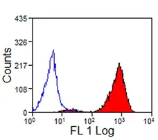
| Flow Cytometry |  |
1/25 | 1/200 |
| Immunohistology - Frozen | |
||
| Immunoprecipitation | |
||
| Western Blotting 1 | |
- 1 Non-reducing conditions required
Where this product has not been tested for use in a particular technique this does not necessarily exclude its use in such procedures. Suggested working dilutions are given as a guide only. It is recommended that the user titrates the product for use in their own system using appropriate negative/positive controls.
- Flow Cytometry
- Use 10ul of the suggested working dilution to label 106 cells in 100ul.
References for MHC Class I antibody
-
Hopkins, J. & Dutia, B.M. (1990) Monoclonal antibodies to the sheep analogues of human CD45 (leucocyte common antigen), MHC class I and CD5. Differential expression after lymphocyte activation in vivo.
Vet Immunol Immunopathol. 24 (4): 331-46. -
Lee, W.C. et al. (1996) The phenotype and phagocytic activity of macrophages during maedi-visna virus infection.
Vet Immunol Immunopathol. 51 (1-2): 113-26. -
Ryan, S. et al. (2000) Infection of dendritic cells by the Maedi-Visna lentivirus.
J Virol. 74 (21): 10096-103. -
Chan, S.S. et al. (2002) Generation and characterization of ovine dendritic cells derived from peripheral blood monocytes.
Immunology. 107: 366-72. -
Wu, C. et al. (2008) Mapping and characterization of visna/maedi virus cytotoxic T-lymphocyte epitopes.
J Gen Virol. 89 (Pt 10): 2586-96. -
Montagnaro, S. et al. (2009) Feline herpesvirus-1 down-regulates MHC class I expression in an homologous cell system.
J Cell Biochem. 106: 179-85.
MCA897GA
If you cannot find the batch/lot you are looking for please contact our technical support team for assistance.
View more products with MHC CLASS I specificity
Please Note: All Products are "FOR RESEARCH PURPOSES ONLY"
View all Anti-Sheep ProductsAlways be the first to know.
When we launch new products and resources to help you achieve more in the lab.
Yes, sign me up