RECA-1 antibody | HIS52
Mouse anti Rat RECA-1
- Product Type
- Monoclonal Antibody
- Clone
- HIS52
- Isotype
- IgG1
- Specificity
- RECA-1


Mouse anti Rat RECA-1 antibody, clone HIS52 (MCA970R) used for the detection of RECA-1 expressing cells by immunofluorescence.
Image caption
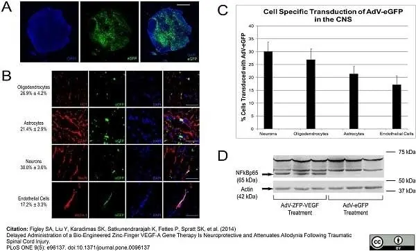
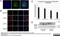
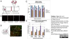
Transduction of AdV-eGFP/AdV-ZFP-VEGF into the spinal cord. (A) Photomicrographs showing a transverse section of rat spinal cord obtained adjacent to the injury site 10 days after spinal cord injury and AdV-eGFP injection. eGFP signal was detected in both the gray matter and white matter. (B) High-power (63X) confocal images show that the AdV-eGFP vector (green) transfected neurons (NeuN), astrocytes (GFAP), oligodendrocytes (CC1) and endothelial cells (RECA-1). Cells have been counter-stained with DAPI (blue) as nuclear marker. (C) Bar graph displays quantification of transduced cell types ± SEM, as identified by the cell-specific markers NeuN, GFAP, RECA-1 and CC1. (D) Evaluation of AdV-ZFP-VEGF gene transfer. Western blot showed that the NFκB p65 rabbit polyclonal antibody recognizes the p65 activation domain in the AdV-ZFP-VEGF treated animals. The higher molecular weight bands are endogenous NFκBp65 fragments, which are also recognized by the antibody; however, these bands are present in both the control and treatment groups. The lower band (arrow) corresponds to the AdV-ZFP-VEGF and was only present in the treated animals. Lower panel shows actin expression as a protein control. Scale bar: 1000 μm for A; 100 μm for B.
From: Figley SA, Liu Y, Karadimas SK, Satkunendrarajah K, Fettes P, et al.
Delayed Administration of a Bio-Engineered Zinc-Finger VEGF-A Gene Therapy Is Neuroprotective and Attenuates Allodynia Following Traumatic Spinal Cord Injury.
PLoS ONE (2014) 9(5): e96137.
doi: 10.1371/journal.pone.0096137.
This image is from an open access article distributed under terms of a Creative Commons Attribution License.
Mouse anti Rat RECA-1 antibody, clone HIS52 (MCA970R) used for the detection of RECA-1 expressing cells by immunofluorescence.
Image caption
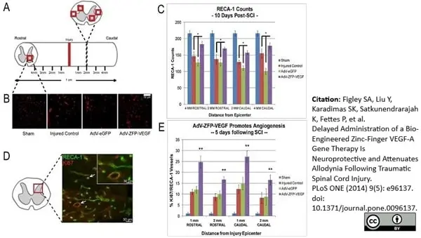
AdV-ZFP-VEGF results in increased vessel counts and angiogenesis. (A) Left panel: Illustration of the area of spinal cord areas used for RECA-1 counting (2 grey matter areas, 2 white matter areas). (B) Representative sections taken 2 mm rostral to the epicenter from a AdV-ZFP-VEGF treated and AdV-eGFP control animal respectively immunostained with RECA-1 at 10 days after SCI; scale 100 μm. An increased number of vessels were observed in the AdV-ZFP-VEGF treated group. (C) Bar graph illustrating the RECA-1 positive cell counts 10 days after SCI. AdV-ZFP-VEGF administration resulted in a significant increase in vascular counts (2 mm and 4 mm away from the epicenter) as compared with the control group. (D) Representative confocal image from an ADV-ZFP-VEGF treated animal at 5 days post-injury. Image was taken at 2 mm rostral from the epicenter, and shows double-labeled cells. Cells were stained for endothelial cells (RECA-1, green) and proliferation (Ki67, red). Scale bar = 50 μm (30 μm for magnified panel). (E) Angiogenesis was assessed by quantifying Ki67/RECA-1 co-labeled vessels. Data is presented at the percentage of RECA-1+ vessels that were also Ki67+, with an overall average increase of 10% vascular proliferation observed in the animals receiving AdV-ZFP-VEGF administration. All data are presented as mean ± SEM, and was analyzed by Two-way ANOVA (Holm-Sidak post-hoc). Angiogenesis data were analyzed by performing an arcsine transformation of the values, prior to Two-way ANOVA and post-hoc testing. *p<0.01, **p<0.001. n = 4/sham and injured control groups, n = 5/AdV-eGFP and AdV-ZFP-VEGF groups.
From: Figley SA, Liu Y, Karadimas SK, Satkunendrarajah K, Fettes P, et al.
Delayed Administration of a Bio-Engineered Zinc-Finger VEGF-A Gene Therapy Is Neuroprotective and Attenuates Allodynia Following Traumatic Spinal Cord Injury.
PLoS ONE (2014) 9(5): e96137.
doi: 10.1371/journal.pone.0096137.
This image is from an open access article distributed under terms of a Creative Commons Attribution License.
Mouse anti Rat RECA-1 antibody, clone HIS52 (MCA970R) used for the detection of RECA-1 expressing cells by immunofluorescence.
Image caption
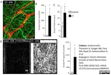
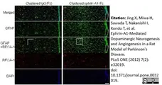
Increased vascular coverage and neuronal projections by angiogenic factors. (A, B) CT treatment of organotypic slice cultures (every 4 days for 2 weeks) retains the vasculature (confocal projection for the pan-endothelial marker RECA-1 and TH), (C) increases the thickness of the striatal portion of the slice, (D,E) promotes the sprouting of TH+ fibers from the S. Nigra section to the striatal section (2-weeks after control (BSA) and CT treatment). [Size bars: 20 μm].
From: Androutsellis-Theotokis A, Rueger MA, Park DM, Boyd JD, Padmanabhan R, et al.
Angiogenic Factors Stimulate Growth of Adult Neural Stem Cells.
PLoS ONE (2010) 5(2): e9414.
10.1371/journal.pone.0009414.
This image is from an open access article distributed under terms of a Creative Commons Attribution License.

Mouse anti Rat RECA-1 antibody, clone HIS52 (MCA970R) used for the detection of RECA-1 expressing cells by immunofluorescence.
Image caption
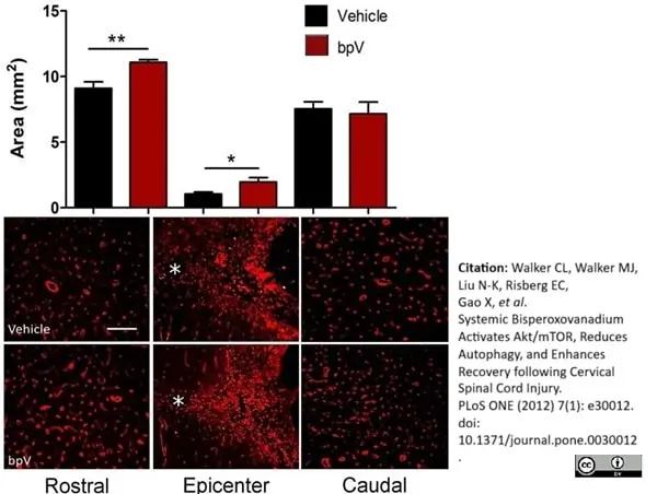
Significant increase in ipsilateral gray matter vasculature rostral and at the epicenter of the injury. bpV(pic)-treated animals demonstrated significantly increased vascular (RECA-1)-positive area in the ipsilateral gray matter 2 mm rostral and at the epicenter of the lesion 6 weeks post-SCI. * = central canal in photomicrographs. No significant difference was observed 2 mm caudal to the epicenter. **, p<0.01; *, p<0.05. n = 4−5. Error bars = SEM. Scale bar (Rostral & Caudal) = 100 μm; (Epicenter) = 150 μm.
From: Walker CL, Walker MJ, Liu N-K, Risberg EC, Gao X, et al.
Systemic Bisperoxovanadium Activates Akt/mTOR, Reduces Autophagy, and Enhances Recovery following Cervical Spinal Cord Injury.
PLoS ONE (2012) 7(1): e30012.
doi: 10.1371/journal.pone.0030012.
This image is from an open access article distributed under terms of a Creative Commons Attribution License.
Mouse anti Rat RECA-1 antibody, clone HIS52 (MCA970R) used for the detection of RECA-1 expressing cells by immunofluorescence.
Image caption
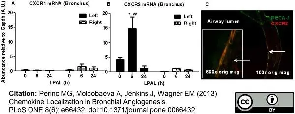
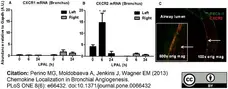
CXCR1(A) and CXCR2 (B) mRNA in left and right bronchi, and (C) co-localization of CXCR2 with RECA-1+ subepithelial blood vessel. Significant changes in CXCR2 were measured only in the left bronchus (*P<0.05 from 0 h and ##P<0.01 from right bronchus). Frozen sections of left bronchus 6 h after LPAL show co-localization of anti-CXCR2 (red) with RECA-1+ subepithelial blood vessels (green: 100× original magnification, and inset 600× original magnification).
From: Perino MG, Moldobaeva A, Jenkins J, Wagner EM
Chemokine Localization in Bronchial Angiogenesis.
PLoS ONE (2013) 8(6): e66432.
This image is from an open access article distributed under terms of a Creative Commons Attribution License.
Mouse anti Rat RECA-1 antibody, clone HIS52 (MCA970GA) used for the detection of RECA-1 expressing cells by immunohistochemistry.
Image caption
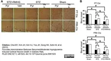
Pyruvate reduces R/M hypoglycemia-induced cortical blood vessel loss. (A) Bright field photomicrographs from coronal sections of cortex demonstrate loss of blood vessels at three days after R/M hypoglycemia by RECA-1 (rat endothelial cell antigen 1) staining. Panels show the progression of blood vessel changes in the parietal (PT Ctx) and perirhinal (PRh Ctx) cortex. After R/M hypoglycemia, blood vessels showed decreased density compared to sham-operated rats. Intraperitoneal injection of pyruvate as an adjuvant to glucose at ten minutes after R/M hypoglycemia reduced blood vessel disappearance. Scale bar = 100 μm. (B) Graph represents the % area of RECA-1 immunoreactivity in the parietal and perirhinal cortex. Data are means ± s.e.m., n=5-6 from each group, *P<0.05.
From: Choi BY, Kim JH, Kim HJ, Yoo JH, Song HK, et al.
Pyruvate Administration Reduces Recurrent/Moderate Hypoglycemia-Induced Cortical Neuron Death in Diabetic Rats.
PLoS One. 2013 Nov 22;8(11):e81523.
doi: 10.1371/journal.pone.0081523
This image is from an open access article distributed under terms of a Creative Commons Attribution License.
Mouse anti Rat RECA-1 antibody, clone HIS52 (MCA970GA) used for the detection of RECA-1 expressing cells by immunofluorescence.
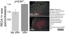
Image caption
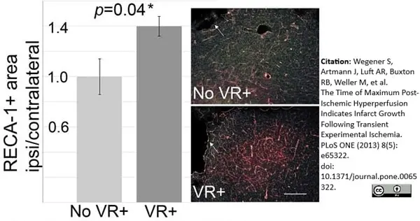
Increased density of blood vessels at perilesional regions with increased d14 vasoreactivity. RECA-1 immunofluorescence staining (red) with DAPI nuclear staining (green) compared in animals without (No VR+) and with (VR+) increased vasoreactivity on d14 (n = 4 per group). Corresponding subcortical regions close to the infarct on d14 showed a higher density of RECA-1 signal. One example per group is shown on the right. White arrows point to the lesion. White line indicates 100 μm.
From: Wegener S, Artmann J, Luft AR, Buxton RB, Weller M, et al.
The Time of Maximum Post-Ischemic Hyperperfusion Indicates Infarct Growth Following Transient Experimental Ischemia.
PLoS ONE (2013) 8(5): e65322.
doi: 10.1371/journal.pone.0065322.
This image is from an open access article distributed under terms of a Creative Commons Attribution License.
Mouse anti Rat RECA-1 antibody, clone HIS52 (MCA970GA) used for the detection of RECA-1 expressing cells by immunofluorescence.
Image caption
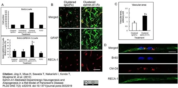
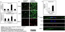
Effect of clustered ephrin-A1-Fc on vascular formation in the rat striatum. (A) Distribution of BrdU(+) endothelial cells. Brain of the unilaterally lesioned rats 6 weeks after infusion of clustered ephrin-A1-Fc were sectioned coronally and stained for BrdU and Rat Endothelial Cell Antigen-1 (RECA-1). Numbers of the BrdU(+) cells and BrdU(+) & RECA-1(+) cells were counted as described in the Materials and Methods. Total numbers of BrdU(+) cells in 8 animals are shown on the top, and percentages of RECA-1(+) cells among BrdU(+) cells are shown on the bottom. Error bars represent SD. *p<0.01 (n = 8) compared to control (IgG[Fc]). (B) Magnified confocal micrographs of insets in Fig S5. Scale bar: 50 μm. (C) Quantification of endothelial cell area. Coronal sections of striatum were stained for RECA-1 as in Fig S5, and the RECA-1 stained area was quantified using an ImageJ computer program (NIH). The areas taken for measurements were as described in the Materials and Methods. The values from 6 animals were analyzed statistically. *p<0.01 (n = 6). Error bars represent SD. (D) Co-localization of BrdU, CM-DiI, and RECA-1 in endothelial cells of the striatum. Sections taken from the rats treated as in Fig. 5B with intraventricular CM-DiI injection were stained for BrdU and RECA-1 and subjected to 3D confocal microscopy. Micrographs are the all-in-focus compilation of 12 confocal micrographs at 0.5 μm intervals.
From: Jing X, Miwa H, Sawada T, Nakanishi I, Kondo T, et al.
Ephrin-A1-Mediated Dopaminergic Neurogenesis and Angiogenesis in a Rat Model of Parkinson's Disease.
PLoS ONE (2012) 7(2): e32019.
doi: 10.1371/journal.pone.0032019.
This image is from an open access article distributed under terms of a Creative Commons Attribution License.
Mouse anti Rat RECA-1 antibody, clone HIS52 (MCA970R) used for the detection of RECA-1 expressing cells by immunofluorescence.
Image caption
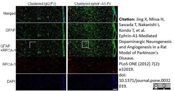
Effect of clustered ephrin-A1-Fc on vascular formation in the rat striatum. Clustered ephrin-A1-Fc was injected into the lesioned side of the lateral ventricle in the unilaterally lesioned rats. Brains taken 6 weeks after injection were sectioned coronally and stained for GFAP (green) and RECA-1 (red) and with DAPI (nuclei; blue). The rectangular insets are shown in Fig. 8B. Scale bar: 100 μm.
From: Jing X, Miwa H, Sawada T, Nakanishi I, Kondo T, et al.
Ephrin-A1-Mediated Dopaminergic Neurogenesis and Angiogenesis in a Rat Model of Parkinson's Disease.
PLoS ONE (2012) 7(2): e32019.
doi: 10.1371/journal.pone.0032019.
This image is from an open access article distributed under terms of a Creative Commons Attribution License.

Mouse anti Rat RECA-1 antibody, clone HIS52 (MCA970R) used for the detection of RECA-1 expressing cells by immunofluorescence.
Image caption
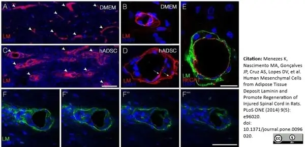
hADSCs led to the appearance of perivascular spaces in between endothelial and astrocytic basement membranes one week after injection. A–D) Confocal images of horizontal sections immunostained with anti-pan-laminin antibody (red) one week after injury. Note that in DMEM animals (A,B) there is no separation between the two membranes whereas in hADSCs–treated animals (C,D) these membranes are separated (arrows in D). E) Confocal images of a horizontal section immunostained with anti-pan-laminin (green) and RECA-1(red). F–F”) Confocal images of sequential optical sections immunostained with anti-pan-laminin (green) and DAPI (blue) showing the extravasation of cells from the blood vessels. Bars: C, F = 50 μm B, D, E = 25 μm.
From: Menezes K, Nascimento MA, Gonçalves JP, Cruz AS, Lopes DV, et al.
Human Mesenchymal Cells from Adipose Tissue Deposit Laminin and Promote Regeneration of Injured Spinal Cord in Rats.
PLoS ONE (2014) 9(5): e96020.
doi: 10.1371/journal.pone.0096020.
This image is from an open access article distributed under terms of a Creative Commons Attribution License.
Mouse anti Rat RECA-1 antibody, clone HIS52 (MCA970R) used for the detection of RECA-1 expressing cells by immunofluorescence.
Image caption
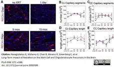
Endothelial cell number and capillary length post radiation. (A) Representative images of sections from the corpus callosum immunostained for rat endothelial cell antigen (RECA) at various time points post radiation. RECA expression declines immediately post radiation but is restored and maintained through 15 months. Stereological estimates of the number of capillary segments in the cortex (B) and corpus callosum (C) and of capillary length in both regions (D, E). (*** p<0.001; ** p<0.01; * p<0.05; ANOVA). Bars = SEM. Scale bar in A corresponds to 100 μm.
From: Panagiotakos G, Alshamy G, Chan B, Abrams R, Greenberg E, et al.
Long-Term Impact of Radiation on the Stem Cell and Oligodendrocyte Precursors in the Brain.
PLoS ONE 2(7): e588.
doi: 10.1371/journal.pone.0000588.
This image is from an open access article distributed under terms of a Creative Commons Attribution License.
Mouse anti Rat RECA-1 antibody, clone HIS52 (MCA970R) used for the detection of RECA-1 expressing cells by immunofluorescence.
Image caption
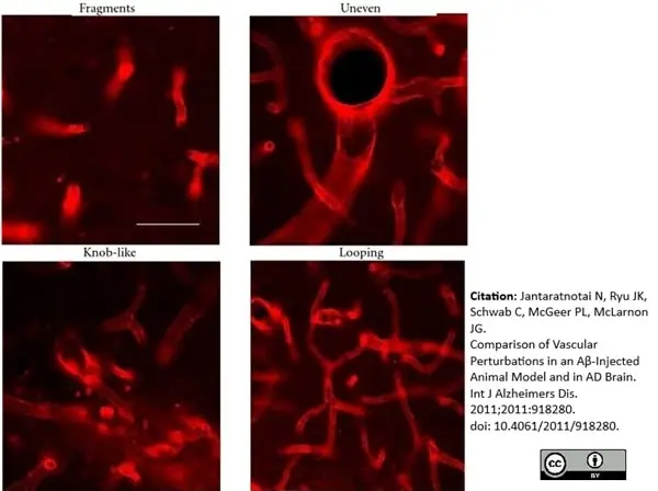
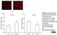
Representative patterns of RECA-1 staining in rat hippocampus. (a) Microvessels in control (PBS) injected rat hippocampus (left panel) and microvessels in Aβ1−42-injected hippocampus (right panel). Scale bar is for 70μm. (b) Bar graph for the number of microvessels/mm2 (n = 5 each). (c) Bar graph for microvessel length (n = 5 each). *P ≤0.05 for A&β1−42 versus PBS.
From: Jantaratnotai N, Ryu JK, Schwab C, McGeer PL, McLarnon JG.
Comparison of Vascular Perturbations in an Aβ-Injected Animal Model and in AD Brain.
Int J Alzheimers Dis. 2011;2011:918280.
doi: 10.4061/2011/918280.
This image is from an open access article distributed under terms of a Creative Commons Attribution License.
Mouse anti Rat RECA-1 antibody, clone HIS52 (MCA970R) used for the detection of RECA-1 expressing cells by immunofluorescence.
Image caption
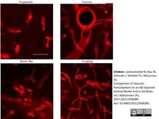
Morphology of microvessels stained with RECA-1 in Aβ 1−42-injected hippocampus. Panels show morphological features including fragments, looping microvessels, and vessels with knob-like and uneven diameters. The scale bar represents 40 μm.
From: Jantaratnotai N, Ryu JK, Schwab C, McGeer PL, McLarnon JG.
Comparison of Vascular Perturbations in an Aβ-Injected Animal Model and in AD Brain.
Int J Alzheimers Dis. 2011;2011:918280.
doi: 10.4061/2011/918280.
This image is from an open access article distributed under terms of a Creative Commons Attribution License.
Mouse anti Rat RECA-1 antibody, clone HIS52 (MCA970R) used for the detection of RECA-1 expressing cells in rat kidney by immunofluorescence.
Image caption:
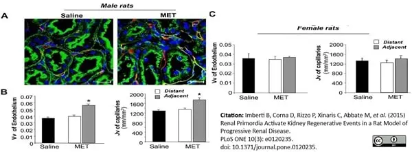
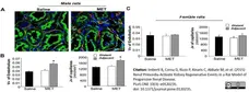
Metanephros effect on endothelium volume density and peritubular capillary length density. (A) Representative images of renal tissue of male MWF rats receiving saline or methanephroi (MET), 6 weeks after transplantation, stained for RECA-1 (red), FITC-labeled WGA lectin (green) and DAPI (blue); Scale bars = 20 μm. (B-C) Endothelial volume density (Vv, on the left), and length density of peritubular capillaries (Jv, on the right), evaluated in renal tissues of male (B) and female (C) animals receiving saline or MET distant from or adjacent to the graft. The results are expressed as mean ± SE. *P < 0.01 vs respective adjacent tissues and vs saline (n = 4 rats/group).
From: Imberti B, Corna D, Rizzo P, Xinaris C, Abbate M, et al. (2015)
Renal Primordia Activate Kidney Regenerative Events in a Rat Model of Progressive Renal Disease.
PLoS ONE 10(3): e0120235.
doi: 10.1371/journal.pone.0120235.
This image is from an open access article distributed under terms of a Creative Commons Attribution License.
Mouse anti Rat RECA-1 antibody, clone HIS52 (MCA970GA) used for the detection of RECA-1 expressing cells in rat kidney by immunofluorescence.
Image caption:
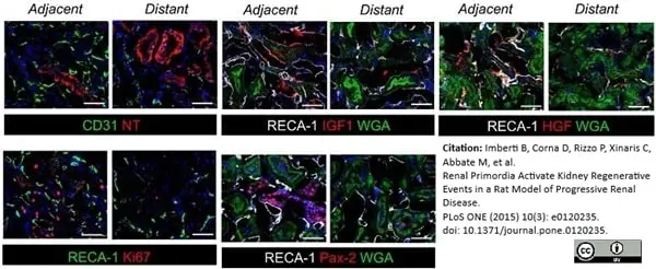
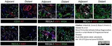
Co-staining of vascular endothelium with markers relevant to kidney regeneration in male MWF rats receiving metanephroi (MET).
Double immunofluorescence staining of endothelial cell markers (RECA-1 or CD31) with oxidative damage marker nitrotyrosine (NT), proliferation marker Ki-67 and HGF, IGF-1 or Pax-2. Improved vascularization in areas adjacent to MET is associated with improvement of oxidative damage and increased expression of the markers associated with regeneration. Renal tissues are labeled with DAPI (blue) and where specified in the picture with WGA-lectin. Scale bars = 100 μm.
From: Imberti B, Corna D, Rizzo P, Xinaris C, Abbate M, et al.
Renal Primordia Activate Kidney Regenerative Events in a Rat Model of Progressive Renal Disease.
PLoS ONE (2015) 10(3): e0120235.
doi: 10.1371/journal.pone.0120235.
This image is from an open access article distributed under terms of a Creative Commons Attribution License.
Mouse anti Rat RECA-1 antibody, clone HIS52 (MCA970R) used for the identification of blood vessels in rat spinal cord following injury by immunofluorescence.
Image caption:
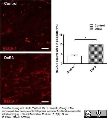
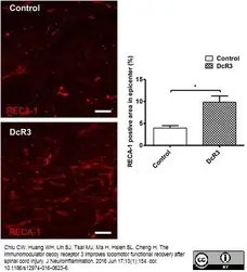
DcR3 treatment promoted angiogenesis at the lesion site. Population of blood vessels (RECA-1-positive, red) at the lesion sites of DcR3.Fc-treated and control SCI rats (left, scale bar: 100 μm). Quantification as the percentage of the RECA-1-positive area related to the total area in three random fields at the lesion site of each section (right, *p <0.05, N = 3). The results are presented as means ± SEM
From: Chiu CW, Huang WH, Lin SJ, Tsai MJ, Ma H, Hsieh SL, Cheng H.
The immunomodulator decoy receptor 3 improves locomotor functional recovery after spinal cord injury.
J Neuroinflammation. 2016 Jun 17;13(1):154.
This image is from an open access article distributed under terms of a Creative Commons Attribution License.
Mouse anti Rat RECA-1 antibody, clone HIS52 (MCA970R) used to identify vascular endothlium in a rat disc angiogenesis assay by immunohistochemistry on paraffin embedded tissue sections
Image caption:
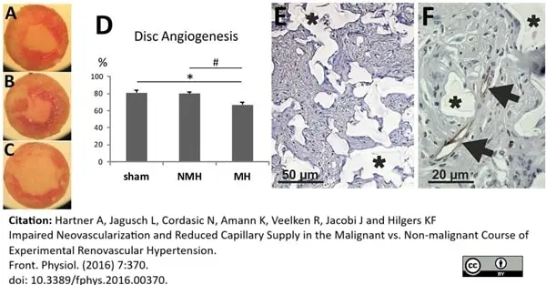
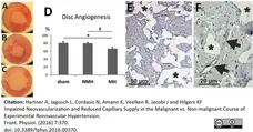
Disc angiogenesis assay. (A) Disc from a control animal (representative for n = 13). (B) Disc from an animal with non-malignant hypertension (representative for n = 9). (C) Disc from an animal with malignant hypertension (representative for n = 9). (D) Evaluation of the disc angiogenesis assay. *p <0.05 vs. sham OP control, #p <0.05 vs. non-malignant hypertension. (E) Exemplary photomicrograph of a PAS stained section of a disc from a control animal showing immigrated cells stained in purple (granulation tissue). Asterisks mark disc matrix. (F) Photomicrograph of αRECA stained section of a disc showing endothelium of blood vessels (arrows). Asterisks mark disc matrix.
From: Hartner A, Jagusch L, Cordasic N, Amann K, Veelken R, Jacobi J and Hilgers KF
Impaired Neovascularization and Reduced Capillary Supply in the Malignant vs. Non-malignant Course of Experimental Renovascular Hypertension.
Front. Physiol. (2016) 7:370.
doi: 10.3389/fphys.2016.00370.
This image is from an open access article distributed under terms of a Creative Commons Attribution License.
Mouse anti Rat RECA-1 antibody, clone HIS52 (MCA970GA) used for the demonstration of endothelial cells in rat brain by immunofluorescence on vibratome sections.
Image caption:
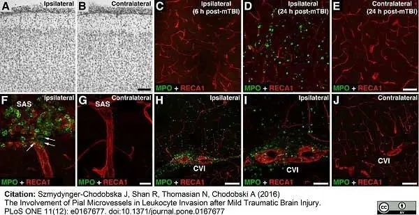
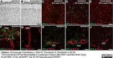
The influx of neutrophils into the injured brain after mTBI.
In these experiments, the depth of brain deformation was set at 1 mm. Double immunostaining of injured brains was performed with anti-myeloperoxidase (MPO) antibody, a neutrophil marker, and an antibody to RECA-1, a marker for rat endothelial cells. (A, B) Histological analysis (cresyl violet staining) of the cerebral cortex at 4 weeks after mTBI. Note no apparent tissue damage with this severity of injury. (C–E) Unlike severe TBI, mTBI was not accompanied by any significant influx of neutrophils into the injured cortex until 24 h post-mTBI. (F, G) In contrast to brain parenchyma, a large number of neutrophils crossed the pial microvessels and invaded the subarachnoid space (SAS) near the injury site at 6 h post-mTBI. Note that some neutrophils entering the SAS appeared to subsequently move along the perivascular space to enter the brain parenchyma (arrows). (H–J) A robust influx of neutrophils into the ipsilateral cistern of velum interpositum (CVI), a slit-shaped cerebrospinal fluid space located above the 3rd ventricle with highly vascularized pia mater, at 6 h post-mTBI. Bars: panels A, B, H, J, 100 μm; panels C–E, I, 50 μm; panels F, G, 20 μm.
From: Szmydynger-Chodobska J, Shan R, Thomasian N, Chodobski A
The Involvement of Pial Microvessels in Leukocyte Invasion after Mild Traumatic Brain Injury.
PLoS ONE (2016) 11(12): e0167677.
doi: 10.1371/journal.pone.0167677.
This image is from an open access article distributed under terms of a Creative Commons Attribution License.
Mouse anti Rat RECA-1 antibody, clone HIS52 (MCA970R) used to demonstrate rat endothelium in rat cerebellum following extended methylmercury exposure by immunohistofluorescence.
Image caption:
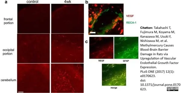
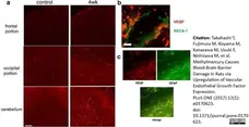
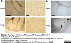
Vascular endothelial growth factor (VEGF) expression associated with methylmercury exposure.
(a) Immunohistochemical staining was performed using rabbit anti-VEGF antibody to detect VEGF expression in the frontal and occipital regions and cerebellum of rats in the control and 4-week MeHg exposure groups (left and right panels, respectively). Scale bar, 50 μm. (b) Representative images of double immunohistochemical staining sections of cerebellum in the 4-week MeHg exposure group. VEGF (red) and rat endothelial cell antigen-1 (RECA-1, a marker of endothelial cells, green) positive cells are shown. Scale bar, 20 μm. (c) Double immunohistochemical staining for VEGF (red) and glial fibrillary acidic protein (GFAP, a marker of astrocytes, green). Scale bar, 30 μm. All experiments were performed in triplicate.
From: Takahashi T, Fujimura M, Koyama M, Kanazawa M, Usuki F, Nishizawa M, et al.
Methylmercury Causes Blood-Brain Barrier Damage in Rats via Upregulation of Vascular Endothelial Growth Factor Expression.
PLoS ONE (2017) 12(1): e0170623.
doi: 10.1371/journal.pone.0170623.
This image is from an open access article distributed under terms of a Creative Commons Attribution License.
Mouse anti Rat RECA-1 antibody, clone HIS52 (MCA970R) used to demonstrate rat endothelium in rat cerebellum following extended methylmercury exposure by immunohistochemistry.
Image caption:
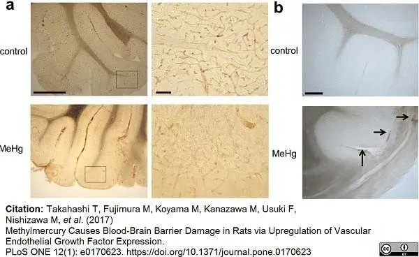
Expression of endothelial cell markers and IgG extravasation in the cerebellum of rats exposed to methylmercury.
(a) Rat cerebellum sections from control and 4-week exposure groups (upper and lower panels, respectively) were stained using an antibody against rat endothelial cell antigen-1 (RECA-1). Low and high (left and right panel, respectively) magnification images are shown. Scale bars, 100 and 10 μm (left and right panel, respectively). (b) Rat cerebellum sections from control and 4-week exposure groups (upper and lower panels, respectively) were stained using an antibody against rat IgG. Vascular hyperpermeability was evaluated by immunostaining of intrinsic IgG outside of vessels in control and 4-week exposure groups. Arrows indicate IgG extravasation in the 4-week MeHg exposure group. No IgG staining outside of vessels was detected in the control group. Scale bar, 25 μm. All experiments were performed in triplicate.
From: Takahashi T, Fujimura M, Koyama M, Kanazawa M, Usuki F, Nishizawa M, et al.
Methylmercury Causes Blood-Brain Barrier Damage in Rats via Upregulation of Vascular Endothelial Growth Factor Expression.
PLoS ONE (2017) 12(1): e0170623.
doi: 10.1371/journal.pone.0170623
This image is from an open access article distributed under terms of a Creative Commons Attribution License.
Mouse anti Rat RECA-1 antibody, clone HIS52 (MCA970GA) used for the demonstration of vasculature in rat tissue sections by immunofluorescence.
Image caption:
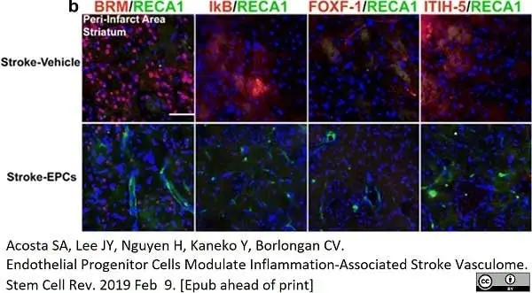
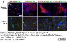
EPC transplantation decreases vasculome/RECA1 ratio at day 7 post-stroke in the cortex peri-infarct area. b Photomicrographs correspond to the coronal sections of the ipsilateral hemisphere showing positive vasculome and blood vessel positive RECA1 staining of the peri-infarct area of the cortex in stroke-vehicle and stroke-EPC treated animals. Arrows indicate co-localization of vasculome with RECA 1 staining for BRM, IKB, Foxf1, and ITIH-5, Scale bar = 50μm. Student t-test, p’s <0.01. Data are expressed as mean ± SEM
From: Acosta SA, Lee JY, Nguyen H, Kaneko Y, Borlongan CV.
Endothelial Progenitor Cells Modulate Inflammation-Associated Stroke Vasculome.
Stem Cell Rev. 2019 15(2):256-75.
doi: 10.1007/s12015-019-9873-x.
This image is from an open access article distributed under the terms of a Creative Commons Attribution License.
Mouse anti Rat RECA-1 antibody, clone HIS52 (MCA970GA) used for the demonstration of vasculature in rat tissue sections by immunofluorescence.
Image caption:
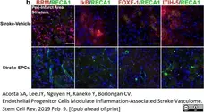
EPC transplantation decreases vasculome/RECA1 ratio at day 7 post-stroke in the striatum peri-infarct area. b Photomicrographs correspond to the coronal sections of the ipsilateral hemisphere showing positive vasculome and brain blood vessel positive (RECA1) expression in the peri-infarct area of the striatum in stroke-vehicle and stroke-EPC treated animals. Arrows indicate co-localization of vasculome with RECA 1 staining for either BRM, IKB, Foxf1, and ITIH-5, Scale bar = 50μm. Student t-test, p’s <0.01. Data are expressed as mean ± SEM
From: Acosta SA, Lee JY, Nguyen H, Kaneko Y, Borlongan CV.
Endothelial Progenitor Cells Modulate Inflammation-Associated Stroke Vasculome.
Stem Cell Rev. 2019 15(2):256-75.
doi: 10.1007/s12015-019-9873-x.
This image is from an open access article distributed under the terms of a Creative Commons Attribution License.
Mouse anti Rat RECA-1 antibody, clone HIS52 (MCA970GA) used for the detection of RECA-1 expressing cells by immunohistochemistry.
Image caption
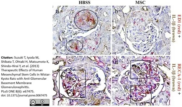
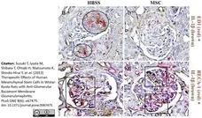
Double immunostaining for ED1 or RECA-1 with IL-1β in the study groups. Kidney sections were stained using two-color immunohistochemistry with ED1 or RECA-1 stained red and IL-1β stained brown in an HBSS-treated rat with nephritis (a,c) and an MSC-treated rat with nephritis (b,d). A large number of ED1+ macrophages shows double staining for IL-1β in the WKY-HBSS rats (circles) (a). RECA-1 is partially double-stained with IL-1β in both the WKY-HBSS rats and the WKY-MSC rats (squares) (c,d). Original magnifications, x1000.
From: Suzuki T, Iyoda M, Shibata T, Ohtaki H, Matsumoto K, et al.
Therapeutic Effects of Human Mesenchymal Stem Cells in Wistar-Kyoto Rats with Anti-Glomerular Basement Membrane Glomerulonephritis.
PLoS ONE (2013) 8(6): e67475.
This image is from an open access article distributed under terms of a Creative Commons Attribution License.
Mouse anti Rat RECA-1 antibody, clone HIS52 (MCA970R) used for the detection of RECA-1 expressing cells by immunofluorescence.
Image caption
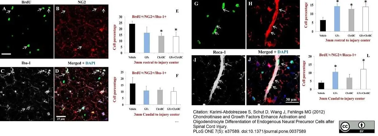
ChABC and GF treatments attenuate the proliferation of microglia/macrophages and promote the generation of new endothelial cells after SCI. (A–D) Representative confocal images of BrdU+/NG2+ macrophages/microglia marked with Iba-1 in the injured spinal cord (arrows). (E–F) Under baseline SCI condition, macrophages/microglia comprised about 25% and 17% of BrdU+/NG2+ cells in rostral and caudal points to the injury center, respectively. After treatment with ChABC and/or GFs, we found a reduction in the number of BrdU+/NG2+/IbA-1+ cells that was statistically significant for ChABC and ChABC+GFs treatment groups relative to the vehicle group. (G–J) Representative confocal images show newly generated endothelial cells marked by Reca-1 and NG2 among BrdU+ cells. Reca-1 positive endothelial cells comprised a subpopulation of proliferating NG2+ cells after SCI (J). (K–L) Quantification of BrdU+/NG2+/Reca-1+ cells showed a significant number of newly generated endothelial cells after treatment with ChABC and/or GFs at both rostral and caudal points to the injury center compared to the vehicle group. *p<0.05, n = 6/group.
From: Karimi-Abdolrezaee S, Schut D, Wang J, Fehlings MG
Chondroitinase and Growth Factors Enhance Activation and Oligodendrocyte Differentiation of Endogenous Neural Precursor Cells after Spinal Cord Injury.
PLoS ONE (2012) 7(5): e37589.
doi: 10.1371/journal.pone.0037589.
This image is from an open access article distributed under terms of a Creative Commons Attribution License.
Mouse anti Rat RECA-1 antibody, clone HIS52 (MCA970) used for the demonstration of endothelial cells in rat brain by immunofluorescence.
Image caption:
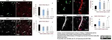
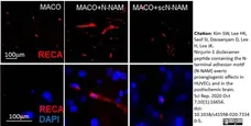
Proangiogenic effects of N-NAM in the postischemic brain. (a) N-NAM (5μg) or scN-NAM (5μg) was administered intranasally three times daily at 4, 5, and 6 days after 60 min of MCAO. (b,c) At 7 days post-MCAO, coronal brain sections were stained using double-fluorescence with anti-rat endothelial cell antigen-1 (RECA-1) antibody and DAPI. Representative images are presented (c), Scale bars, 100 μm.
From: Kim SW, Lee HK, Seol SI, Davaanyam D, Lee H, Lee JK.
Ninjurin 1 dodecamer peptide containing the N-terminal adhesion motif (N-NAM) exerts proangiogenic effects in HUVECs and in the postischemic brain.
Sci Rep. 2020 Oct 7;10(1):16656.
doi: 10.1038/s41598-020-73340-5.
This image is from an open access article distributed under terms of a Creative Commons Attribution License.
Mouse anti Rat RECA-1 antibody, clone HIS52 (MCA970R) used to label endothelial cells by immunohistochemistry on formalin fixed cryosections.
Image caption:
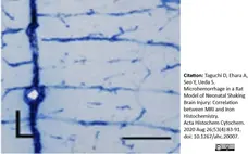
Histochemical and/or immunohistochemical staining of coronal sections though the medial prefrontal cortex on P7 (L) for non-shaken rats. Endothelial cells are stained by RECA-1 immunohistochemistry. Penetrating blood vessels in the cerebral cortex are evident. Bar = 200 μm.
From: Taguchi D, Ehara A, Seo Y, Ueda S.
Microhemorrhage in a Rat Model of Neonatal Shaking Brain Injury: Correlation between MRI and Iron Histochemistry.
Acta Histochem Cytochem. 2020 Aug 26;53(4):83-91.
doi: 10.1267/ahc.20007.
This image is from an open access article distributed under terms of a Creative Commons Attribution License.
Mouse anti Rat RECA-1 antibody, clone HIS52 (MCA970R) used for the detection of RECA-1 expressing cells in rat kidney by immunofluorescence.
Image caption:
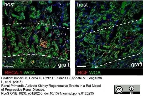
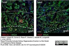
Low magnification immunofluorescence staining (red) for RECA-1 and HGF on male MWF renal tissues.
Sections are labeled with WGA-lectin (green) and DAPI (blue). Dashed lines divide the graft from the host, while dotted lines divide the area adjacent to and distant from the graft. Scale bars = 100 μm.
From: Imberti B, Corna D, Rizzo P, Xinaris C, Abbate M, et al.
Renal Primordia Activate Kidney Regenerative Events in a Rat Model of Progressive Renal Disease.
PLoS ONE (2015) 10(3): e0120235.
This image is from an open access article distributed under terms of a Creative Commons Attribution License.
Mouse anti Rat RECA-1 antibody, clone HIS52 (MCA970R) used to identify blood vessels in rat brain tissue sections by immunofluorescence
Image caption:
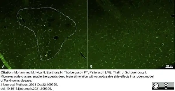
Immunofluorescent staining of RECA, visualizing blood vessel density, at the site of the electrode cluster (A), and in an adjacent area about 500 μm from the border of the electrode cluster (B). The dashed line in (A) indicates the approximate electrode cluster area and corresponds to the area encircled in Fig.8A.No obvious changes in blood vessel density could be observed throughout the cluster area (A) or compared to areas outside the cluster (B).
From: Mohammed M, Ivica N, Bjartmarz H, Thorbergsson PT, Pettersson LME, Thelin J, Schouenborg J.
Microelectrode clusters enable therapeutic deep brain stimulation without noticeable side-effects in a rodent model of Parkinson's disease.
J Neurosci Methods. 2021 Oct 22:109399.
doi: 10.1016/j.jneumeth.2021.109399.
This image is from an open access article distributed under terms of a Creative Commons Attribution License.

Mouse anti Rat RECA-1 antibody, clone HIS52 (MCA970R) used to identify blood vessels in rat brain tissue sections by immunofluorescence
Image caption:
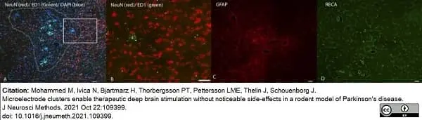
Immunofluorescent staining of tissue reactions around the microelectrode cluster array. (A) Visualization of NeuN (neurons; red), ED1 (activated microglia; green) and DAPI (all nuclei; blue) staining in the implantation area. Dashed line encircles the approximate electrode cluster area. Rectangular box delineates the area enlarged in B. (B) High magnification image showing NeuN and ED1 staining near a single microelectrode in an area with high neuronal density (DAPI was omitted from this panel for enhanced clarity of the microglial and neuronal density). (C) GFAP (astrocytes) and (D) RECA (blood vessel)-staining of the same area in a section 80 μm below the section shown in A and B. All scale bars are 50 μm.
From: Mohammed M, Ivica N, Bjartmarz H, Thorbergsson PT, Pettersson LME, Thelin J, Schouenborg J.
Microelectrode clusters enable therapeutic deep brain stimulation without noticeable side-effects in a rodent model of Parkinson's disease.
J Neurosci Methods. 2021 Oct 22:109399.
doi: 10.1016/j.jneumeth.2021.109399.
This image is from an open access article distributed under terms of a Creative Commons Attribution License.
Mouse anti Rat RECA-1 antibody, clone HIS52 (MCA970R) used to stain blood vessels in rat substantia nigra by immunofluorescence.
Image caption:
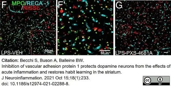
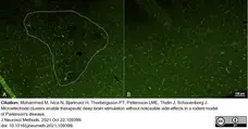
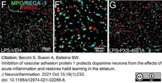
VAP-1 inhibition reduces the inflammatory response in the SN. F, G Immunostaining for MPO, RECA-1-positive blood vessels and Nissl in the SN 24 h after LPS insult. F’ Enlargement of F: MPO-positive cells mainly localized outside blood vessels.
From: Becchi S, Buson A, Balleine BW.
Inhibition of vascular adhesion protein 1 protects dopamine neurons from the effects of acute inflammation and restores habit learning in the striatum.
J Neuroinflammation. 2021 Oct 15;18(1):233.
doi: 10.1186/s12974-021-02288-8.
This image is from an open access article distributed under terms of a Creative Commons Attribution License.
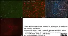
Mouse anti Rat RECA-1 antibody, clone HIS52 (MCA970R) used to label microvasculature in rat brain by immunofluorescence on cryostat sections.
Image caption:
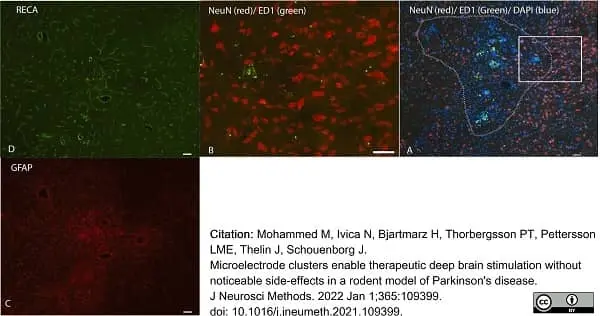
Immunofluorescent staining of tissue reactions around the microelectrode cluster array. (A) Visualization of NeuN (neurons; red), ED1 (activated microglia; green) and DAPI (all nuclei; blue) staining in the implantation area. Dashed line encircles the approximate electrode cluster area. Rectangular box delineates the area enlarged in B. (B) High magnification image showing NeuN and ED1 staining near a single microelectrode in an area with high neuronal density (DAPI was omitted from this panel for enhanced clarity of the microglial and neuronal density). (C) GFAP (astrocytes) and (D) RECA (blood vessel)-staining of the same area in a section 80μm below the section shown in A and B. All scale bars are 50μm.
From: Mohammed M, Ivica N, Bjartmarz H, Thorbergsson PT, Pettersson LME, Thelin J, Schouenborg J.
Microelectrode clusters enable therapeutic deep brain stimulation without noticeable side-effects in a rodent model of Parkinson's disease.
J Neurosci Methods. 2022 Jan 1;365:109399.
doi: 10.1016/j.jneumeth.2021.109399.
This image is from an open access article distributed under terms of a Creative Commons Attribution License.
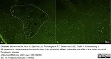
Mouse anti Rat RECA-1 antibody, clone HIS52 (MCA970R) used to label microvasculature in rat brain by immunofluorescence on cryostat sections.
Image caption:
Immunofluorescent staining of RECA, visualizing blood vessel density, at the site of the electrode cluster(A), and in an adjacent area about 500μm from the border of the electrode cluster(B). The dashed line in (A) indicates the approximate electrode cluster area and corresponds to the area encircled in Fig.8A. No obvious changes in blood vessel density could be observed throughout the cluster area (A) or compared to areas outside the cluster (B).
From: Mohammed M, Ivica N, Bjartmarz H, Thorbergsson PT, Pettersson LME, Thelin J, Schouenborg J.
Microelectrode clusters enable therapeutic deep brain stimulation without noticeable side-effects in a rodent model of Parkinson's disease.
J Neurosci Methods. 2022 Jan 1;365:109399.
doi: 10.1016/j.jneumeth.2021.109399.
This image is from an open access article distributed under terms of a Creative Commons Attribution License.
Mouse anti Rat RECA-1 antibody, clone HIS52 (MCA970R) used to label endothelial cells in regenerating nerve tissue following insult and subsequent excision of the chitosan conduit, by immunofluorescence.
Image caption:
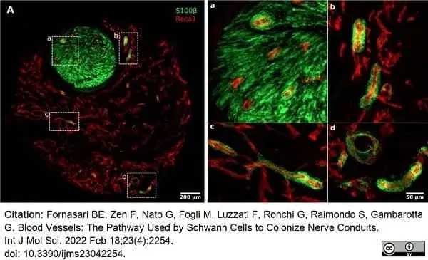
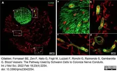
High magnification pictures of a regenerating nerve 7 days after the injury and repair. (A) A 50μm thick section of the distal portion of the conduit double-labeled with Reca1 (red, endothelial cell marker) and S100β (green, Schwann cell marker); (a–d) inserts: high magnification details of migrating Schwann cells closely associated with endothelial cells.
From: Fornasari BE, Zen F, Nato G, Fogli M, Luzzati F, Ronchi G, Raimondo S, Gambarotta G.
Blood Vessels: The Pathway Used by Schwann Cells to Colonize Nerve Conduits.
Int J Mol Sci. 2022 Feb 18;23(4):2254.
doi: 10.3390/ijms23042254.
This image is from an open access article distributed under terms of a Creative Commons Attribution License.

Mouse anti Rat RECA-1 antibody, clone HIS52 (MCA970R) used to demonstrate RECA-1 expression in rat spinal cord by immunofluorescence.
Image caption:
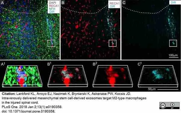
DiR hotspots not detected in other cell types at the lesion site.
Selected regions contused spinal cords 24 hours after infusion of DiR MSCexos (A-G) or PBS with free DiR (G-H), showing close proximity between DiR hotspots and some endothelial cells (A-C) within the central scar tissue at the contusion site and paucity of DiR fluorescence signal in DiR only infused animal. A-C: Region near the border of the lesion with antibody staining directed against neurofilament (NDH green), the endothelial cell marker RECA-1 (red) (A), RECA-1 (red) and DiR wavelengths (B), or DiR fluorescence only (cyan) (C). A1-C1, B2: enlarged and rotated boxed region shown in A-C. Note that DiR hot spots are very close to the endothelial cells, but not within them.
From: Lankford KL, Arroyo EJ, Nazimek K, Bryniarski K, Askenase PW, Kocsis JD.
Intravenously delivered mesenchymal stem cell-derived exosomes target M2-type macrophages in the injured spinal cord.
PLoS One. 2018 Jan 2;13(1):e0190358.
doi: 10.1371/journal.pone.0190358.
This image is from an open access article distributed under terms of a Creative Commons Attribution License.
Mouse anti Rat RECA-1 antibody, clone HIS52 (MCA970R) used to demonstrate RECA-1 expression in rat spinal cord by immunofluorescence.
Image caption:
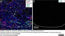
DiR hotspots not detected in other cell types at the lesion site.
H-I: Representative region of the lesion in a contused rat infused with DiR only, without exosomes and stained with antibodies directed against RECA-1 (red), and neurofilament (green) and counterstained with DAPI (G) and the same regions showing DiR wavelength fluorescence only. Dashed lines in A-C and H, I indicate the approximate lesion boundary with the lesioned area above and intact spinal cord tissue below. Scale bars in I = 100μm.
From: Lankford KL, Arroyo EJ, Nazimek K, Bryniarski K, Askenase PW, Kocsis JD.
Intravenously delivered mesenchymal stem cell-derived exosomes target M2-type macrophages in the injured spinal cord.
PLoS One. 2018 Jan 2;13(1):e0190358.
doi: 10.1371/journal.pone.0190358.
This image is from an open access article distributed under terms of a Creative Commons Attribution License.
Mouse anti Rat RECA-1 antibody, clone HIS52 (MCA970R) used to stain endothelial cells by immunofluorescence.
Image caption:
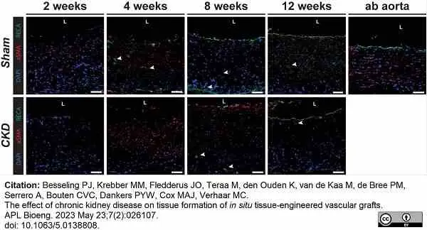
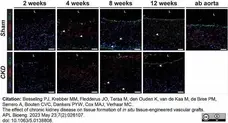
Vascular tissue formation. Grafts explanted from Sham and CKD animals at 2, 4, 8, and 12 weeks. Stained with rat endothelial marker, RECA (green) and smooth muscle cell marker, αSMA (red), with cell nuclei in blue (DAPI). Arrows indicate vascular infiltration. Lumen is indicated by L. Scale bars represent 50 μm.
From: Besseling PJ, Krebber MM, Fledderus JO, Teraa M, den Ouden K, van de Kaa M, de Bree PM, Serrero A, Bouten CVC, Dankers PYW, Cox MAJ, Verhaar MC.
The effect of chronic kidney disease on tissue formation of in situ tissue-engineered vascular grafts.
APL Bioeng. 2023 May 23;7(2):026107.
doi: 10.1063/5.0138808.
This image is from an open access article distributed under terms of a Creative Commons Attribution License.
Mouse anti Rat RECA-1 antibody, clone HIS52 (MCA970R) used to label endothelial cells in rat cauda equina by immunofluorescence.
Image caption:
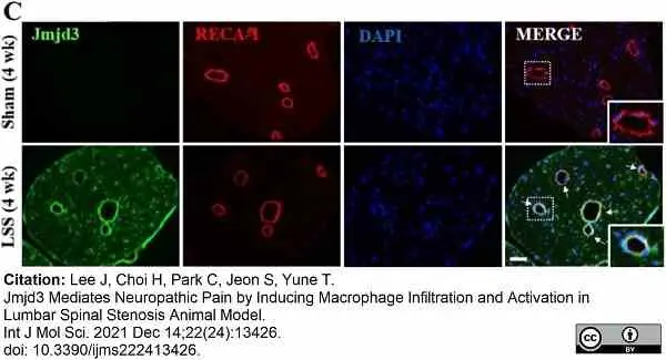
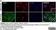
Jmjd3 expression and activation are increased in the cauda equina after compression injury in an LSS model.
(C) Double immunofluorescence staining for Jmjd3 and RECA-1 (endothelial cell marker) in Sham (4 weeks) and injured cauda equina (4 weeks after compression). Arrows indicate double-positive cells. The right, bottom panels show a higher magnification of the boxed area. Scale bar, 50 μm.
From: Lee J, Choi H, Park C, Jeon S, Yune T.
Jmjd3 Mediates Neuropathic Pain by Inducing Macrophage Infiltration and Activation in Lumbar Spinal Stenosis Animal Model.
Int J Mol Sci. 2021 Dec 14;22(24):13426.
doi: 10.3390/ijms222413426. .
This image is from an open access article distributed under terms of a Creative Commons Attribution License.

Mouse anti Rat RECA-1 antibody, clone HIS52 (MCA970R) used to label vasculature in rat spinal cord by immunofluorescence.
Image caption:
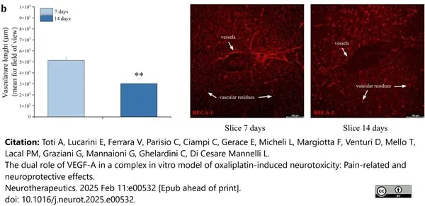
b) Immunohistochemical analysis and representative images of RECA-1 (blood vessels marker, red), performed after 7 and 14 days of slices maturation (40X magnification). Scale bar = 100 μM ∗∗P <0.01 vs slice 7 days.
From: Toti A, Lucarini E, Ferrara V, Parisio C, Ciampi C, Gerace E, Micheli L, Margiotta F, Venturi D, Mello T, Lacal PM, Graziani G, Mannaioni G, Ghelardini C, Di Cesare Mannelli L.
The dual role of VEGF-A in a complex in vitro model of oxaliplatin-induced neurotoxicity: Pain-related and neuroprotective effects.
Neurotherapeutics. 2025 Feb 11:e00532.
doi: 10.1016/j.neurot.2025.e00532.
This image is from an open access article distributed under terms of a Creative Commons Attribution License.
Mouse anti Rat RECA-1 antibody, clone HIS52 (MCA970) used to label endothelium in a rat spinal cord injury model by immunofluorescence.
Image caption:
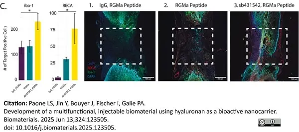
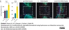
Immunocytochemical staining of key biomarker panels in a 2-week C4/5 hemisection injury model in female Sprague Dawley rats. (A,i) Schematic to show all three conditions of HA nanocarriers, (A,ii) injected in a C4/5 hemisection injury model. (A,iii) Image of PluHA injection at the site of injury prior to suturing the dura. (B–D) From left to right: Raw number of total positive cells in the injury area across three conditions of PluHA-RGMa-IgG, PluHA-RGMa, and PluHA-RGMa-sb431542. A one factor ANOVA was performed to demonstrate significance (n = 3). Representative confocal imaging at 4× magnification with ROI outlined to show representation of data. Scale = 500μm.
From: Paone LS, Jin Y, Bouyer J, Fischer I, Galie PA.
Development of a multifunctional, injectable biomaterial using hyaluronan as a bioactive nanocarrier.
Biomaterials. 2025 Jun 13;324:123505.
doi: 10.1016/j.biomaterials.2025.123505.
This image is from an open access article distributed under terms of a Creative Commons Attribution License.
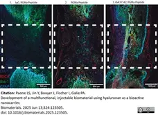
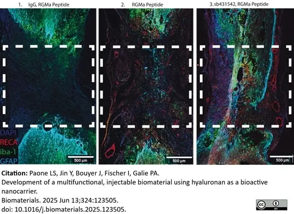
Mouse anti Rat RECA-1 antibody, clone HIS52 (MCA970) used to label endothelium in a rat spinal cord injury model by immunofluorescence (Enlargement from previous image)
Image caption:
Immunocytochemical staining of key biomarker panels in a 2-week C4/5 hemisection injury model in female Sprague Dawley rats. (A,i) Schematic to show all three conditions of HA nanocarriers, (A,ii) injected in a C4/5 hemisection injury model. (A,iii) Image of PluHA injection at the site of injury prior to suturing the dura. (B–D) From left to right: Raw number of total positive cells in the injury area across three conditions of PluHA-RGMa-IgG, PluHA-RGMa, and PluHA-RGMa-sb431542. A one factor ANOVA was performed to demonstrate significance (n = 3). Representative confocal imaging at 4× magnification with ROI outlined to show representation of data. Scale = 500μm.
From: Paone LS, Jin Y, Bouyer J, Fischer I, Galie PA.
Development of a multifunctional, injectable biomaterial using hyaluronan as a bioactive nanocarrier.
Biomaterials. 2025 Jun 13;324:123505.
doi: 10.1016/j.biomaterials.2025.123505.
This image is from an open access article distributed under terms of a Creative Commons Attribution License.
Filter by Application:
IF C P Reset| Mouse anti Rat RECA-1 antibody, clone HIS52 recognizes RECA-1, a cell surface antigen which is expressed by all rat endothelial cells. The endothelium is the thin layer of cells that line the interior surface of blood vessels, forming an interface between circulating blood in the lumen and the rest of the vessel wall. Endothelial cells line the entire circulatory system, from the heart to the smallest capillary. |
- Target Species
- Rat
- Species Cross-Reactivity
-
Target Species Cross Reactivity Goat Chicken Guinea Pig Sheep Mouse Rabbit Pig - N.B. Antibody reactivity and working conditions may vary between species.
- Product Form
- Purified IgG - liquid
- Preparation
- Purified IgG prepared by affinity chromatography on Protein G from tissue culture supernatant
- Buffer Solution
- Phosphate buffered saline
- Preservative Stabilisers
- <0.1% Sodium Azide (NaN3)
- Immunogen
- Stromal cells from rat lymph node.
- Approx. Protein Concentrations
- IgG concentration 0.5 mg/ml
- Fusion Partners
- Spleen cells from immunized mice were fused with cells of the SP2/0 mouse myeloma cell line.
- Regulatory
- For research purposes only
- Guarantee
- 12 months from date of despatch
This product is shipped at ambient temperature. It is recommended to aliquot and store at -20°C on receipt. When thawed, aliquot the sample as needed. Keep aliquots at 2-8°C for short term use (up to 4 weeks) and store the remaining aliquots at -20°C.
Avoid repeated freezing and thawing as this may denature the antibody. Storage in frost-free freezers is not recommended.
Avoid repeated freezing and thawing as this may denature the antibody. Storage in frost-free freezers is not recommended.
This product has been reported to work in the following applications. This information is derived from testing within our laboratories, peer-reviewed publications or personal communications from the originators. Please refer to references indicated for further information. For general protocol recommendations, please visit the antibody protocols page.
| Application Name | Verified | Min Dilution | Max Dilution |
|---|---|---|---|
| Immunofluorescence |  |
||
| Immunohistology - Frozen | |
Where this antibody has not been tested for use in a particular technique this does not necessarily exclude its use in such procedures. The suggested working dilution is given as a guide only. It is recommended that the user titrates the antibody for use in their own system using appropriate negative/positive controls.
References for RECA-1 antibody
-
Duijvestijn, A.M. et al. (1992) Antibodies defining rat endothelial cells: RECA-1, a pan-endothelial cell-specific monoclonal antibody.
Lab Invest. 66 (4): 459-66. -
Wada, Y. et al. (2002) Impairment of vascular regeneration precedes progressive glomerulosclerosis in anti-Thy 1 glomerulonephritis.
Kidney Int. 61 (2): 432-43. -
Simard, M. et al. (2003) Signaling at the gliovascular interface.
J Neurosci. 23: 9254-62. -
Tikkanen, J.M. et al. (2006) Role of platelet-derived growth factor and vascular endothelial growth factor in obliterative airway disease.
Am J Respir Crit Care Med. 174: 1145-52. -
Konno, T. et al. (2007) Pregnancy in the brown Norway rat: a model for investigating the genetics of placentation.
Biol Reprod. 76 (4): 709-18. -
Alam, S.M. et al. (2008) Decidual cells produce a heparin-binding prolactin family cytokine with putative intrauterine regulatory actions.
J Biol Chem. 283 (27): 18957-68. -
Valable, S. et al. (2009) MRI assessment of hemodynamic effects of angiopoietin-2 overexpression in a brain tumor model.
Neuro Oncol. 11: 488-502. -
Bexell, D. et al. (2009) Bone marrow multipotent mesenchymal stroma cells act as pericyte-like migratory vehicles in experimental gliomas.
Mol Ther. 17: 183-90.
View The Latest Product References
-
Cattaruzza, F. et al. (2009) Endothelin-converting enzyme 1 promotes re-sensitization of neurokinin 1 receptor-dependent neurogenic inflammation.
Br J Pharmacol. 156: 730-9. -
March, S. et al. (2009) Microenvironmental regulation of the sinusoidal endothelial cell phenotype in vitro.
Hepatology. 50: 920-8. -
Benton RL et al. (2009) Transcriptional activation of endothelial cells by TGFβ coincides with acute microvascular plasticity following focal spinal cord ischaemia/reperfusion injury.
ASN Neuro. 1 (3): pii: e00015. -
Androutsellis-Theotokis, A. et al. (2010) Angiogenic factors stimulate growth of adult neural stem cells.
PLoS One. 5 (2): e9414. -
Schödel, J. et al. (2010) Factor inhibiting HIF limits the expression of hypoxia-inducible genes in podocytes and distal tubular cells.
Kidney Int. 78 (9): 857-67. -
Szmydynger-Chodobska, J. et al. (2011) Multiple sites of vasopressin synthesis in the injured brain.
J Cereb Blood Flow Metab. 31: 47-51. -
Hamdi, H. et al. (2011) Epicardial adipose stem cell sheets results in greater post-infarction survival than intramyocardial injections.
Cardiovasc Res. 91 (3): 483-91. -
Morin-Brureau, M. et al. (2011) Epileptiform activity induces vascular remodeling and zonula occludens 1 downregulation in organotypic hippocampal cultures: role of VEGF signaling pathways.
J Neurosci. 31: 10677-88 -
Hawthorne, A.L. et al. (2011) The unusual response of serotonergic neurons after CNS Injury: lack of axonal dieback and enhanced sprouting within the inhibitory environment of the glial scar.
J Neurosci. 31: 5605-16. -
Jantaratnotai, N. et al. (2011) Comparison of Vascular Perturbations in an Aβ-Injected Animal Model and in AD Brain.
Int J Alzheimers Dis. 2011: 918280. -
Miya, M. et al. (2012) Age-related decline in label-retaining tubular cells: implication for reduced regenerative capacity after injury in the aging kidney.
Am J Physiol Renal Physiol. 302: F694-702. -
Nakamura, K. et al. (2014) Soluble thrombomodulin attenuates sinusoidal obstruction syndrome in rat through suppression of high mobility group box 1.
Liver Int. 34 (10): 1473-87. -
Mitkari, B. et al. (2014) Human bone marrow mesenchymal stem/stromal cells produce efficient localization in the brain and enhanced angiogenesis after intra-arterial delivery in rats with cerebral ischemia, but this is not translated to behavioral recovery.
Behav Brain Res. 259: 50-9. -
Wong, K. et al. (2015) Restoration of sensory dysfunction following peripheral nerve injury by the polysaccharide from culinary and medicinal mushroom, Hericium erinaceus. (Bull.: Fr.) Pers. through its neuroregenerative action
Food Sci Technol. 35 (4): 712-21. -
Hu, J. et al. (2015) 3D angioarchitecture changes after spinal cord injury in rats using synchrotron radiation phase-contrast tomography.
Spinal Cord. 53 (8): 585-90. -
Walker, C.L. et al. (2015) Biphasic bisperoxovanadium administration and Schwann cell transplantation for repair after cervical contusive spinal cord injury.
Exp Neurol. 264: 163-72. -
Ramessur Chandran, S. et al. (2015) Spleen tyrosine kinase contributes to acute renal allograft rejection in the rat.
Int J Exp Pathol. 96 (1): 54-62. -
Van Slooten, A.R. et al. (2015) L-NIO as a novel mechanism for inducing focal cerebral ischemia in the adult rat brain.
J Neurosci Methods. 245: 44-57. -
Nakahara, T. et al. (2015) Structural and functional changes in retinal vasculature induced by retinal ischemia-reperfusion in rats.
Exp Eye Res. 135: 134-45. -
Wang, L. et al. (2015) Lobe-specific expression of phosphodiesterase 5 in rat prostate.
Urology. 85 (3): 703.e7-13. -
Garbelli, R. et al. (2015) PDGFRβ(+) cells in human and experimental neuro-vascular dysplasia and seizures.
Neuroscience. 306: 18-27. -
Syrjälä, S.O. et al. (2015) Donor Heart Treatment With COMP-Ang1 Limits Ischemia-Reperfusion Injury and Rejection of Cardiac Allografts.
Am J Transplant. 15 (8): 2075-84. -
Imberti, B. et al. (2015) Renal Primordia Activate Kidney Regenerative Events in a Rat Model of Progressive Renal Disease.
PLoS One. 10 (3): e0120235. -
Cantaluppi V et al. (2015) Endothelial progenitor cell-derived extracellular vesicles protect from complement-mediated mesangial injury in experimental anti-Thy1.1 glomerulonephritis.
Nephrol Dial Transplant. 30 (3): 410-22. -
Valiente-Soriano, F.J. et al. (2015) BDNF rescues RGCs but not ipRGCs in ocular hypertensive albino rat retinas.
Invest Ophthalmol Vis Sci. pii: IOVS-15-16454. -
Matsushita T et al. (2015) Diffuse and persistent blood-spinal cord barrier disruption after contusive spinal cord injury rapidly recovers following intravenous infusion of bone marrow mesenchymal stem cells.
Exp Neurol. 267: 152-64. -
Wang, J. et al. (2016) Nafamostat mesilate protects against acute cerebral ischemia via blood-brain barrier protection.
Neuropharmacology. 105: 398-410. -
Nakano, A. et al. (2016) Short-term treatment with VEGF receptor inhibitors induces retinopathy of prematurity-like abnormal vascular growth in neonatal rats.
Exp Eye Res. 143: 120-31. -
Nozawa-Inoue, K. et al. (2016) Contribution of synovial lining cells to synovial vascularization of the rat temporomandibular joint.
J Anat. 228 (3): 520-9. -
Cutiongco, M.F. et al. (2016) Planar and tubular patterning of micro and nano-topographies on poly(vinyl alcohol) hydrogel for improved endothelial cell responses.
Biomaterials. 84: 184-95. -
Alves-Sampaio, A. et al. (2016) Biofunctionalized PEDOT-coated microfibers for the treatment of spinal cord injury.
Biomaterials. 89: 98-113. -
Zhang, W. et al. (2016) Omega-3 polyunsaturated fatty acids mitigate blood-brain barrier disruption after hypoxic-ischemic brain injury.
Neurobiol Dis. 91: 37-46. -
Wakamatsu, A. et al. (2016) Role of calcineurin (CN) in kidney glomerular podocyte: CN inhibitor ameliorated proteinuria by inhibiting the redistribution of CN at the slit diaphragm.
Physiol Rep. 4 (6): pii: e12679. -
Liberini, C.G. et al. (2016) The satiating hormone amylin enhances neurogenesis in the area postrema of adult rats.
Mol Metab. 5 (10): 834-43. -
Badner A et al. (2016) Early Intravenous Delivery of Human Brain Stromal Cells Modulates Systemic Inflammation and Leads to Vasoprotection in Traumatic Spinal Cord Injury.
Stem Cells Transl Med. 5 (8): 991-1003. -
Hartner, A. et al. (2016) Impaired Neovascularization and Reduced Capillary Supply in the Malignant vs. Non-malignant Course of Experimental Renovascular Hypertension.
Front Physiol. 7: 370. -
Szmydynger-Chodobska, J. et al. (2016) The Involvement of Pial Microvessels in Leukocyte Invasion after Mild Traumatic Brain Injury.
PLoS One. 11 (12): e0167677. -
Morita, T. et al. (2016) Intravenous infusion of mesenchymal stem cells promotes functional recovery in a model of chronic spinal cord injury.
Neuroscience. 335: 221-31. -
Takahashi, Y. et al. (2017) Rituximab protects podocytes and exerts anti-proteinuric effects in rat adriamycin-induced nephropathy independent of B-lymphocytes.
Nephrology (Carlton). 22 (1): 49-57. -
Okumura, S. et al. (2017) Liver graft preservation using perfluorocarbon improves the outcomes of simulated donation after cardiac death liver transplantation in rats.
Liver Transpl. 23 (9): 1171-85. -
Raissadati, A. et al. (2017) Vascular Endothelial Growth Factor-B Overexpressing Hearts Are Not Protected From Transplant-Associated Ischemia-Reperfusion Injury.
Exp Clin Transplant. 15 (2): 203-12. -
Takahashi, T. et al. (2017) Methylmercury Causes Blood-Brain Barrier Damage in Rats via Upregulation of Vascular Endothelial Growth Factor Expression.
PLoS One. 12 (1): e0170623. -
Becchi, S. et al. (2017) Inhibition of semicarbazide-sensitive amine oxidase/vascular adhesion protein-1 reduces lipopolysaccharide-induced neuroinflammation.
Br J Pharmacol. 174 (14): 2302-17. -
Lankford, K.L. et al. (2018) Intravenously delivered mesenchymal stem cell-derived exosomes target M2-type macrophages in the injured spinal cord.
PLoS One. 13 (1): e0190358. -
Harrell, C.S. et al. (2018) High-fructose diet during adolescent development increases neuroinflammation and depressive-like behavior without exacerbating outcomes after stroke.
Brain Behav Immun. 73: 340-351. -
Acosta, S.A. et al. (2019) Endothelial Progenitor Cells Modulate Inflammation-Associated Stroke Vasculome.
Stem Cell Rev Rep. 15 (2): 256-75. -
Nakazaki, M. et al. (2019) Intravenous infusion of mesenchymal stem cells improves impaired cognitive function in a cerebral small vessel disease model.
Neuroscience. 408: 361-77. -
Kim, S.W. et al. (2020) Ninjurin 1 dodecamer peptide containing the N-terminal adhesion motif (N-NAM) exerts proangiogenic effects in HUVECs and in the postischemic brain.
Sci Rep. 10 (1): 16656. -
Menezes, K. et al. (2020) Human mesenchymal stromal/stem cells recruit resident pericytes and induce blood vessels maturation to repair experimental spinal cord injury in rats.
Sci Rep. 10 (1): 19604. -
Taguchi, D. et al. (2020) Microhemorrhage in a Rat Model of Neonatal Shaking Brain Injury: Correlation between MRI and Iron Histochemistry.
Acta Histochem Cytochem. 53 (4): 83-91. -
Nakano, A. et al. (2020) Changes in components of the neurovascular unit in the retina in a rat model of retinopathy of prematurity.
Cell Tissue Res. 379 (3): 473-86. -
van Vliet, E.A. et al. (2020) Long-lasting blood-brain barrier dysfunction and neuroinflammation after traumatic brain injury.
Neurobiol Dis. 145: 105080. -
Takeuchi, H. et al. (2021) The efficacy of combining a vascularized biogenic conduit and a decellularized nerve graft in the treatment of peripheral nerve defects: An experimental study using the rat sciatic nerve defect model.
Microsurgery. Dec 25 [Epub ahead of print]. -
Mori, A. et al. (2021) Impairment of endothelium-dependent vasodilator function of retinal blood vessels in adult rats with a history of retinopathy of prematurity
J Pharmacol Sci.146 (4): 233-43. -
Mohammed, M. et al. (2021) Microelectrode clusters enable therapeutic deep brain stimulation without noticeable side-effects in a rodent model of Parkinson's disease.
J Neurosci Methods. 2021: 109399. -
Becchi, S. et al. (2021) Inhibition of vascular adhesion protein 1 protects dopamine neurons from the effects of acute inflammation and restores habit learning in the striatum.
J Neuroinflammation. 18 (1): 233. -
Liu, X. et al. (2022) A novel peptide ligand-coated nano-siRNA-lipoplex technology for kidney targeted gene therapy.
Am J Transl Res. 14 (10): 7362-77. -
Mohammed, M. et al. (2022) Microelectrode clusters enable therapeutic deep brain stimulation without noticeable side-effects in a rodent model of Parkinson's disease.
J Neurosci Methods. 365: 109399. -
Fornasari, B.E. et al. (2022) Blood Vessels: The Pathway Used by Schwann Cells to Colonize Nerve Conduits.
Int J Mol Sci. 23 (4): 2254. -
Grandizoli, P.S. et al. (2024) Tau Phosphorylation Patterns in the Rat Cerebral Cortex After Traumatic Brain Injury and Sodium Selenate Effects: An Epibios4rx Project 2 Study.
J Neurotrauma. 41 (1-2): 222-43. -
Besseling, P.J. et al. (2023) The effect of chronic kidney disease on tissue formation of in situ tissue-engineered vascular grafts.
APL Bioeng. 7 (2): 026107. -
Lee, J. et al. (2021) Jmjd3 Mediates Neuropathic Pain by Inducing Macrophage Infiltration and Activation in Lumbar Spinal Stenosis Animal Model.
Int J Mol Sci. 22 (24): 13426. -
Seo, Y. et al. (2024) Optimal timing for drug delivery into the hippocampus by focused ultrasound: A comparison of hydrophilic and lipophilic compounds
Heliyon. : e29480. -
David, B.T. et al. (2023) Temporary induction of hypoxic adaptations by preconditioning fails to enhance Schwann cell transplant survival after spinal cord injury.
Glia. 71 (3): 648-66. -
Grandizoli Saletti, P. et al. (2024) Tau Phosphorylation Patterns in the Rat Cerebral Cortex After Traumatic Brain Injury and Sodium Selenate Effects: An Epibios4rx Project 2 Study.
J Neurotrauma. 41 (1-2): 222-43. -
Toti, A. et al. (2025) The dual role of VEGF-A in a complex in vitro model of oxaliplatin-induced neurotoxicity: Pain-related and neuroprotective effects.
Neurotherapeutics. : e00532. -
Paone, L.S. et al. (2025) Development of a multifunctional, injectable biomaterial using hyaluronan as a bioactive nanocarrier.
Biomaterials. 324: 123505. -
Wiriyabanditkul, W. et al. (2026) Amniotic fluid stem cell therapy improves erectile function in a diabetic rat model.
Andrology. 14 (2): 603-610. -
Deguchi, S. et al. (2026) Anti-Vascular Endothelial Growth Factor Drugs Restore Glial-Vascular Interaction in a Rat Model of Retinopathy of Prematurity.
Curr Eye Res. : 1-7. -
Krebber, M.M. et al. (2026) Chronic kidney disease and stromal cell-derived factor 1α peptide-functionalization impact on polycarbonate-bisurea based in situ tissue-engineered vascular graft dynamics.
Sci Rep. Jun 23 [Epub ahead of print].
- RRID
- AB_323297
Request a different product with this specificity
Please Note: All Products are "FOR RESEARCH PURPOSES ONLY"
View all Anti-Rat ProductsAlways be the first to know.
When we launch new products and resources to help you achieve more in the lab.
Yes, sign me up