CD61 antibody | JM2E5
Mouse anti Pig CD61
- Product Type
- Monoclonal Antibody
- Clone
- JM2E5
- Isotype
- IgG1
- Specificity
- CD61

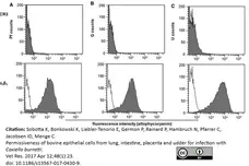
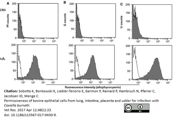
Mouse anti Pig CD61 antibody, clone JM2E5 (MCA2263) used for evaluation of expression of CD61 on bovine epithelial cells by flow cytometry.
Image caption:
Determination of receptor distribution on bovine epithelial cells.
Uninfected epithelial cell (Pf [panel A], G [B], U [C]) were analyzed by flow cytometry for expression of CR3 and αVβ3 on their surface. Grey shaded curves depict detection of the respective antigens and black lines represent secondary antibody control (representative results of two technical replicates in two independent experiments).
From: Sobotta K, Bonkowski K, Liebler-Tenorio E, et al.
Permissiveness of bovine epithelial cells from lung, intestine, placenta and udder for infection with Coxiella burnetii.
Vet Res. 2017;48(1):23.
doi: 10.1186/s13567-017-0430-9.
This image is from an open access article distributed under terms of a Creative Commons Attribution License.
Filter by Application:
F Reset| Mouse anti Pig CD61 antibody, clone JM2E5 recognizes the porcine CD61 cell surface antigen, also known as platelet glycoprotein IIIa or integrin beta. CD61 is present on the megakaryocyte/platelet lineage, granulocytes, cells from the monocyte/macrophage lineage and endothelial cells (Moreno et al. 2002). CD61 is also broadly expressed on tissues, such as epithelial cells from tubules in the kidney (Piriou-Guzylack et al., 2008), spleen, intestinal mucosa and Leydig cells in testis (Moreno et al. 2002). Mouse anti Pig CD61 antibody, clone JM2E5 detects a band of approximately 85 kDa in porcine platelet lysates by western blotting. The epitope recognized by this antibody is not sensitive to EDTA. |
- Target Species
- Pig
- Species Cross-Reactivity
-
Target Species Cross Reactivity Dog Human Bovine Horse - N.B. Antibody reactivity and working conditions may vary between species.
- Product Form
- Purified IgG - liquid
- Preparation
- Purified IgG prepared by affinity chromatography on Protein A from tissue culture supernatant
- Buffer Solution
- Phosphate buffered saline
- Preservative Stabilisers
- 0.09% sodium azide (NaN3)
- Carrier Free
- Yes
- Immunogen
- Porcine peripheral blood mononuclear cells.
- Approx. Protein Concentrations
- IgG concentration 1.0 mg/ml
- Fusion Partners
- Spleen cells from immunized Balb/c mice were fused with cells of the mouse SP2/0 - Ag14 myeloma cell line.
- Regulatory
- For research purposes only
- Guarantee
- 12 months from date of despatch
This product is shipped at ambient temperature. It is recommended to aliquot and store at -20°C on receipt. When thawed, aliquot the sample as needed. Keep aliquots at 2-8°C for short term use (up to 4 weeks) and store the remaining aliquots at -20°C.
Avoid repeated freezing and thawing as this may denature the antibody. Storage in frost-free freezers is not recommended.
Avoid repeated freezing and thawing as this may denature the antibody. Storage in frost-free freezers is not recommended.
This product has been reported to work in the following applications. This information is derived from testing within our laboratories, peer-reviewed publications or personal communications from the originators. Please refer to references indicated for further information. For general protocol recommendations, please visit the antibody protocols page.
| Application Name | Verified | Min Dilution | Max Dilution |
|---|---|---|---|
| Flow Cytometry |  |
1/50 | 1/100 |
| Immunohistology - Frozen | |
||
| Immunohistology - Paraffin 1 | |
||
| Immunoprecipitation | |
||
| Western Blotting | |
- 1 This product requires antigen retrieval using heat treatment methods prior to staining of paraffin sections. Sodium citrate buffer pH6.0 is recommended for this purpose.
Where this product has not been tested for use in a particular technique this does not necessarily exclude its use in such procedures. Suggested working dilutions are given as a guide only. It is recommended that the user titrates the product for use in their own system using appropriate negative/positive controls.
- Flow Cytometry
- Use 10μl of the suggested working dilution to label 106 cells in 100μl
| Description | Product Code | Applications | Pack Size | List Price | Your Price | Quantity | |
|---|---|---|---|---|---|---|---|
| Mouse IgG1 Negative Control | MCA928 | F | 100 Tests |

|
Log in | ||
| List Price | Your Price | ||||||
|
|
Log in | ||||||
| Description | Mouse IgG1 Negative Control | ||||||
References for CD61 antibody
-
Pérez de la Lastra, J.M. et al. (1997) Characterization of the porcine homologue to human platelet glycoprotein IIb-IIIa (CD41/CD61) by a monoclonal antibody.
Tissue Antigens. 49 (6): 588-94. -
Arce, C et al. (2001) Expression of CD61 (beta 3 integrin subunit) on canine cells.
Platelets 12:69-73. -
Moreno, A. et al. (2002) Immunohistochemical analysis of beta3 integrin (CD61): expression in pig tissues and human tumors.
Histol Histopathol. 17 (2): 347-52. -
Zhang, J.L. et al. (2007) Up-regulated expression of beta3 integrin induced by dengue virus serotype 2 infection associated with virus entry into human dermal microvascular endothelial cells.
Biochem Biophys Res Commun. 356: 763-8. -
Campos, E. et al. (2004) In vitro effect of classical swine fever virus on a porcine aortic endothelial cell line.
Vet Res. 35: 625-33. -
Sobotta, K. et al. (2017) Permissiveness of bovine epithelial cells from lung, intestine, placenta and udder for infection with Coxiella burnetii.
Vet Res. 48 (1): 23. -
Arenal, Á. et al. (2022) Effects of Cardiac Stem Cell on Postinfarction Arrhythmogenic Substrate.
Int J Mol Sci. 23 (24): 16211. -
Batchinsky, A.I. et al. (2023) Intravenous Autologous Bone-Marrow-derived Mesenchymal Stromal Cells Delay Acute Respiratory Distress Syndrome in Swine.
Am J Respir Crit Care Med. Oct 05 [Epub ahead of print].
Further Reading
-
Piriou-Guzylack, L. (2008) Membrane markers of the immune cells in swine: an update.
Vet Res. 39: 54.
- Synonyms
- Integrin Beta 3 Chain
- RRID
- AB_11152935
- UniProt
- Q95JH1
MCA2263GA
If you cannot find the batch/lot you are looking for please contact our technical support team for assistance.
View more products with CD61 specificity
Please Note: All Products are "FOR RESEARCH PURPOSES ONLY"
View all Anti-Pig ProductsAlways be the first to know.
When we launch new products and resources to help you achieve more in the lab.
Yes, sign me up