WIPI2 antibody | 2A2
Mouse anti WIPI2
- Product Type
- Monoclonal Antibody
- Clone
- 2A2
- Isotype
- IgG1
- Specificity
- WIPI2


Mouse anti Human WIPI2 antibody, clone 2A2 (MCA5780) used to evaluate expression of the shortened isoform of human WIPI2, WIPI2B by western blotting.
Image caption:
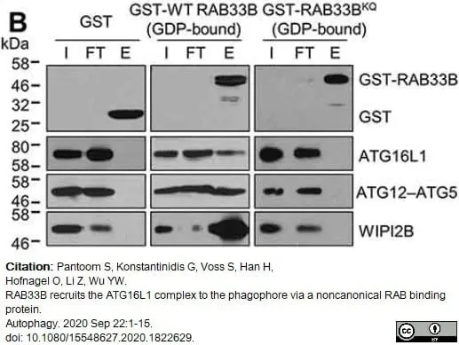
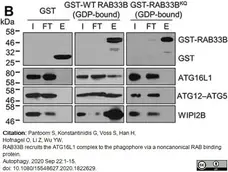
Recombinant wild-type or mutant GST-RAB33B were incubated with cell lysate from HeLa cells previously incubated in EBSS for 2 h as indicated. Samples were subjected to affinity isolation using glutathione beads and immunoblot analysis. WIPI2B binding was trapped by crosslinking.
I: Input, FT: Flow-through, W: Wash, E: Elution.
From: Pantoom S, Konstantinidis G, Voss S, Han H, Hofnagel O, Li Z, Wu YW.
RAB33B recruits the ATG16L1 complex to the phagophore via a noncanonical RAB binding protein.
Autophagy. 2020 Sep 22:1-15.
doi: 10.1080/15548627.2020.1822629.
This image is from an open access article distributed under terms of a Creative Commons Attribution License
Mouse anti Human WIPI2 antibody, clone 2A2 (MCA5780GA) used to demonstrate WIPI2 expression in HeLa cells by immunofluorescence.
Image caption:
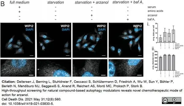
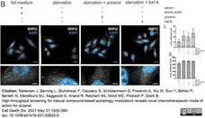
Arzanol induces colocalization of ATG16L1 and LC3 and inhibits autophagic flux, but does not alter WIPI2 localization. (B) Shown are representative microscopy images of wild-type HeLa cells immunofluorescently labelled for endogenous WIPI2. Cells were grown in full medium or starved in serum- and amino acid-free medium for 2 h while incubated with 3μM arzanol or 10 nM bafilomycin A1. Scale bars in upper panels are 15.5μm, scale bars in magnifications are 3.875μm. (i.) Data show average number of dots per cell as mean ± SEM (n=5). Digits in bars show total number of cells quantified using ImageJ software. (ii.) Data show average diameter of dots in nm as mean ± SEM. Statistical analysis was performed usingordinary oneway ANOVA with Tukey's multiple comparison test.
From: Deitersen J, Berning L, Stuhldreier F, Ceccacci S, Schlütermann D, Friedrich A, Wu W, Sun Y, Böhler P, Berleth N, Mendiburo MJ, Seggewiß S, Anand R, Reichert AS, Monti MC, Proksch P, Stork B.
High-throughput screening for natural compound-based autophagy modulators reveals novel chemotherapeutic mode of action for arzanol.
Cell Death Dis. 2021 May 31;12(6):560.
doi: 10.1038/s41419-021-03830-5.
This image is from an open access article distributed under terms of a Creative Commons Attribution License.
Mouse anti Human WIPI2 antibody, clone 2A2 (MCA5780GA) used to evaluate WI{PI2 expression in HK cell lysates by western blotting.
Image caption:
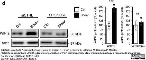
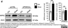
Shear-stress-induced PI3P synthesis at the PC depends on PI3KC2α
Western blot analysis and quantification of WIPI2 protein levels in lysates of polarized siCTRL or siPI3KC2α HK2 cells, upon static (ctrl) and shear-stress (96 h) conditions. Bar graph denotes average protein levels normalized to actin (mean ± SEM, from three independent experiments). NS: not significant, ***p <0.001 in two-tailed Student’s t test..
From: Boukhalfa A, Nascimbeni AC, Ramel D, Dupont N, Hirsch E, Gayral S, Laffargue M, Codogno P, Morel E.
PI3KC2α-dependent and VPS34-independent generation of PI3P controls primary cilium-mediated autophagy in response to shear stress.
Nat Commun. 2020 Jan 15;11(1):294.
doi: 10.1038/s41467-019-14086-1.
This image is from an open access article distributed under terms of a Creative Commons Attribution License.
Mouse anti Human WIPI2 antibody, clone 2A2 (MCA5780GA) used to assess WIPI2 expression in MCF7 cells by western blotting and immunofluorescence.
Image caption:
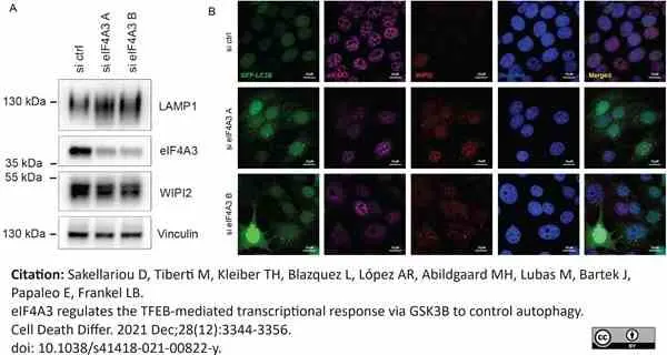
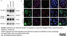
(A) Western blot of LAMP1, WIPI2 and eIF4A3 in MCF‐7 GFP‐LC3B cells after 72h transfection with indicated siRNAs. A representative experiment is shown (n = 3). (B) Representative immunofluorescent images of eIF4A3 levels, WIPI2 puncta and GFP-LC3B puncta in MCF‐7 GFP-LC3B cells 72h after transfection with ctrl and eIF4A3 siRNAs. Scale bars 20 μm.
From: Sakellariou D, Tiberti M, Kleiber TH, Blazquez L, López AR, Abildgaard MH, Lubas M, Bartek J, Papaleo E, Frankel LB.
eIF4A3 regulates the TFEB-mediated transcriptional response via GSK3B to control autophagy.
Cell Death Differ. 2021 28 (12): 3344-56.
doi: 10.1038/s41418-021-00822-y.
This image is from an open access article distributed under terms of a Creative Commons Attribution License.
Filter by Application:
WB IF Reset| Mouse anti Human WIPI2 antibody, clone 2A2 recognies WD repeat domain phosphoinositide-interacting protein 2 (WIPI-2), also known as WIPI49-like protein 2. WIPI2 is a 454 amino acid ~54 kDa autophagosomal marker containing three WD repeats. WIPI2 is a mammalian orthologue of the yeast protein Atg18 and is similarly recruited to early autophagosomal structures and is required for their maturation into mature autophagosomes (Polson et al. 2010). Human WIPI2 exists in multiple isoforms including WIPI2A, the canonical 454 amino acid isoform and WIPI2B with deletions towards both the N and C terminal regions. Mouse anti Human WIPI2 antibody, clone 2A2 was generated using a C-terminal sequence and recognizes both WIPI2A and WIPI2B by western blotting (Pantoom et al. 2020) Mouse anti Human WIPI2 antibody, clone 2A2 has been used for the immunofluorescent detection of WIPI2 in the human retinal epithelial cell line RPE1 (MacVicar and Lane 2014). |
- Target Species
- Human
- Species Cross-Reactivity
-
Target Species Cross Reactivity Mouse - N.B. Antibody reactivity and working conditions may vary between species.
- Product Form
- Purified IgG - liquid
- Preparation
- Antibody purified from tissue culture supernatant
- Buffer Solution
- Phosphate buffered saline
- Preservative Stabilisers
- 0.09% Sodium Azide (NaN3)
- Carrier Free
- Yes
- Immunogen
- Synthetic peptide corresponding to the C-terminus of WIPI2b (CSALRLDEDSEHPPMILRTD)
- Approx. Protein Concentrations
- IgG concentration 1.0 mg/ml
- Regulatory
- For research purposes only
- Guarantee
- 12 months from date of despatch
This product is shipped at ambient temperature. It is recommended to aliquot and store at -20°C on receipt. When thawed, aliquot the sample as needed. Keep aliquots at 2-8°C for short term use (up to 4 weeks) and store the remaining aliquots at -20°C.
Avoid repeated freezing and thawing as this may denature the antibody. Storage in frost-free freezers is not recommended.
Avoid repeated freezing and thawing as this may denature the antibody. Storage in frost-free freezers is not recommended.
This product has been reported to work in the following applications. This information is derived from testing within our laboratories, peer-reviewed publications or personal communications from the originators. Please refer to references indicated for further information. For general protocol recommendations, please visit the antibody protocols page.
| Application Name | Verified | Min Dilution | Max Dilution |
|---|---|---|---|
| Immunofluorescence |  |
||
| Immunohistology - Paraffin | |
||
| Immunoprecipitation | |
||
| Western Blotting | |
Where this product has not been tested for use in a particular technique this does not necessarily exclude its use in such procedures. Suggested working dilutions are given as a guide only. It is recommended that the user titrates the product for use in their own system using the appropriate negative/positive controls.
Source Reference
-
Polson, H.E. et al. (2010) Mammalian Atg18 (WIPI2) localizes to omegasome-anchored phagophores and positively regulates LC3 lipidation.
Autophagy. 6 (4): 506-22.
References for WIPI2 antibody
-
Dooley, H.C. et al. (2014) WIPI2 links LC3 conjugation with PI3P, autophagosome formation, and pathogen clearance by recruiting Atg12-5-16L1.
Mol Cell. 55 (2): 238-52. -
MacVicar, T.D. and Lane, J.D. (2014) Impaired OMA1-dependent cleavage of OPA1 and reduced DRP1 fission activity combine to prevent mitophagy in cells that are dependent on oxidative phosphorylation.
J Cell Sci. 127: 2313-25. -
Karanasios, E. et al. (2014) Imaging autophagy.
Curr Protoc Cytom. 69: 12.34.1-12.34.16. -
Gomez-Sanchez, J.A. et al. (2015) Schwann cell autophagy, myelinophagy, initiates myelin clearance from injured nerves.
J Cell Biol. 210 (1): 153-68. -
Kjos, I. et al. (2017) Rab7b modulates autophagic flux by interacting with Atg4B.
EMBO Rep. 18 (10): 1727-39. -
Nascimbeni, A.C. et al. (2017) ER-plasma membrane contact sites contribute to autophagosome biogenesis by regulation of local PI3P synthesis.
EMBO J. 36 (14): 2018-33. -
Pantoom, S. et al. (2020) RAB33B recruits the ATG16L1 complex to the phagophore via a noncanonical RAB binding protein.
Autophagy. : 1-15. -
Boukhalfa, A. et al. (2020) PI3KC2α-dependent and VPS34-independent generation of PI3P controls primary cilium-mediated autophagy in response to shear stress.
Nat Commun. 11 (1): 294.
View The Latest Product References
-
Deitersen, J. et al. (2021) High-throughput screening for natural compound-based autophagy modulators reveals novel chemotherapeutic mode of action for arzanol.
Cell Death Dis. 12 (6): 560. -
Sakellariou, D. et al. (2021) eIF4A3 regulates the TFEB-mediated transcriptional response via GSK3B to control autophagy.
Cell Death Differ. 28 (12): 3344-56.
- RRID
- AB_10845951
- UniProt
- Q9Y4P8
- Q80W47
- Entrez Gene
- Wipi2
- WIPI2
- GO Terms
- GO:0000045 autophagic vacuole assembly
- GO:0005829 cytosol
- GO:0032266 phosphatidylinositol-3-phosphate binding
- GO:0034045 pre-autophagosomal structure membrane
- GO:0080025 phosphatidylinositol-3,5-bisphosphate binding
- GO:0043234 protein complex
MCA5780GA
If you cannot find the batch/lot you are looking for please contact our technical support team for assistance.
Request a different product with this specificity
Please Note: All Products are "FOR RESEARCH PURPOSES ONLY"
View all Anti-Human ProductsAlways be the first to know.
When we launch new products and resources to help you achieve more in the lab.
Yes, sign me up