CD59 antibody | MEM-43
Mouse anti Human CD59
- Product Type
- Monoclonal Antibody
- Clone
- MEM-43
- Isotype
- IgG2a
- Specificity
- CD59

Figure B. FITC conjugated Mouse anti Human CD3 antibody, clone UCHT1 (MCA463F) and RPE conjugated Mouse anti Human CD59 antibody, clone MEM-43 (MCA1054PE). All experiments performed on human peripheral blood lymphocytes in the presence of human SeroBlock (BUF070A).
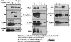
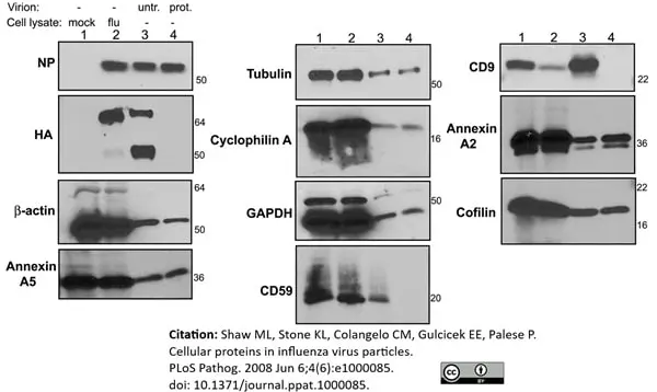
Mouse anti Human CD59 antibody, clone MEM-43 (MCA1054) used for the detection of CD59 in virus infected cell lysates by western blotting.
Image caption:
The effect of protease treatment on influenza virion associated host proteins. Purified influenza A/WSN/33 virus was either mock treated or subjected to overnight digestion with subtilisin followed by concentration through a sucrose cushion. 10 ug of mock infected cell lysate (lane 1) or influenza infected cell lysate (lane 2) and 2 μg of untreated influenza virions (lane 3) or protease treated influenza virions (lane 4) were then analyzed by western blot with antibodies against the indicated proteins. Numbers to the right are molecular weight markers.
From: Shaw ML, Stone KL, Colangelo CM, Gulcicek EE, Palese P (2008)
Cellular Proteins in Influenza Virus Particles.
PLoS Pathog 4(6): e1000085.
This image is from an open access article distributed under terms of a Creative Commons Attribution License.
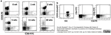
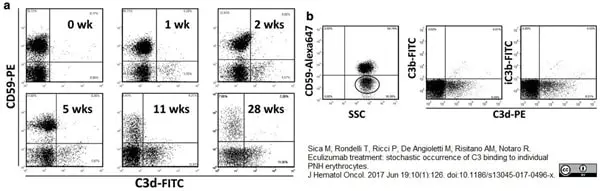
R-Phycoerythrin and Alexa Fluor® 647 conjugated Mouse anti Human CD59 antibodies, clone MEM-43 (MCA1054PE and MCA1054A647) used for the evaluation of CD59 expression on human erythrocytes by flow cytometry.
Image caption:
In vivo C3 binding on red cells of PNH patients on eculizumab. a. Kinetics of C3 binding on red cells from a representative PNH patient during eculizumab treatment. Before treatment (0 week), no red cell binds C3, whereas starting from 1 week of treatment three populations of red cells are displayed: one population of normal red cells (CD59 + C3−) and two distinct populations of PNH (CD59-negative) red cells, one with (C3+) and one without (C3−) fragments bound on their surface. b. Characterization of C3 fragment bound to erythrocytes from PNH patient on eculizumab. The left panel shows the normal (CD59-positive) and PNH (CD59-negative) populations (gated by the elliptic mark). The PNH (CD59-negative) population (see gate in the left panel) has been analyzed with anti-C3d and either anti-C3b (middle panel) or anti-iC3b (left panel). SSC: side scatter
From: Sica M, Rondelli T, Ricci P, De Angioletti M, Risitano AM, Notaro R.
Eculizumab treatment: stochastic occurrence of C3 binding to individual PNH erythrocytes.
J Hematol Oncol. 2017 Jun 19;10(1):126.
doi: 10.1186/s13045-017-0496-x.
This image is from an open access article distributed under terms of a Creative Commons Attribution License.

Filter by Application:
F WB P Reset| Mouse anti Human CD59 antibody, clone MEM-43 recognizes CD59, a glycosyl-phosphatidylinositol (GPI) anchored membrane protein also known as membrane attack complex inhibition factor. CD59 blocks the formation of the complement membrane attack complex (MAC) by binding of C8a and C9. CD59 is found on all types of leucocytes including platelets and is also expressed on many non-haematopoietic cells. The epitope recognized by Mouse anti Human CD59 antibody, clone MEM-43 is lost after reduction therefore, non-reducing conditions are required for western blotting techniques. |
- Target Species
- Human
- Product Form
- Purified IgG - liquid
- Preparation
- Purified IgG prepared by affinity chromatography on Protein A from tissue culture supernatant
- Buffer Solution
- Phosphate buffered saline
- Preservative Stabilisers
- 0.09% sodium azide (NaN3)
- Immunogen
- Thymocytes and T lymphocytes.
- Approx. Protein Concentrations
- IgG concentration 1 mg/ml
- Regulatory
- For research purposes only
- Guarantee
- 12 months from date of despatch
This product is shipped at ambient temperature. It is recommended to aliquot and store at -20°C on receipt. When thawed, aliquot the sample as needed. Keep aliquots at 2-8°C for short term use (up to 4 weeks) and store the remaining aliquots at -20°C.
Avoid repeated freezing and thawing as this may denature the antibody. Storage in frost-free freezers is not recommended.
Avoid repeated freezing and thawing as this may denature the antibody. Storage in frost-free freezers is not recommended.
This product has been reported to work in the following applications. This information is derived from testing within our laboratories, peer-reviewed publications or personal communications from the originators. Please refer to references indicated for further information. For general protocol recommendations, please visit the antibody protocols page.
| Application Name | Verified | Min Dilution | Max Dilution |
|---|---|---|---|
| ELISA |  |
||
| Flow Cytometry | |
1/25 | 1/100 |
| Immuno-electron Microscopy | |
||
| Immunofluorescence | |
||
| Immunohistology - Frozen | |
||
| Immunohistology - Paraffin 1 | |
||
| Immunoprecipitation | |
||
| Western Blotting 2 | |
- 1This product requires antigen retrieval using heat treatment prior to staining of paraffin sections.Sodium citrate buffer pH 6.0 is recommended for this purpose.
- 2This product recognizes CD59 under non-reducing conditions.
Where this antibody has not been tested for use in a particular technique this does not necessarily exclude its use in such procedures. Suggested working dilutions are given as a guide only. It is recommended that the user titrates the antibody for use in their own system using appropriate negative/positive controls.
- Flow Cytometry
- Use 10μl of the suggested working dilution to label 106 cells in 100μl
- Histology Positive Control Tissue
- Tonsil
| Description | Product Code | Applications | Pack Size | List Price | Your Price | Quantity | |
|---|---|---|---|---|---|---|---|
| Mouse IgG2a Negative Control | MCA929 | F | 100 Tests |

|
Log in | ||
| List Price | Your Price | ||||||
|
|
Log in | ||||||
| Description | Mouse IgG2a Negative Control | ||||||
References for CD59 antibody
-
Horejsí, V. et al. (1988) Monoclonal antibodies against human leucocyte antigens. II. Antibodies against CD45 (T200), CD3 (T3), CD43, CD10 (CALLA), transferrin receptor (T9), a novel broadly expressed 18-kDa antigen (MEM-43) and a novel antigen of restricted expression (MEM-74).
Folia Biol (Praha). 34 (1): 23-34. -
Stefanová, I. et al. (1989) Characterization of a broadly expressed human leucocyte surface antigen MEM-43 anchored in membrane through phosphatidylinositol.
Mol Immunol. 26 (2): 153-61. -
Stefanová, I. et al. (1989) in Leucocyte Typing IV: White cell differentiation antigens.
Ed. Knapp, W. et al. Oxford University Press pp 678-97. -
Stefanová, I. & Horejsí, V. (1991) Association of the CD59 and CD55 cell surface glycoproteins with other membrane molecules.
J Immunol. 147 (5): 1587-92. -
Tandon, N. et al. (1994) Expression and function of multiple regulators of complement activation in autoimmune thyroid disease.
Immunology. 81 (4): 643-7. -
Vanderplasschen, A. et al. (1997) Extracellular enveloped vaccinia virus is resistant to complement because of incorporation of host complement control proteins into its envelope.
Proc Natl Acad Sci U S A. 95: 7544-9. -
Cowan, P.J. et al. (1998) High-level endothelial expression of human CD59 prolongs heart function in an ex vivo model of xenograft rejection.
Transplantation. 65: 826-31. -
Chong, Y.H. and Lee, M.J. (2000) Expression of complement inhibitor protein CD59 in human neuronal and glial cell lines treated with HIV-1 gp41 peptides.
J Neurovirol. 6: 51-60.
View The Latest Product References
-
Shamri, R. et al. (2002) Chemokine stimulation of lymphocyte alpha 4 integrin avidity but not of leukocyte function-associated antigen-1 avidity to endothelial ligands under shear flow requires cholesterol membrane rafts.
J Biol Chem. 277: 40027-35. -
Zhang, J. et al. (2002) Early complement activation and decreased levels of glycosylphosphatidylinositol-anchored complement inhibitors in human and experimental diabetic retinopathy.
Diabetes. 51: 3499-504. -
Donin, N. et al. (2003) Complement resistance of human carcinoma cells depends on membrane regulatory proteins, protein kinases and sialic acid.
Clin Exp Immunol. 131: 254-63. -
Gendek-Kubiak, H. and Gendek, E.G. (2004) Immunolocalization of protectin (CD59) and macrophages in polymyositis and dermatomyositis.
J Neuroimmunol. 149: 187-94. -
Jolly, C, and Sattentau. Q.J. (2005) Human Immunodeficiency Virus Type 1 Virological Synapse Formation in T Cells Requires Lipid Raft Integrity
J Virol. 79: 12088-94. -
Ohyama, M. et al. (2006) Characterization and isolation of stem cell-enriched human hair follicle bulge cells.
J Clin Invest. 116: 249-60. -
Ellison, B.S. et al. (2007) Complement susceptibility in glutamine deprived breast cancer cells.
Cell Div. 2007 2: 20. -
Takemoto, M. et al. (2007) Human herpesvirus 7 infection increases the expression levels of CD46 and CD59 in target cells.
J Gen Virol. 88: 1415-22. -
Shaw, M.L. et al. (2008) Cellular proteins in influenza virus particles.
PLoS Pathog. 4: e1000085. -
Bonnon, C. et al. (2010) Selective export of human GPI-anchored proteins from the endoplasmic reticulum.
J Cell Sci. 123: 1705-15. -
Sadallah, S. et al. (2011) Microparticles (ectosomes) shed by stored human platelets downregulate macrophages and modify the development of dendritic cells.
J Immunol. 186: 6543-52. -
Rondelli, T. et al. (2013) The frequency of granulocytes with spontaneous somatic mutations: a wide distribution in a normal human population.
PLoS One. 8 (1): e54046. -
Abe, Y. et al. (2017) Glycan region of GPI anchored-protein is required for cytocidal oligomerization of an anticancer parasporin-2, Cry46Aa1 protein, from Bacillus thuringiensis strain A1547.
J Invertebr Pathol. 142: 71-81. -
Sica, M. et al. (2017) Eculizumab treatment: stochastic occurrence of C3 binding to individual PNH erythrocytes.
J Hematol Oncol. 10 (1): 126. -
Gullipalli, D. et al. (2018) Antibody Inhibition of Properdin Prevents Complement-Mediated Intravascular and Extravascular Hemolysis.
J Immunol. 201 (3): 1021-1029. -
Ueda, M. et al. (2019) Endovascular trophoblast expresses CD59 to evade complement-dependent cytotoxicity.
Mol Cell Endocrinol. 490: 57-67.
- Synonyms
- HRF
- Protectin
- RRID
- AB_321508
- UniProt
- P13987
- Entrez Gene
- CD59
- GO Terms
- GO:0005515 protein binding
- GO:0007596 blood coagulation
- GO:0005576 extracellular region
- GO:0005624 membrane fraction
- GO:0007166 cell surface receptor linked signaling pathway
- GO:0031362 anchored to external side of plasma membrane
View more products with CD59 specificity
Please Note: All Products are "FOR RESEARCH PURPOSES ONLY"
View all Anti-Human ProductsAlways be the first to know.
When we launch new products and resources to help you achieve more in the lab.
Yes, sign me up