CD55 antibody | 67
Mouse anti Human CD55
- Product Type
- Monoclonal Antibody
- Clone
- 67
- Isotype
- IgG1
- Specificity
- CD55

Mouse anti Human CD55 antibody, clone 67 (MCA1614) used for the identification of CD55 expressing cells in rheumatoid arthritis synovial tissue by immunohistochemistry on cryostat sections.
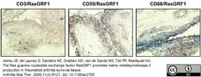
Image caption:
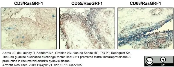
Representative double staining of rheumatoid arthritis synovial tissue with antibodies against RasGRF1 and cell-specific markers. Synovial tissue sections were stained overnight with antibodies against Ras guanine nucleotide-releasing factor 1(RasGRF1), followed by antibodies against CD3, CD55, and CD68. After biotin tyramide enhancement, staining was developed with amino-ethylcarbazole (red, RasGRF1) and Fast blue (blue, cell-specific markers). Magnification x 100.
From: Abreu JR, de Launay D, Sanders ME, Grabiec AM, van de Sande MG, Tak PP, Reedquist KA.
The Ras guanine nucleotide exchange factor RasGRF1 promotes matrix metalloproteinase-3 production in rheumatoid arthritis synovial tissue.
Arthritis Res Ther. 2009;11(4):R121.
This is from an open access article distributed under the terms of the Creative Commons Attribution License.
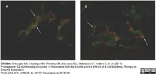
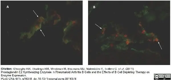
Mouse anti CD55 antibofdy, clone 67 (MCA1614) used for the demonstration of CD55 and MPGES1 in fibroblasts from sinovial lining by immunofluorescence.
Image caption:
MPGES1 expression in synovial lining fibroblasts before and 16 weeks after initiation of rituximab therapy.
Double immunofluorescence pictures show the presence of MPGES1 (red) expression in CD55 positive fibroblasts (green) in the rheumatoid tissue before rituximab initiation (A) and 16 weeks later (B). Original magnification 500x. Arrows point to double stained cells
From: Gheorghe KR, Thurlings RM, Westman M, Boumans MJ, Malmström V, Trollmo C, et al. (2011)
Prostaglandin E2 Synthesizing Enzymes in Rheumatoid Arthritis B Cells and the Effects of B Cell Depleting Therapy on Enzyme Expression.
PLoS ONE 6(1): e16378.
doi: 10.1371/journal.pone.0016378
This image is from an open access article distributed under the terms of the Creative Commons Attribution License.

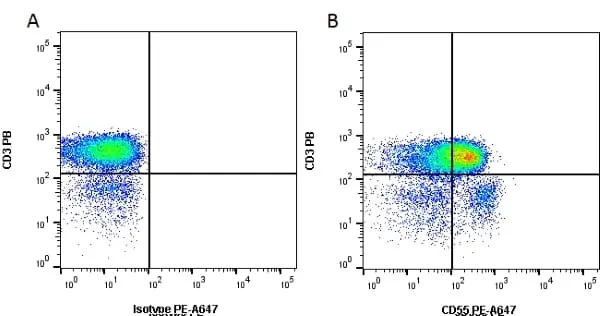
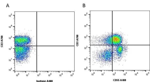
Figure B. Pacific Blue® conjugated Mouse anti Human CD3 antibody, clone UCHT1 (MCA463PB) and RPE-Alexa Fluor® 647 conjugated Mouse anti Human CD55 antibody, clone 67 (MCA1614P647). All experiments performed on red cell lysed human blood gated on lymphoid cells in the presence of 10% human serum.
Data acquired on the ZE5 Cell Analyzer.
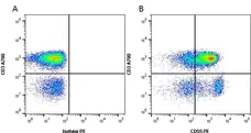
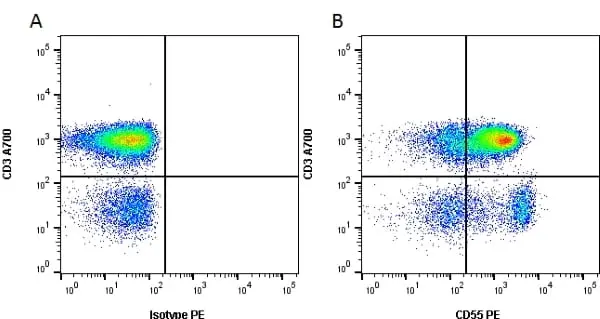
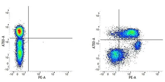
Figure B. Alexa Fluor® 700 conjugated Mouse anti Human CD3 antibody, clone UCHT1 (MCA463A700) and RPE conjugated Mouse anti Human CD55 antibody, clone 67 (MCA1614PE). All experiments performed on red cell lysed human blood gated on lymphoid cells in the presence of 10% human serum.
Data acquired on the ZE5 Cell Analyzer.
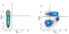
Figure B. Alexa Fluor® 700 conjugated Mouse anti Human CD3 antibody, clone UCHT1 (MCA463A700) and RPE conjugated Mouse anti Human CD55 antibody, clone 67 (MCA1614PE). All experiments performed on red cell lysed human blood gated on lymphoid cells in the presence of 10% human serum.
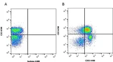
Figure B. Alexa Fluor® 700 conjugated Mouse anti Human CD3 antibody, clone UCHT1 (MCA463A700) and Alexa Fluor® 488 conjugated Mouse anti Human CD55 antibody, clone 67 (MCA1614A488). All experiments performed on red cell lysed human blood gated on lymphocytes in the presence of Human Seroblock (BUF070A).
Data acquired on the ZE5 Cell Analyzer.
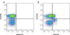
Figure B. Alexa Fluor® 700 conjugated Mouse anti Human CD3 antibody, clone UCHT1 (MCA463A700) and FITC conjugated Mouse anti Human CD55 antibody, clone 67 (MCA1614F). All experiments performed on red cell lysed human blood gated on lymphocytes in the presence of Human Seroblock (BUF070A).
Data acquired on the ZE5 Cell Analyzer.
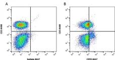
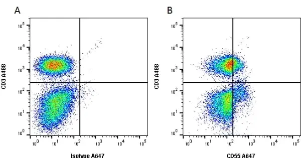
Figure B. Alexa Fluor® 488 conjugated Mouse anti Human CD3 antibody, clone UCHT1 (MCA463A488) and Alexa Fluor® 647 conjugated Mouse anti Human CD55 antibody, clone 67 (MCA1614A647). All experiments performed on red cell lysed human blood gated on lymphocytes in the presence of Human Seroblock (BUF070A).
Data acquired on the ZE5 Cell Analyzer.
Filter by Application:
C IF F Reset| Mouse anti Human CD55 antibody, clone 67 recognizes the human CD55 cell surface antigen, a GPI linked molecule also known as decay accelerating factor (DAF). CD55 is expressed by a wide range of cell types. CD55 is the complement regulatory protein, decay accelerating factor (DAF) (Lublin and Atkinson 1989). Human CD55 is a ~70 kDa glycoprotein (in erythrocytes) anchored in the membrane by glycosylphosphatidylinositol tail. In other cells the apparent molecular weight is somewhat larger. It has a substantial content of O-glycans, and also on N-glycan. DAF binds to activated C4b or C3b complement fragments on the cell surface, preventing the assembly and accelerating the decay of both classical and alternative pathways. DAF carries the DAF has a wide distribution on cells in non-haematopoietic tissues, particularly epithelium and is found at the fetal-maternal interface in placenta (Holmes et al. 1990 and Yang et al. 2009). Soluble forms of DAF are found, for example, in plasma, saliva and urine ( |
- Target Species
- Human
- Product Form
- Purified IgG - liquid
- Preparation
- Purified IgG prepared by affinity chromatography on Protein A from tissue culture supernatant
- Buffer Solution
- Phosphate buffered saline
- Preservative Stabilisers
- 0.09% sodium azide (NaN3)
- Carrier Free
- Yes
- Immunogen
- K562 cells
- Approx. Protein Concentrations
- IgG concentration 1.0 mg/ml
- Regulatory
- For research purposes only
- Guarantee
- 12 months from date of despatch
This product is shipped at ambient temperature. It is recommended to aliquot and store at -20°C on receipt. When thawed, aliquot the sample as needed. Keep aliquots at 2-8°C for short term use (up to 4 weeks) and store the remaining aliquots at -20°C.
Avoid repeated freezing and thawing as this may denature the antibody. Storage in frost-free freezers is not recommended.
Avoid repeated freezing and thawing as this may denature the antibody. Storage in frost-free freezers is not recommended.
This product has been reported to work in the following applications. This information is derived from testing within our laboratories, peer-reviewed publications or personal communications from the originators. Please refer to references indicated for further information. For general protocol recommendations, please visit the antibody protocols page.
| Application Name | Verified | Min Dilution | Max Dilution |
|---|---|---|---|
| Flow Cytometry |  |
1/10 | 1/25 |
| Immunohistology - Frozen | |
1/100 | 1/1000 |
| Western Blotting | |
Where this product has not been tested for use in a particular technique this does not necessarily exclude its use in such procedures. Suggested working dilutions are given as a guide only. It is recommended that the user titrates the product for use in their own system using appropriate negative/positive controls.
- Flow Cytometry
- Use 10μl of the suggested working dilution to label 106 cells in 100μl. Please note: Bio-Rad do not recommend the use of this reagent to stain erythrocytes
- Histology Positive Control Tissue
- Human tonsil
| Description | Product Code | Applications | Pack Size | List Price | Your Price | Quantity | |
|---|---|---|---|---|---|---|---|
| Mouse IgG1 Negative Control | MCA928 | F | 100 Tests |

|
Log in | ||
| List Price | Your Price | ||||||
|
|
Log in | ||||||
| Description | Mouse IgG1 Negative Control | ||||||
References for CD55 antibody
-
Hadam, M.R. (1989) In Leucocyte Typing IV: White Cell Differentiation Antigens.
Edited by Knapp, W. et al. Oxford University Press pp 694-697. -
Holmes, C.H. et al. (1990) Preferential expression of the complement regulatory protein decay accelerating factor at the fetomaternal interface during human pregnancy.
J Immunol. 144 (8): 3099-105. -
O'Brien, D.P. et al. (2009) Regulation of the Helicobacter pylori cellular receptor decay-accelerating factor.
J Biol Chem. 283: 23922-30. -
Kraan, M.C. et al. (2004) T cells, fibroblast-like synoviocytes, and granzyme B+ cytotoxic cells are associated with joint damage in patients with recent onset rheumatoid arthritis.
Ann Rheum Dis. 63: 483-8. -
van Holten, J. et al. (2005) A multicentre, randomised, double blind, placebo controlled phase II study of subcutaneous interferon beta-1a in the treatment of patients with active rheumatoid arthritis.
Ann Rheum Dis. 64 (1): 64-9. -
Yang, P. et al. (2009) Expression and modulation of RPE cell membrane complement regulatory proteins.
Invest Ophthalmol Vis Sci. 50: 3473-81. -
van de Sande, M.G. et al. (2011) Different stages of rheumatoid arthritis: features of the synovium in the preclinical phase.
Ann Rheum Dis. 70: 772-7. -
Araten, D.J. et al. (2005) A quantitative measurement of the human somatic mutation rate.
Cancer Res. 65: 8111-7.
View The Latest Product References
-
Mo, B. et al. (2006) ECC-1 cells: a well-differentiated steroid-responsive endometrial cell line with characteristics of luminal epithelium.
Biol Reprod. 75: 387-94. -
Vos, K. et al. (2007) Early effects of rituximab on the synovial cell infiltrate in patients with rheumatoid arthritis.
Arthritis Rheum. 56 (3): 772-8. -
de Launay, D. et al. (2010) Silencing the expression of Ras family GTPase homologues decreases inflammation and joint destruction in experimental arthritis.
Am J Pathol. 177: 3010-24. -
Gheorghe, K.R. et al. (2011) Prostaglandin E2 synthesizing enzymes in rheumatoid arthritis B cells and the effects of B cell depleting therapy on enzyme expression.
PLoS One. ;6: e16378. -
Abreu, J.R. et al. (2009) The Ras guanine nucleotide exchange factor RasGRF1 promotes matrix metalloproteinase-3 production in rheumatoid arthritis synovial tissue.
Arthritis Res Ther.11(4):R121. -
Thurlings, R.M. et al. (2008) Synovial tissue response to rituximab: mechanism of action and identification of biomarkers of response.
Ann Rheum Dis. 67 (7): 917-25. -
Edginton S et al. (2016) Effects of Rituximab and Infliximab Treatment on Carboxypeptidase B and Its Substrates in RA Synovium.
J Rheumatol. 43 (5): 846-54.
Further Reading
-
Lublin, D.M. & Atkinson, J.P. (1989) Decay-accelerating factor: biochemistry, molecular biology, and function.
Annu Rev Immunol. 7: 35-58. -
Daniels, G. (1989) Cromer-related antigens--blood group determinants on decay-accelerating factor.
Vox Sang. 56 (4): 205-11.
- Synonyms
- DAF
- RRID
- AB_321499
- UniProt
- P08174
- Entrez Gene
- CD55
- GO Terms
- GO:0005576 extracellular region
- GO:0005887 integral to plasma membrane
- GO:0005625 soluble fraction
- GO:0006958 complement activation, classical pathway
- GO:0007204 elevation of cytosolic calcium ion concentration
- GO:0031225 anchored to membrane
- GO:0045087 innate immune response
- GO:0045121 membrane raft
- GO:0045730 respiratory burst
- View More GO Terms
MCA1614GA
MCA1614
If you cannot find the batch/lot you are looking for please contact our technical support team for assistance.
View more products with CD55 specificity
Please Note: All Products are "FOR RESEARCH PURPOSES ONLY"
View all Anti-Human ProductsAlways be the first to know.
When we launch new products and resources to help you achieve more in the lab.
Yes, sign me up