CD53 antibody | MEM-53
Mouse anti Human CD53
- Product Type
- Monoclonal Antibody
- Clone
- MEM-53
- Isotype
- IgG1
- Specificity
- CD53


Figure B. RPE-Alexa Fluor® 647 tandem conjugated Mouse anti Human CD3 antibody, clone UCHT1 (MCA463P647) and FITC conjugated Mouse anti Human CD53 antibody, clone MEM-53 (MCA723F). All experiments performed on human Peripheral blood lymphocytes in the presence of human SeroBlock (BUF070A).
Mouse anti Human CD53 antibody, clone MEM-53 (MCA723G) used for the evaluation of CD53 expression on white blood cell subsets by flow cytometry.
Image caption:
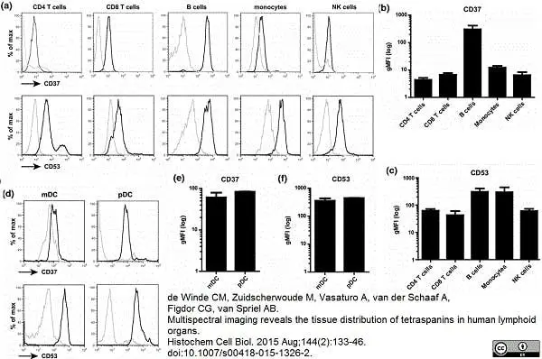
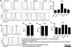
Expression of CD37 and CD53 on immune cell subsets in blood. a Flow cytometry analysis of expression of CD37 or CD53 (black line) on CD4 and CD8 T cells, B cells, monocytes and NK cells versus isotype control (gray line). Gating strategy is presented in Supplementary Figure 1. Expression levels of CD37 (b) and CD53 (c) were normalized for isotype staining by background subtraction. Experiments were performed with PBLs from three healthy donors. Data present mean ± SD. d Flow cytometry analysis of expression of CD37 or CD53 (black line) on mDCs (BDCA1+CD19−) or pDCs (BDCA2+) versus isotype control (gray line). Expression levels of CD37 (e) and CD53 (f) were normalized for isotype staining by background subtraction. Experiments were performed with PBLs from two healthy donors. Data present mean ± SD
From: de Winde CM, Zuidscherwoude M, Vasaturo A, van der Schaaf A, Figdor CG, van Spriel AB.
Multispectral imaging reveals the tissue distribution of tetraspanins in human lymphoid organs.
Histochem Cell Biol. 2015 Aug;144(2):133-46.
doi: 10.1007/s00418-015-1326-2.
This image is from an open access article distributed under terms of a Creative Commons Attribution License.

Mouse anti Human CD53 antibody, clone MEM-53 (MCA723G) used for the evaluation of CD53 distribution on monocytes using immunofluorescence.
Image caption:
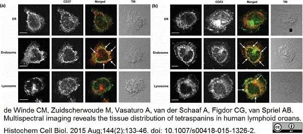
Subcellular localization of CD37 and CD53. Localization of a CD37 or b CD53 (green) in monocytes was studied by dual staining with calreticulin (ER), syntaxin 13 (endosomes) or Lamp1 (lysosomes) (red). Merge: co-localization in yellow (white arrows). Scale bar 5 μm
From: de Winde CM, Zuidscherwoude M, Vasaturo A, van der Schaaf A, Figdor CG, van Spriel AB.
Multispectral imaging reveals the tissue distribution of tetraspanins in human lymphoid organs.
Histochem Cell Biol. 2015 Aug;144(2):133-46.
doi: 10.1007/s00418-015-1326-2.
This image is from an open access article distributed under terms of a Creative Commons Attribution License.

Mouse anti Human CD53 antibody, clone MEM-53 (MCA723G) used for the evaluation of CD53 expression on erythroid cultures by flow cytometry.
Image caption:
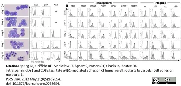
Erythroid culture characterization and expression of tetraspanins and integrins during terminal maturation. A. Temporal expression of erythroid-specific markers, Kell, GPA and AE1 and morphology of the culture at the same time points. AE1 was tested from day 5 onwards. B. Tetraspanin and integrin profile of the same cultures as shown in A. Results are depicted from one culture where directly conjugated antibodies were used (days 3 and 4) and a second culture with indirectly labeled antibodies (day 5 onwards). The y-axis scale is linear to 350 counts; the x-axis is logarithmic to 104. Images were captured on a Leica DM750 microscope, x20 magnification, using Image-Pro Express 6.0 software.
From: Spring FA, Griffiths RE, Mankelow TJ, Agnew C, Parsons SF, Chasis JA, et al. (2013)
Tetraspanins CD81 and CD82 Facilitate α4β1-Mediated Adhesion of Human Erythroblasts to Vascular Cell Adhesion Molecule-1.
PLoS ONE 8(5): e62654
doi: 10.1371/journal.pone.0062654.
This image is from an open access article distributed under terms of a Creative Commons Attribution License.
Mouse anti Human CD53 antibody, clone MEM-53 (MCA723G) used for the evaluation of CD53 expression on erythroid cultures by immunoprecipitation.
Image caption:
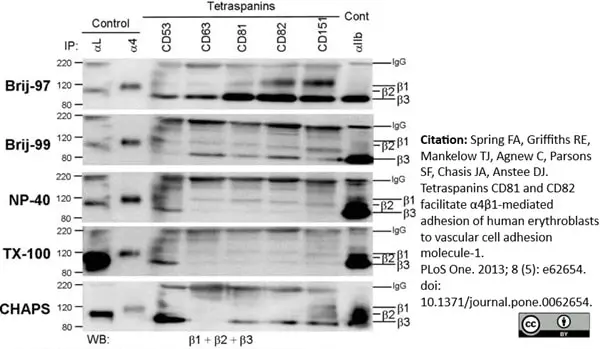
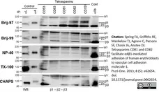
Several anti-tetraspanin antibodies co-precipitate β1 integrins from HEL cells solubilised in Brij-97. Precipitates were prepared from HEL cells solubilised in different detergents in the presence of Mn2+. CD53, MEM-53; CD63, MEM-259; CD81, 454720; CD82, TS82b; CD151, IIG5a; α4, HP2/1; αL, TS1/22; αIIb, PAB-1. Precipitates were run on 7.5% non-reduced gels.
From: Spring FA, Griffiths RE, Mankelow TJ, Agnew C, Parsons SF, Chasis JA, et al. (2013) Tetraspanins CD81 and CD82 Facilitate α4β1-Mediated Adhesion of Human Erythroblasts to Vascular Cell Adhesion Molecule-1.
PLoS ONE 8(5): e62654.
doi: 10.1371/journal.pone.0062654.
This image is from an open access article distributed under terms of a Creative Commons Attribution License.
Mouse anti Human CD53 antibody, clone MEM-53 (MCA723) used for the evaluation of CD53 expression on erythroid cultures by immunoprecipitation and western blotting.
Image caption:
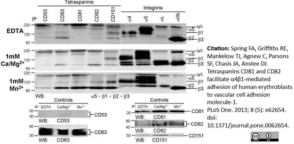
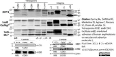
Co-precipitation of β1 and β3 integrins by tetraspanins with different cations from HEL cells. Precipitates were prepared from HEL cells solubilised in Brij-97 in the presence of EDTA or cations. β1 is co-precipitated by CD63, CD81, CD82 and CD151 while β3 is co-precipitated by all tetraspanins in the presence of Mn2+. CD151 co-precipitates β1 under all conditions. Integrins were separated on 7.5% gels, tetraspanin controls on 12% gels, both non-reducing conditions. CD53, MEM-53; CD63, MEM-259; CD81, 454720; CD82, TS82b; CD151, IIG5a; α4, HP2/1; αL, TS1/22; αIIb, PAB-1.
From: Spring FA, Griffiths RE, Mankelow TJ, Agnew C, Parsons SF, Chasis JA, et al. (2013)
Tetraspanins CD81 and CD82 Facilitate α4β1-Mediated Adhesion of Human Erythroblasts to Vascular Cell Adhesion Molecule-1.
PLoS ONE 8(5): e62654.
doi: 10.1371/journal.pone.0062654.
This image is from an open access article distributed under terms of a Creative Commons Attribution License.
Filter by Application:
F IF IP WB Reset| Mouse anti Human CD53 antibody, clone MEM-53 recognizes the human CD53 cell surface antigen, also known as Tetraspanin-25. CD53 is a 219 amino acid multi pass transmembrane glycoprotein of 32-42 kDa. CD53 is expressed by all leucocytes, but is absent from erythrocytes and platelets. |
- Target Species
- Human
- Product Form
- Purified IgG - liquid
- Preparation
- Purified IgG prepared by affinity chromatography on Protein A from tissue culture supernatant
- Buffer Solution
- Phosphate buffered saline
- Preservative Stabilisers
0.09% Sodium Azide - Approx. Protein Concentrations
- IgG concentration 1 mg/ml
- Regulatory
- For research purposes only
- Guarantee
- 12 months from date of despatch
This product is shipped at ambient temperature. It is recommended to aliquot and store at -20°C on receipt. When thawed, aliquot the sample as needed. Keep aliquots at 2-8°C for short term use (up to 4 weeks) and store the remaining aliquots at -20°C.
Avoid repeated freezing and thawing as this may denature the antibody. Storage in frost-free freezers is not recommended.
Avoid repeated freezing and thawing as this may denature the antibody. Storage in frost-free freezers is not recommended.
This product has been reported to work in the following applications. This information is derived from testing within our laboratories, peer-reviewed publications or personal communications from the originators. Please refer to references indicated for further information. For general protocol recommendations, please visit the antibody protocols page.
| Application Name | Verified | Min Dilution | Max Dilution |
|---|---|---|---|
| Flow Cytometry |  |
1/10 | 1/25 |
| Immunohistology - Frozen | |
||
| Immunoprecipitation | |
||
| Western Blotting 1 | |
- 1Clone MEM-53 recognizes human CD53 under non-reducing conditions.
Where this antibody has not been tested for use in a particular technique this does not necessarily exclude its use in such procedures. It is recommended that the user titrates the antibody for use in their own system using appropriate negative/positive controls.
- Flow Cytometry
- Use 10ul of the suggested working dilution to label 106 cells in 100ul.
| Description | Product Code | Applications | Pack Size | List Price | Your Price | Quantity | |
|---|---|---|---|---|---|---|---|
| Mouse IgG1 Negative Control | MCA928 | F | 100 Tests |

|
Log in | ||
| List Price | Your Price | ||||||
|
|
Log in | ||||||
| Description | Mouse IgG1 Negative Control | ||||||
References for CD53 antibody
-
Bazil, V. et al. (1989) Monoclonal antibodies against human leucocyte antigens. III. Antibodies against CD45R, CD6, CD44 and two newly described broadly expressed glycoproteins MEM-53 and MEM-102.
Folia Biol (Praha). 35 (5): 289-97. -
Angelisová P et al. (1990) The human leucocyte surface antigen CD53 is a protein structurally similar to the CD37 and MRC OX-44 antigens.
Immunogenetics. 32 (4): 281-5. -
Mollinedo, F. et al. (1997) Recurrent infectious diseases in human CD53 deficiency.
Clin Diagn Lab Immunol. 4: 229-31. -
Matsumoto K et al. (1999) Functional expression of transmembrane 4 superfamily molecules on human eosinophils.
Int Arch Allergy Immunol. 120 Suppl 1: 38-44. -
Spring, F.A. et al. (2013) Tetraspanins CD81 and CD82 facilitate α4β1-mediated adhesion of human erythroblasts to vascular cell adhesion molecule-1.
PLoS One. 8 (5): e62654. -
Tippett, E. et al. (2013) Characterization of tetraspanins CD9, CD53, CD63, and CD81 in monocytes and macrophages in HIV-1 infection.
J Leukoc Biol. 93 (6): 913-20. -
de Winde, C.M. et al. (2015) Multispectral imaging reveals the tissue distribution of tetraspanins in human lymphoid organs.
Histochem Cell Biol. 144 (2): 133-46. -
Zuidscherwoude, M. et al. (2015) The tetraspanin web revisited by super-resolution microscopy.
Sci Rep. 5: 12201.
View The Latest Product References
-
Dunlock, V.E. et al. (2022) Tetraspanin CD53 controls T cell immunity through regulation of CD45RO stability, mobility, and function.
Cell Rep. 39 (13): 111006.
- RRID
- AB_2075724
- UniProt
- P19397
- Entrez Gene
- CD53
- GO Terms
- GO:0005886 plasma membrane
- GO:0016021 integral to membrane
- GO:0007165 signal transduction
MCA723G
If you cannot find the batch/lot you are looking for please contact our technical support team for assistance.
Request a different product with this specificity
Please Note: All Products are "FOR RESEARCH PURPOSES ONLY"
View all Anti-Human ProductsAlways be the first to know.
When we launch new products and resources to help you achieve more in the lab.
Yes, sign me up