CD43 antibody | DFT-1
Mouse anti Human CD43
- Product Type
- Monoclonal Antibody
- Clone
- DFT-1
- Isotype
- IgG1
- Specificity
- CD43


Filter by Application:
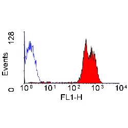
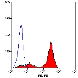
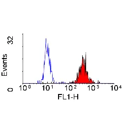
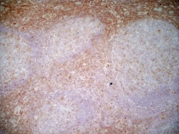
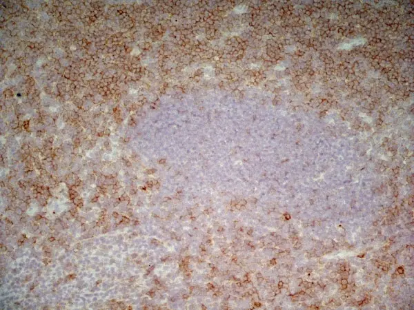
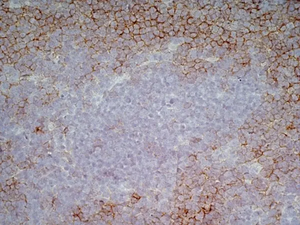
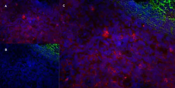
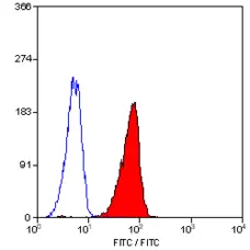
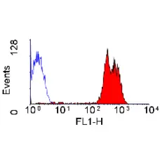
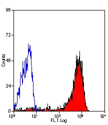
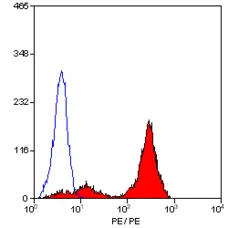
F C IF Reset| Mouse anti Human CD43 antibody, clone DFT-1 recognizes human CD43 also known as leukosialin or sialophorin. CD43 is a 381 amino acid single pass type I transmembrane glycoprotein present on the cell surface of multiple cell types including thymocytes, T-lymphocytes, neutrophils, plasma cells and platelets. It is lacking on the surface of most B-lymphocytes. CD34 is a major membrane sialoglycoprotein expressed on T lymphocytes as two predominant glycoforms, a ~115 kDa form present on all T-cells and a ~130 kDa form indicative of T-cell activation (Barren et al. 1997) and acts as a T-cell counter-receptor for CD169/Siglec-1 (van den Berg et al. 2001). Mouse anti Human CD43 antibody, clone DFT-1 binds to both glycoforms of human CD43 (Kyoizumi et al. 2004) CD34 expression is deficient and/or defective in patients suffering from the X-linked chromosomal immunodeficiency condition Wiskott-Aldrich syndrome and acts as a ligand for CD54 (Rosenstein et al. 1991). Mouse anti human CD43, clone DFT-1 has been used successfully for the evaluation of CD43 expression in patients with Myelodysplastic syndromes (Kyriakou et al. 2006). |
- Target Species
- Human
- Product Form
- Purified IgG - liquid
- Preparation
- Purified IgG prepared by affinity chromatography on Protein A from tissue culture supernatant
- Buffer Solution
- Phosphate buffered saline
- Preservative Stabilisers
0.09% Sodium Azide - Carrier Free
- Yes
- Immunogen
- KG-1 cells.
- Approx. Protein Concentrations
- IgG concentration 1 mg/ml
- Regulatory
- For research purposes only
- Guarantee
- 12 months from date of despatch
This product is shipped at ambient temperature. It is recommended to aliquot and store at -20°C on receipt. When thawed, aliquot the sample as needed. Keep aliquots at 2-8°C for short term use (up to 4 weeks) and store the remaining aliquots at -20°C.
Avoid repeated freezing and thawing as this may denature the antibody. Storage in frost-free freezers is not recommended.
Avoid repeated freezing and thawing as this may denature the antibody. Storage in frost-free freezers is not recommended.
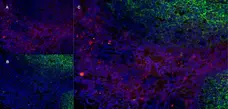
This product has been reported to work in the following applications. This information is derived from testing within our laboratories, peer-reviewed publications or personal communications from the originators. Please refer to references indicated for further information. For general protocol recommendations, please visit the antibody protocols page.
| Application Name | Verified | Min Dilution | Max Dilution |
|---|---|---|---|
| Flow Cytometry |  |
1/100 | |
| Immunofluorescence | |
||
| Immunohistology - Frozen | |
||
| Immunohistology - Paraffin | |
1/10 | 1/100 |
Where this antibody has not been tested for use in a particular technique this does not necessarily exclude its use in such procedures. Suggested working dilutions are given as a guide only. It is recommended that the user titrates the antibody for use in their own system using appropriate negative/positive controls.
- Flow Cytometry
- Use 10ul of the suggested working dilution to label 106 cells in 100ul.
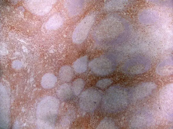
- Immunohistology
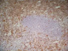
- This product does not require protein digestion pre-treatment of paraffin sections however, staining of paraffin embedded formalin fixed tissues may be improved in some cases by enzyme pretreatment using Trypsin.
- Histology Positive Control Tissue
- Tonsil
| Description | Product Code | Applications | Pack Size | List Price | Your Price | Quantity | |
|---|---|---|---|---|---|---|---|
| Mouse IgG1 Negative Control | MCA928 | F | 100 Tests |

|
Log in | ||
| List Price | Your Price | ||||||
|
|
Log in | ||||||
| Description | Mouse IgG1 Negative Control | ||||||
References for CD43 antibody
-
Stross, W.P. et al. (1989) Molecule detected in formalin fixed tissue by antibodies MT1, DF-T1, and L60 (Leu-22) corresponds to CD43 antigen.
J Clin Pathol. 42 (9): 953-61. -
Kojima N et al. (1994) Role of cell surface O-linked oligosaccharides in adhesion of HL60 cells to fibronectin: regulation of integrin-dependent cell adhesion by O-linked oligosaccharide elongation.
Exp Cell Res. 214 (2): 537-42. -
Matsuo, A. et al. (1996) Expression of CD43 in human microglia and its downregulation in Alzheimer's disease.
J Neuroimmunol. 71 (1-2): 81-6. -
Anzai, N. et al. (1999) Modulation of integrin function in hematopoietic progenitor cells by CD43 engagement: possible involvement of protein tyrosine kinase and phospholipase C-gamma.
Blood. 93 (10): 3317-26. -
Nonomura, C. et al. (2008) CD43, but not P-selectin glycoprotein ligand-1, functions as an E-selectin counter-receptor in human pre-B-cell leukemia NALL-1.
Cancer Res. 68 (3): 790-9. -
Woroniecka, R. et al. (2015) Cytogenetic and flow cytometry evaluation of Richter syndrome reveals MYC, CDKN2A, IGH alterations with loss of CD52, CD62L and increase of CD71 antigen expression as the most frequent recurrent abnormalities.
Am J Clin Pathol. 143 (1): 25-35.
- Synonyms
- Leukosialin
- RRID
- AB_321702
- UniProt
- P16150
- Entrez Gene
- SPN
- GO Terms
- GO:0042742 defense response to bacterium
- GO:0007596 blood coagulation
- GO:0004888 transmembrane receptor activity
- GO:0005887 integral to plasma membrane
- GO:0005615 extracellular space
- GO:0006935 chemotaxis
- GO:0006955 immune response
- GO:0006968 cellular defense response
- GO:0007162 negative regulation of cell adhesion
- View More GO Terms
- GO:0007163 establishment or maintenance of cell polarity
- GO:0042535 positive regulation of tumor necrosis factor biosynthetic process
- GO:0050900 leukocyte migration
- GO:0051635 bacterial cell surface binding
MCA555
If you cannot find the batch/lot you are looking for please contact our technical support team for assistance.
View more products with CD43 specificity
Please Note: All Products are "FOR RESEARCH PURPOSES ONLY"
View all Anti-Human ProductsAlways be the first to know.
When we launch new products and resources to help you achieve more in the lab.
Yes, sign me up