CD166 antibody | 3A6
Mouse anti Human CD166
- Product Type
- Monoclonal Antibody
- Clone
- 3A6
- Isotype
- IgG1
- Specificity
- CD166

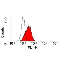
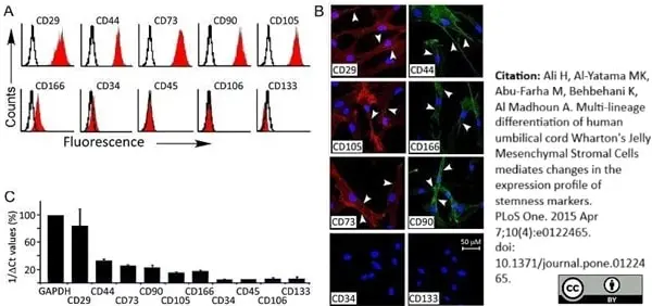
Mouse anti Human CD166 antibody, clone 3A6 (MCA1926) used for the evaluation of CD166 expression on umbilical cord derived mesenchymal stem cells by flow cytometry.
Image caption:
Flow cytometry, Immunofluorescence and qRT-PCR of WJ-MSCs. (A). Representative flow cytometry of WJ-MSCs (n = 3). Cells express CD29, CD44, CD73, CD90, CD105, and are negative for the hematopoietic (CD34 and CD45) and endothelial (CD106 and CD133) markers. Black open histogram indicates controls signal; red shaded histogram represents positive reactivity with the indicated antibody. (B) Confocal laser images of Immunofluorescence using APEX-labeling system for conjugating primary antibodies; CD29-Alexa Fluor 594, CD34-, CD44-, CD90- and CD133- Alexa Fluor 488. CD73-PE and CD105-PE were manufacturer labeled. 600X magnifications (C) qRT-PCR of the prospective markers for RNA isolated from undifferentiated WJ-MSCs cells, values were expressed as a percentage relative to 1/dCt of GAPDH gene.
From: Ali H, Al-Yatama MK, Abu-Farha M, Behbehani K, Al Madhoun A (2015)
Multi-Lineage Differentiation of Human Umbilical Cord Wharton’s Jelly Mesenchymal Stromal Cells Mediates Changes in the Expression Profile of Stemness Markers.
PLoS ONE 10(4): e0122465.
This image is from an open access article distributed under the terms of the Creative Commons Attribution License.

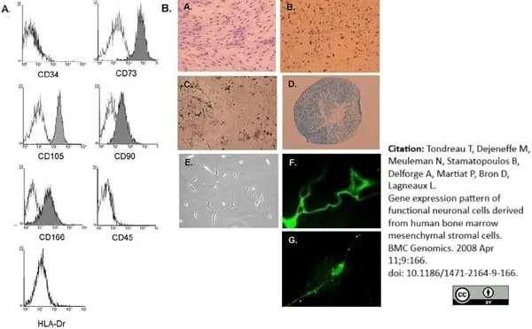
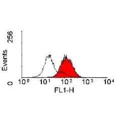
Mouse anti Human CD166 antibody, clone 3A6 (MCA1926) used for the evaluation of CD166 expression on mesenchymal stromal cells by flow cytometry.
Image caption:
Characterization and differentiation of BM-MSC. (A) Representative flow cytometric analysis of cultured mesenchymal stromal cells. Solid white represents the isotype control. (B) A. BM-MSC, displayed an homogeneous morphology of fibroblastic cells. Cells were stained with May Grunwald Giemsa staining (40×). Under specific induction BM-MSC were differentiated into (B) adipocytes (lipid vacuoles were colored by Oil Red O, ×40), (C) osteocytes (calcium deposits were revealed by Von Kossa method, ×40), (D) chondrocytes (cell pellet was sectioned and stained by toluidine blue, ×4), and (E) neuron-like cells derived from BM-MSC upon treatment with neurogenic medium (×40). Expression of nestin and MAP-2 after 10 days induction, detected by immunofluorescence (F and G, 100×).
From: Tondreau T, Dejeneffe M, Meuleman N, Stamatopoulos B, Delforge A, Martiat P, Bron D, Lagneaux L. Gene expression pattern of functional neuronal cells derived from human bone marrow mesenchymal stromal cells.
BMC Genomics. 2008 Apr 11;9:166.
This image is from an open access article distributed under the terms of the Creative Commons Attribution License.
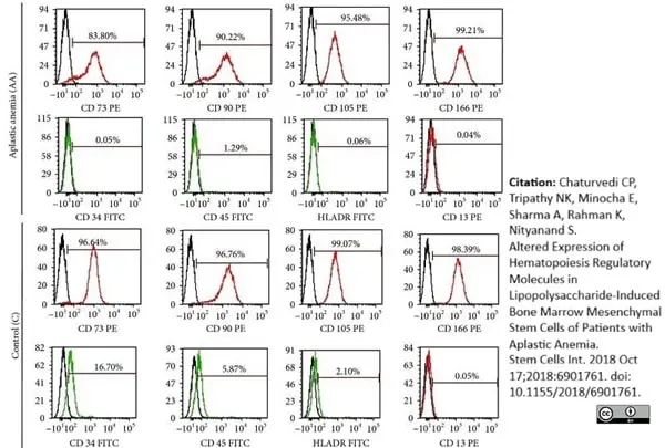
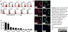
Phycoerythrin conjugated Mouse anti Human CD166 antibody, clone 3A6 (MCA1926PE) used to evaluate CD73 expression on bone-marrow derived mesenchymal stem cells by flow cytometry
Image caption:
Immunophenotypic and differentiation characterization of BM-MSCs of aplastic anemia (AA) patients. (a) Representative flow cytometric histograms showing immunophenotype of BM-MSCs of AA patients and controls.
From: Chaturvedi CP, Tripathy NK, Minocha E, Sharma A, Rahman K, Nityanand S.
Altered Expression of Hematopoiesis Regulatory Molecules in Lipopolysaccharide-Induced Bone Marrow Mesenchymal Stem Cells of Patients with Aplastic Anemia.
Stem Cells Int. 2018 Oct 17;2018:6901761.
doi: 10.1155/2018/6901761.
From: Insert article citation with space after colon and Hyperlink to PubMed record.
This image is from an open access article distributed under the terms of the Creative Commons Attribution License.
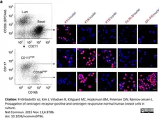
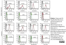
Alexa Fluor® 488 conjugated Mouse anti Human CD166 antibody, clone 3A6 (MCA1926A488) used for the evaluation of ALCAM expression on human breast epithelial cells (HBECs) by flow cytometry.
Image caption:
ERpos cells are purified and tracked by sequential CD326/CD271–CD166/CD117 FACS followed by multicolour staining and qRT–PCR.
Multicolour flow cytometry of uncultured HBECs incubated with CD326/CD271/CD166/CD117 and visualized pairwise (left diagrams) to recover luminal cells (CD326high) and basal cells (CD271high) and from the luminal gate CD166high and CD117high cells. Smears of sorted cells were stained (right panel) with either of the markers against basal cells, cytokeratin K14; luminal cells, cytokeratin K18; luminal progenitors, cytokeratin K15; Ks20.8 or ER–PR and counterstained with DAPI nuclear stain. Hormone receptor-positive cells are observed primarily among CD166high cells. Scale bar, 50 μm.
From: Fridriksdottir AJ, Kim J, Villadsen R, Klitgaard MC, Hopkinson BM, Petersen OW, Rønnov-Jessen L.
Propagation of oestrogen receptor-positive and oestrogen-responsive normal human breast cells in culture.
Nat Commun. 2015 Nov 13;6:8786.
This image is from an open access article distributed under the terms of the Creative Commons Attribution License.
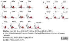
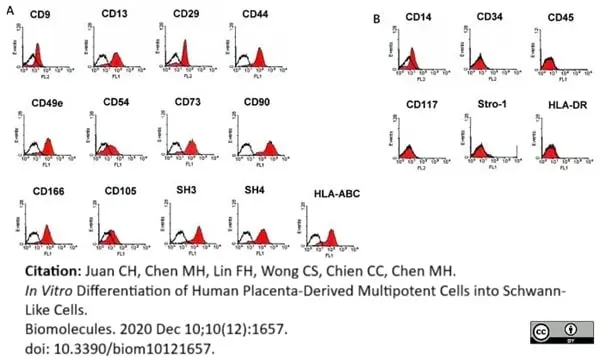
FITC conjugated Mouse anti Human CD166 antibody, clone 3A6 (MCA1926F) used to evaluate CD166 (ALCAM) expression on human placenta derived mesenchymal stem cells by flow cytometry.
Image caption:
Phenotypic expression of placenta-derived multipotent stem cells (PDMCs). The results of flow cytometry revealed HLA-ABC+, CD29+, CD44+, CD9+, CD90+, CD105+, SH3+, SH4+, CD166+, CD13+, CD49e+, and CD54+ and HAL-DR-, CD45-, CD34-, Stro-1-, and CD117-. (A,B) markers for MSCs (mesenchymal stem cell) and hematopoietic cells, respectively.
From: Juan CH, Chen MH, Lin FH, Wong CS, Chien CC, Chen MH.
In Vitro Differentiation of Human Placenta-Derived Multipotent Cells into Schwann-Like Cells.
Biomolecules. 2020 Dec 10;10(12):E1657.
doi: 10.3390/biom10121657.
This image is from an open access article distributed under terms of a Creative Commons Attribution License.
Filter by Application:
F Reset| Mouse anti Human CD166 antibody, clone 3A6 recognizes the 100 kDa adhesion molecule CD166, also known as ALCAM. CD166 is a member of the Ig superfamily and is expressed on activated T-cells, B cells and other cells including thymic epithelial cells, fibroblasts, keratinocytes and neurons. CD6 has been identified as a receptor for ALCAM (Skonier et al. 1996). Mouse anti Human CD166 antibody, clone 3A6 is reported to cross-react with CD166 on ovine tissues and provides a useful tool for the identification and characterization of ovine mesenchymal stem cells in conjunction with CD44 which is expressed by this cell lineage and the hematopoietic cell marker CD45 which is not expressed on mesenchymal stem cells (Sanjurjo-Rodríguez et al. 2017). |
- Target Species
- Human
- Species Cross-Reactivity
-
Target Species Cross Reactivity Sheep - N.B. Antibody reactivity and working conditions may vary between species.
- Product Form
- Purified IgG - liquid
- Preparation
- Purified IgG prepared by affinity chromatography on Protein A from tissue culture supernatant
- Buffer Solution
- Phosphate buffered saline
- Preservative Stabilisers
- 0.09% sodium azide (NaN3)
- Carrier Free
- Yes
- Immunogen
- Human thymic epithelial cells.
- Approx. Protein Concentrations
- IgG concentration 1.0mg/ml
- Fusion Partners
- Spleen cells from immunized mice were fused with cells of the P3X63 Ag8 myeloma cell line.
- Regulatory
- For research purposes only
- Guarantee
- 12 months from date of despatch
This product is shipped at ambient temperature. It is recommended to aliquot and store at -20°C on receipt. When thawed, aliquot the sample as needed. Keep aliquots at 2-8°C for short term use (up to 4 weeks) and store the remaining aliquots at -20°C.
Avoid repeated freezing and thawing as this may denature the antibody. Storage in frost-free freezers is not recommended.
Avoid repeated freezing and thawing as this may denature the antibody. Storage in frost-free freezers is not recommended.
This product has been reported to work in the following applications. This information is derived from testing within our laboratories, peer-reviewed publications or personal communications from the originators. Please refer to references indicated for further information. For general protocol recommendations, please visit the antibody protocols page.
| Application Name | Verified | Min Dilution | Max Dilution |
|---|---|---|---|
| ELISA |  |
||
| Flow Cytometry | |
1/10 | 1/50 |
| Immunohistology - Frozen | |
||
| Immunoprecipitation | |
Where this product has not been tested for use in a particular technique this does not necessarily exclude its use in such procedures. Suggested working dilutions are given as a guide only. It is recommended that the user titrates the product for use in their own system using appropriate negative/positive controls.
- Flow Cytometry
- Use 10μl of the suggested working dilution to label 106 cells in 100μl
| Description | Product Code | Applications | Pack Size | List Price | Your Price | Quantity | |
|---|---|---|---|---|---|---|---|
| Mouse IgG1 Negative Control | MCA928 | F | 100 Tests |

|
Log in | ||
| List Price | Your Price | ||||||
|
|
Log in | ||||||
| Description | Mouse IgG1 Negative Control | ||||||
References for CD166 antibody
-
Patel, D. D. et al. (1997) CD166 Workshop: Tissue distribution and functional analysis of antibodies reactive for CD166, a ligand for CD6.
In Leukocyte Typing IV. Kishimoto, T. et al. eds Garland publishing Inc. New York p. 461-4. -
Wang, D. et al. (2004) Proteomic profiling of bone marrow mesenchymal stem cells upon transforming growth factor beta1 stimulation.
J Biol Chem. 279 (42): 43725-34. -
Yeh, S.P. et al. (2005) Mesenchymal stem cells can be easily isolated from bone marrow of patients with various haematological malignancies but the surface antigens expression may be changed after prolonged ex vivo culture.
Leukemia. 19: 1505-7. -
Tondreau, T. et al. (2008) Gene expression pattern of functional neuronal cells derived from human bone marrow mesenchymal stromal cells.
BMC Genomics. 9:166. -
Srouji, S. et al. (2009) The Schneiderian membrane contains osteoprogenitor cells: in vivo and in vitro study.
Calcif Tissue Int. 84 (2): 138-45. -
Agha-Hosseini, F. et al. (2010) In vitro isolation of stem cells derived from human dental pulp.
Clin Transplant. 24: E23-8. -
Bhattacharya, S. et al. (2010) Toponome imaging system: in situ protein network mapping in normal and cancerous colon from the same patient reveals more than five-thousand cancer specific protein clusters and their subcellular annotation by using a three symbol code.
J Proteome Res. 9: 6112-25. -
Katsube, Y. et al. (2010) Restoration of cellular function of mesenchymal stem cells from a hypophosphatasia patient.
Gene Ther. 17 (4): 494-502.
View The Latest Product References
-
Brune, J.C. et al. (2011) Mesenchymal stromal cells from primary osteosarcoma are non-malignant and strikingly similar to their bone marrow counterparts.
Int J Cancer. 129 (2): 319-30. -
Green, L.R. et al. (2011) Cooperative role for tetraspanins in adhesin-mediated attachment of bacterial species to human epithelial cells.
Infect Immun. 79 (6): 2241-9. -
Ali, H. et al. (2015) Multi-Lineage Differentiation of Human Umbilical Cord Wharton's Jelly Mesenchymal Stromal Cells Mediates Changes in the Expression Profile of Stemness Markers.
PLoS One. 10 (4): e0122465. -
Fridriksdottir, A.J. et al. (2015) Propagation of oestrogen receptor-positive and oestrogen-responsive normal human breast cells in culture.
Nat Commun. 6: 8786. -
Holmannova, D. et al. (2017) Effects of conventional CPB and mini-CPB on neutrophils CD162, CD166 and CD195 expression.
Perfusion. 32 (2): 141-50. -
Prins, H.J. et al. (2016) Bone Regeneration Using the Freshly Isolated Autologous Stromal Vascular Fraction of Adipose Tissue in Combination With Calcium Phosphate Ceramics.
Stem Cells Transl Med. 5 (10): 1362-1374. -
Chen, F. et al. (2018) Bone morphogenetic protein 7-transduced human dermal-derived fibroblast cells differentiate into osteoblasts and form bone in vivo.
Connect Tissue Res. 59 (3): 223-232. -
Juan, C.H. et al. (2020) In Vitro Differentiation of Human Placenta-Derived Multipotent Cells into Schwann-Like Cells.
Biomolecules. 10 (12) Dec 10 [Epub ahead of print]. -
Hidalgo, L. et al. (2023) Switchable CAR T cell strategy against osteosarcoma.
Cancer Immunol Immunother. 72 (8): 2623-33. -
Kohler, K.T. et al. (2024) Oncogene activated human breast luminal progenitors contribute basally located myoepithelial cells.
Breast Cancer Res. 26 (1): 183.
- Synonyms
- ALCAM
- RRID
- AB_323338
- UniProt
- Q13740
- Entrez Gene
- ALCAM
- GO Terms
- GO:0005102 receptor binding
- GO:0007155 cell adhesion
- GO:0016021 integral to membrane
- GO:0007165 signal transduction
Request a different product with this specificity
Please Note: All Products are "FOR RESEARCH PURPOSES ONLY"
View all Anti-Human ProductsAlways be the first to know.
When we launch new products and resources to help you achieve more in the lab.
Yes, sign me up