CD10 antibody | 56C6
Mouse anti Human CD10
- Product Type
- Monoclonal Antibody
- Clone
- 56C6
- Isotype
- IgG1
- Specificity
- CD10


Filter by Application:
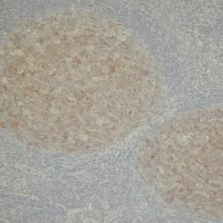
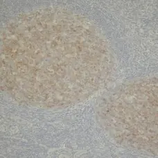
P Reset| Mouse anti Human CD10 antibody, clone 56C6 recognizes human Neprilysin, also known as CD10, Atriopeptidase, Common acute lymphocytic leukemia antigen (CALLA), Enkephalinase, Neutral endopeptidase 24.11, Neutral endopeptidase or Skin fibroblast elastase. CD10 is a 749 amino acid, ~100 kDa single pass type II transmembrane glycoprotein, expressed on a variety of cell types including immature and germinal centre B cells, lymphoblastic leukaemia cells and also on the brush border of kidney epithelial cells. Mutations in the MME gene (encoding CD10) are involved in the development of Charcot-Marie-Tooth disease 2T (CMT2T), a disorder of the peripheral nervous system, demonstrating progerssive peripheral muscle weakness and atrophy (Higuchi et al. 2016). Mutations of the MME gene can also lead to Spinocerebellar ataxia 43 (SCA43) characterized by a progressive loss of gait coordination along with hand, speech and eye coordination. Degeneration of the cerebellum, with variable involvement of the brain stem and spinal cord are apparent in this slow progressive autosomal dominant form of the disease (Depondt et al. 2016). |
- Target Species
- Human
- Species Cross-Reactivity
-
Target Species Cross Reactivity Cynomolgus monkey - N.B. Antibody reactivity and working conditions may vary between species.
- Product Form
- Tissue culture supernatant - liquid
- Preservative Stabilisers
- <0.1% sodium azide (NaN3)
- Immunogen
- Recombinant human CD10.
- Fusion Partners
- Spleen cells from immunised mice were fused with cells of the mouse p3-NS1-Ag4-1 myeloma cell line
- Regulatory
- For research purposes only
- Guarantee
- 12 months from date of despatch
This product is shipped at ambient temperature. It is recommended to aliquot and store at -20°C on receipt. When thawed, aliquot the sample as needed. Keep aliquots at 2-8°C for short term use (up to 4 weeks) and store the remaining aliquots at -20°C.
Avoid repeated freezing and thawing as this may denature the antibody. Storage in frost-free freezers is not recommended.
Avoid repeated freezing and thawing as this may denature the antibody. Storage in frost-free freezers is not recommended.
This product has been reported to work in the following applications. This information is derived from testing within our laboratories, peer-reviewed publications or personal communications from the originators. Please refer to references indicated for further information. For general protocol recommendations, please visit the antibody protocols page.
| Application Name | Verified | Min Dilution | Max Dilution |
|---|---|---|---|
| Immunohistology - Frozen |  |
1/80 | |
| Immunohistology - Paraffin 1 | |
1/80 |
- 1This product requires antigen retrieval using heat treatment prior to staining of paraffin sections.Sodium citrate buffer pH 6.0 is recommended for this purpose.
Where this product has not been tested for use in a particular technique this does not necessarily exclude its use in such procedures. Suggested working dilutions are given as a guide only. It is recommended that the user titrates the product for use in their own system using appropriate negative/positive controls.
- Histology Positive Control Tissue
- Human tonsil, intestine or kidney
References for CD10 antibody
-
Angelin-Duclos, C. et al. (2000) Commitment of B lymphocytes to a plasma cell fate is associated with Blimp-1 expression in vivo.
J Immunol. 165 (10): 5462-71. -
Carvounis, E.E. et al. (2003) Undifferentiated carcinoma with osteoclast-like giant cells of the pancreas.
Int J Gastrointest Cancer. 33: 103-6. -
Matsumoto, T. et al. (2005) Increase of bone marrow-derived secretory lineage epithelial cells during regeneration in the human intestine.
Gastroenterology. 128: 1851-67. -
Ince, T.A. et al. (2007) Transformation of different human breast epithelial cell types leads to distinct tumor phenotypes.
Cancer Cell. 12 (2): 160-70. -
Tasaki K et al. (2007) CD5-positive mucosa-associated lymphoid tissue (MALT) lymphoma of ocular adnexal origin: usefulness of fluorescence in situ hybridization for distinction between mantle cell lymphoma and MALT lymphoma.
Pathol Int. 57 (2): 101-7. -
de Moraes Schenka, N.G. et al. (2007) Use of p63 and CD10 in the differential diagnosis of papillary neoplasms of the breast.
Breast J. 14: 68-75. -
Scholzen, T.E. et al. (2007) Terminating the stress: peripheral peptidolysis of proopiomelanocortin-derived regulatory hormones by the dermal microvascular endothelial cell extracellular peptidases neprilysin and angiotensin-converting enzyme.
Endocrinology. 148: 2793-805. -
Rasini, V. et al. (2013) Mesenchymal stromal/stem cells markers in the human bone marrow.
Cytotherapy. 15 (3): 292-306.
View The Latest Product References
-
Mizowaki, T. et al. (2015) STAT3 activation is associated with cerebrospinal fluid interleukin-10 (IL-10) in primary central nervous system diffuse large B cell lymphoma.
J Neurooncol. 124 (2): 165-74. -
Nishimoto-Kakiuchi, A. et al. (2016) Characteristics of histologically confirmed endometriosis in cynomolgus monkeys.
Hum Reprod. 31 (10): 2352-9.
- Synonyms
- CALLA
- RRID
- AB_323793
- UniProt
- P08473
- Entrez Gene
- MME
- GO Terms
- GO:0005515 protein binding
- GO:0004222 metalloendopeptidase activity
- GO:0005887 integral to plasma membrane
- GO:0046872 metal ion binding
- GO:0006508 proteolysis
- GO:0007267 cell-cell signaling
View more products with CD10 specificity
Please Note: All Products are "FOR RESEARCH PURPOSES ONLY"
View all Anti-Human ProductsAlways be the first to know.
When we launch new products and resources to help you achieve more in the lab.
Yes, sign me up