IHC Antibody Tips - Step 18: Counterstain

- On This Page
- Counterstain
- Related resources
- Support
Counterstains provide contrast as well as knowledge about localization within the tissue sample (for instance by visualizing nuclei or filamentous actin).
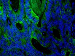
Fig. 3. PureBlu™ Nuclear Fluorescent Dye Hoechst 33342. Paraffin section of human colon adenocarcinoma (heat induced antigen retrieval with citrate buffer
(pH 6), blocked with 10% FCS) stained with Mouse Anti-Human Cytokeratin 18 Antibody (MCA1864H, green). Nuclei were counterstained with PureBlu Dye Hoechst 33342 (1351304, blue).
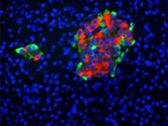
Fig. 4. PureBlu Dye DAPI. Paraffin section of human pancreas stained with Guinea Pig Anti-Pig Insulin Antibody (5330-0104G, red), Mouse Anti-Human Chromogranin A Antibody (MCA4773, green) and PureBlu Dye DAPI (1351303, blue).
What to consider when selecting counterstains?
- Ensure that the counterstain can be easily spectrally distinguished from your precipitate color or fluorescent label. For example, when using red emitting fluorophores, select a blue nuclear counterstain such as DAPI or Hoechst 33342 (Tables 4 and 5)
- To save time, consider using pre-made mounting media already containing counterstains
Table 4: Commonly used chromogenic counterstains.
|
Chromogenic Counterstains |
Colors |
Counterstain for |
|
Eosin |
Red |
Proteins containing cationic groups (Kim et al. 2016) |
|
Fast Red/Kernechtrot |
Red |
Nuclei |
|
Hematoxylin (4 types - Harris’s, Mayer’s, Carazzi’s, and Gill’s) |
Blue |
Nuclei |
|
Methylene blue |
Blue |
Nuclei |
|
Methylene green |
Blue/green |
Nuclei |
|
Toluidine blue |
Blue |
Nuclei |
Adapted from Kim et al. 2016 and Paul no date. Fast red/Kernechtrot has been included in the table according to IHCWorld (no date c).
Table 5: Commonly used fluorescent counterstains.
|
Fluorescent Counterstains |
Colors |
Counterstain for |
| DAPI | Blue | Nuclei |
| DRAQ5 | Red | Nuclei |
| Hoechst 33258/33342 | Blue | Nuclei |
| Propidium iodide | Red | Nuclei |
| SYTOX Green | Green | Nuclei |
| Phalloidin | Conjugate dependent | Filamentous actin |


