DAPI - An easy-to-use Nuclear Dye
Bio-Rad's PureBlu™ DAPI Dye can be used for routine nuclear staining in fluorescence microscopy and cell imaging applications as well as cell viability and cell cycle analysis in flow cytometry. PureBlu DAPI Dye is available as a ready-to-reconstitute, highly pure powder form, with only one dilution step required to obtain a ready-to-use solution.
DAPI Structure
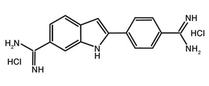
DAPI (4',6-diamidino-2-phenylindole) (Figure 1) is a well-characterized blue-emitting fluorescent compound widely utilized for nuclear staining. DAPI permeates cell membranes and binds to the minor groove of A/T-rich dsDNA sequences, thus preferentially staining nuclei.

Fig. 1. Molecular structure of PureBlu DAPI Nuclear Staining Dye.
DAPI Excitation / Emission Spectra
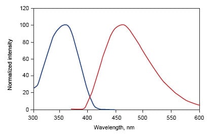
When bound to dsDNA, PureBlu DAPI Dye has a maximum excitation wavelength in the ultraviolet range (359 nm), and the dye can be optimally detected in the blue channel with an emission maximum of 461 nm (Figure 2).

Fig. 2. DAPI excitation and emission spectra.
The characteristic Stokes shift between excitation and emission wavelengths is fairly wide for DAPI, making this dye an optimal choice when good spectral separation is desired to reduce fluorescence interference, for example, in chromatin counterstaining for immunofluorescence microscopy.

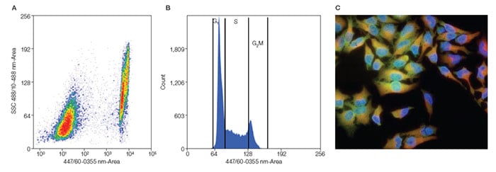
Fig.3. Analysis of Jurkat and HeLa cells. A, flow cytometric analysis of Jurkat cell viability. Jurkat cells were heat shocked at 65°C for 5 min. Dead cells show positive staining with PureBlu DAPI. B, analysis of DNA content of Jurkat cells. Cultured cells were harvested, washed in phosphate buffered saline (PBS), and fixed in cold 70% ethanol for 2 hr at 4°C. The Jurkat cells were then washed in Bio-Rad Staining Buffer (#BUF073) and stained with PureBlu DAPI Dye containing 0.1% Triton X-100 for 30 min. Cells were first gated to discriminate single cells and then plotted into histograms discriminating G1, S, and G2/M populations. All flow cytometry was carried out on a Bio-Rad ZE5 Cell Analyzer (#12004279). C, HeLa cells, formaldehyde fixed and permeabilized, stained with Rat Anti-Alpha Tubulin Antibody (red) (#MCA78G), Mouse Anti-Human CD49a Antibody, green (#MCA1133), and PureBlu DAPI Dye (blue).
Alternative for unfixed samples
As an alternative to DAPI we also have PureBlu Hoechst 33342 Nuclear Staining Dye in a similar 'ready to reconstitute' format suitable for unfixed samples.
Related documentation
- PureBlu DAPI Nuclear Staining Dye 取扱説明書
- PureBlu DAPI Nuclear Staining Dye Product Insert (inc. protocol)
To determine cell viability we also offer the non membrane permeable DNA-binding cell staining dyes Readidrop 7-AAD and Readidrop propidium iodide, in ready-to-use formulations and fixable VivaFix™ Cell Viability Assays in a range of excitation and emission spectra to enable easy incorporation into multi-color Flow Cytometry panels.


