CD4 antibody | GK1.5
Rat anti Mouse CD4
- Product Type
- Monoclonal Antibody
- Clone
- GK1.5
- Isotype
- IgG2b
- Specificity
- CD4


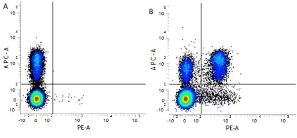
Figure B. APC conjugated Rat anti Mouse CD3 antibody, clone KT3 (MCA500APC) and RPE conjugated Rat anti Mouse CD4 antibody, clone GK1.5 (MCA4635PE).
All experiments performed on murine splenocytes in the presence of murine SeroBlock (BUF041A).
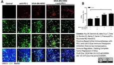
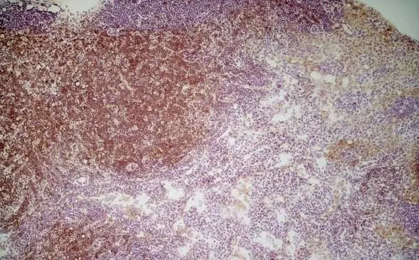
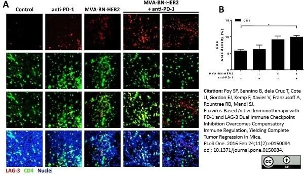
Rat anti Mouse CD4 antibody, clone GK1.5 (MCA4635GA) used for the identification of CD4 expressing cells in tumors by immunofluorescence.
Image caption:
LAG-3 and CD4 expression in the tumor microenvironment after MVA-BN-HER2 and anti-PD-1 therapy.
(A) Tumors from mice treated with MVA-BN-HER2 and/or anti-PD-1 were collected on day 16 and stained for LAG-3 (red), CD4 (green), and nuclei (DAPI, blue). (B) The area density for CD8+ T cells.* p<0.05. n = 3–4 mice/group.
From: Foy SP, Sennino B, dela Cruz T, Cote JJ, Gordon EJ, Kemp F, et al. (2016)
Poxvirus-Based Active Immunotherapy with PD-1 and LAG-3 Dual Immune Checkpoint Inhibition Overcomes Compensatory Immune Regulation, Yielding Complete Tumor Regression in Mice.
PLoS ONE 11(2): e0150084.
This image is from an open access article distributed under terms of a Creative Commons Attribution License.
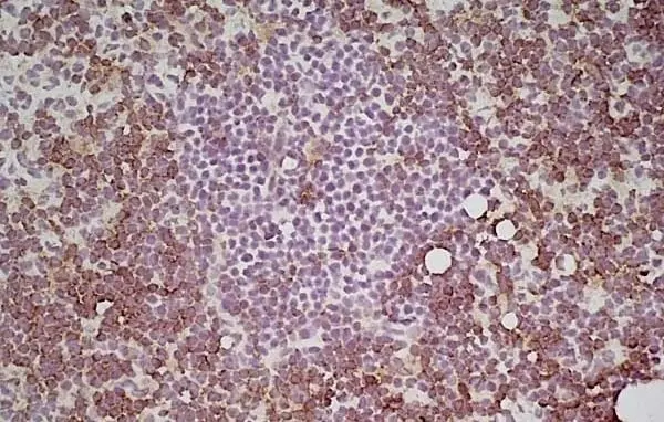
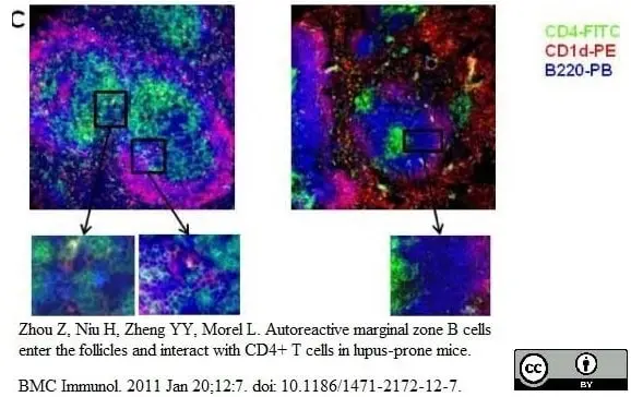
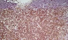
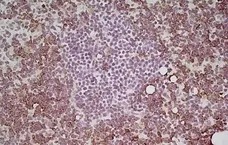
FITC conjugated Rat anti Mouse CD4 antibody, clone GK1.5 (MCA4635F) used to identify CD4 expressing cells in mouse spleen cryosections by immunofluorescence.
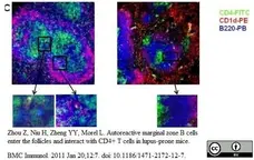
Published customer image:
Intrafollicular location of MZB cells in B6.TC spleens. C. Representative spleen sections from 10 mo old B6.TC (left) and B6 (right) mice stained with CD4-FITC, CD1d-PE and B220-PB. Boxed areas show multiple contacts between green B6.TC CD4 T cells and pink MZB cells, but not in the B6 spleen. Original magnification: 200X.
From: Zhou Z, Niu H, Zheng YY, Morel L.
Autoreactive marginal zone B cells enter the follicles and interact with CD4+ T cells in lupus-prone mice.
BMC Immunol. 2011 Jan 20;12:7.
doi: 10.1186/1471-2172-12-7.
This image is from an open access article distributed under terms of a Creative Commons Attribution License.
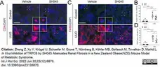
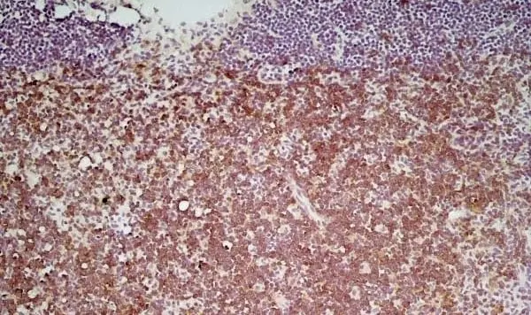
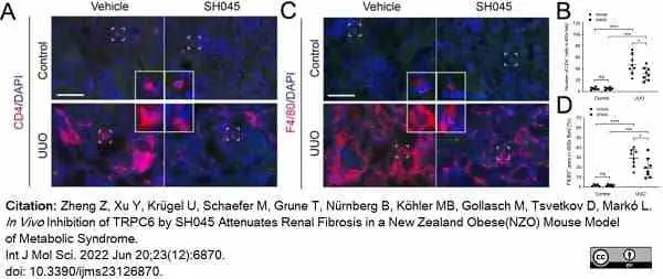
Rat anti Mouse CD4 antibody, clone GK1.5 (MCA4635) used to label lymphocytes in murine renal tissue by immunofluorescence.
Image caption:
SH045 impact on renal inflammatory cell accumulation after UUO. (A) Representative images of control and UUO-injured kidneys stained with CD4+ T cells (magnification: 400×). Rectangles represent single-cell magnifications. Scale bars are 50 μm. (B) Quantification in renal infiltration of CD4+ T cells (control n = 6, UUO n = 8). (C) Representative images of control and UUO-injured kidneys stained with F4/80+ macrophages (magnification: 400×). Rectangles represent single-cell magnifications. Scale bars are 50 μm. (D) Quantification in renal infiltration of F4/80+ macrophages (control n = 6, UUO n = 8). Data expressed as means ± SD. Two-way ANOVA followed by Sidak’s multiple comparisons post hoc test. * p <0.05, *** p <0.001, and **** p <0.0001 defined as significant. ns, not statistically significant. AU, arbitrary units.
From: Zheng Z, Xu Y, Krügel U, Schaefer M, Grune T, Nürnberg B, Köhler MB, Gollasch M, Tsvetkov D, Markó L.
In Vivo Inhibition of TRPC6 by SH045 Attenuates Renal Fibrosis in a New Zealand Obese (NZO) Mouse Model of Metabolic Syndrome.
Int J Mol Sci. 2022 Jun 20;23(12):6870.
doi: 10.3390/ijms23126870.
This image is from an open access article distributed under terms of a Creative Commons Attribution License.
Filter by Application:
F C IF Reset| Rat anti Mouse CD4 antibody, clone GK1.5 recognizes mouse CD4, a ~55 kDa protein also known as Ly-4 and L3T4. CD4 is a single chain transmembraneous glycoprotein which belongs to the immunoglobulin superfamily, and is primarily expressed on T helper cells, peripheral blood monocytes and tissue macrophages. CD4 is also expressed on a subpopulation of regulatory T cells (CD4+ CD25+), which play a key role in the maintenance of self tolerance. Rat anti Mouse CD4 antibody, clone GK1.5 has been reported to block CD4+ T-cell activation. It blocks class II MHC antigen-specific binding, thereby inhibiting functions such as class II MHC antigen-specific proliferation and the release of lymphokines. It may also be used for in vivo and in vitro cell depletion of CD4+ T-cells. |
- Target Species
- Mouse
- Product Form
- Purified IgG - liquid
- Preparation
- Purified IgG prepared by affinity chromatography on Protein G from tissue culture supernatant
- Buffer Solution
- Phosphate buffered saline
- Preservative Stabilisers
0.09% Sodium Azide - Carrier Free
- Yes
- Immunogen
- Murine CD4.
- Approx. Protein Concentrations
- IgG concentration 1.0 mg/ml
- Fusion Partners
- Spleen cells from immunised Lewis rats were fused with cells of the SP2/0 myeloma cell line.
- Regulatory
- For research purposes only
- Guarantee
- 12 months from date of despatch
This product is shipped at ambient temperature. It is recommended to aliquot and store at -20°C on receipt. When thawed, aliquot the sample as needed. Keep aliquots at 2-8°C for short term use (up to 4 weeks) and store the remaining aliquots at -20°C.
Avoid repeated freezing and thawing as this may denature the antibody. Storage in frost-free freezers is not recommended.
Avoid repeated freezing and thawing as this may denature the antibody. Storage in frost-free freezers is not recommended.
This product has been reported to work in the following applications. This information is derived from testing within our laboratories, peer-reviewed publications or personal communications from the originators. Please refer to references indicated for further information. For general protocol recommendations, please visit the antibody protocols page.
| Application Name | Verified | Min Dilution | Max Dilution |
|---|---|---|---|
| ELISA |  |
||
| Flow Cytometry | |
1/10 | 1/200 |
| Immunofluorescence | |
||
| Immunohistology - Frozen | |
||
| Immunoprecipitation | |
||
| Western Blotting | |
Where this antibody has not been tested for use in a particular technique this does not necessarily exclude its use in such procedures. Suggested working dilutions are given as a guide only. It is recommended that the user titrates the antibody for use in their own system using appropriate negative/positive controls.
- Flow Cytometry
- Use 10ul of the suggested working dilution to label 1x10⁶ cells in 100ul.
References for CD4 antibody
-
Dialynas, D.P. et al. (1983) Characterization of the murine T cell surface molecule, designated L3T4, identified by monoclonal antibody GK1.5: similarity of L3T4 to the human Leu-3/T4 molecule.
J Immunol. 131 (5): 2445-51. -
Wilde, D.B. et al. (1983) Evidence implicating L3T4 in class II MHC antigen reactivity; monoclonal antibody GK1.5 (anti-L3T4a) blocks class II MHC antigen-specific proliferation, release of lymphokines, and binding by cloned murine helper T lymphocyte lines.
J Immunol. 131 (5): 2178-83. -
Näher, H. et al. (1985) Dynamics of T cells of L3T4 and Ly 2 phenotype within granulomas in murine listeriosis.
Clin Exp Immunol. 60 (3): 559-64. -
Ye, X. et al. (2000) Transient depletion of CD4 lymphocyte improves efficacy of repeated administration of recombinant adenovirus in the ornithine transcarbamylase deficient sparse fur mouse.
Gene Ther. 7 (20): 1761-7. -
Chu, N.R. et al. (2000) Immunotherapy of a human papillomavirus (HPV) type 16 E7-expressing tumour by administration of fusion protein comprising Mycobacterium bovis bacille Calmette-Guérin (BCG) hsp65 and HPV16 E7.
Clin Exp Immunol. 121:216-25 -
Zhou, Z. et al. (2011) Autoreactive marginal zone B cells enter the follicles and interact with CD4+ T cells in lupus-prone mice.
BMC Immunol. 12: 7. -
Pletinckx, K. et al. (2015) Immature dendritic cells convert anergic nonregulatory T cells into Foxp3- IL-10+ regulatory T cells by engaging CD28 and CTLA-4.
Eur J Immunol. 45 (2): 480-91. -
Foy, S.P. et al. (2016) Poxvirus-Based Active Immunotherapy with PD-1 and LAG-3 Dual Immune Checkpoint Inhibition Overcomes Compensatory Immune Regulation, Yielding Complete Tumor Regression in Mice.
PLoS One. 11 (2): e0150084.
View The Latest Product References
-
Steinl, D.C. et al. (2016) Noninvasive Contrast-Enhanced Ultrasound Molecular Imaging Detects Myocardial Inflammatory Response in Autoimmune Myocarditis.
Circ Cardiovasc Imaging. 9 (8): . -
Olesen, M. N. et al. (2018) CD4 T cells react to local increase of α-synuclein in a pathology-associated variant-dependent manner and modify brain microglia in absence of brain pathology
Heliyon. 4 (1): e00513. -
Jalili, R.B. et al. (2018) Fibroblast cell-based therapy prevents induction of alopecia areata in an experimental model.
Cell Transplant. 27 (6): 994-1004. -
Zheng, Z. et al. (2022) In Vivo. Inhibition of TRPC6 by SH045 Attenuates Renal Fibrosis in a New Zealand Obese (NZO) Mouse Model of Metabolic Syndrome.
Int J Mol Sci. 23(12):6870.
Further Reading
-
Dialynas, D.P. et al. (1983) Characterization of the murine antigenic determinant, designated L3T4a, recognized by monoclonal antibody GK1.5: expression of L3T4a by functional T cell clones appears to correlate primarily with class II MHC antigen-reactivity.
Immunol Rev. 74: 29-56.
- Synonyms
- L3T4 Antigen
- LY-4
- RRID
- AB_1898233
- UniProt
- P06332
- Entrez Gene
- Cd4
- GO Terms
- GO:0001816 cytokine production
- GO:0007155 cell adhesion
- GO:0016021 integral to membrane
- GO:0005788 endoplasmic reticulum lumen
- GO:0005789 endoplasmic reticulum membrane
- GO:0006955 immune response
- GO:0007166 cell surface receptor linked signaling pathway
- GO:0009897 external side of plasma membrane
- GO:0045058 T cell selection
- View More GO Terms
- GO:0045121 membrane raft
- GO:0050731 positive regulation of peptidyl-tyrosine phosphorylation
- GO:0050850 positive regulation of calcium-mediated signaling
- GO:0050870 positive regulation of T cell activation
- GO:0051789 response to protein stimulus
View more products with CD4 specificity
Please Note: All Products are "FOR RESEARCH PURPOSES ONLY"
View all Anti-Mouse ProductsAlways be the first to know.
When we launch new products and resources to help you achieve more in the lab.
Yes, sign me up