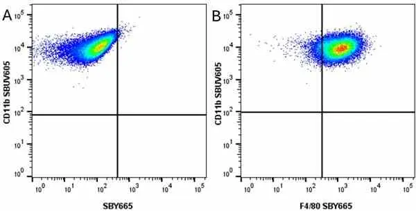
CD11b antibody | 5C6
Rat anti Mouse CD11b
- Product Type
- Monoclonal Antibody
- Clone
- 5C6
- Isotype
- IgG2b
- Specificity
- CD11b


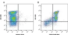
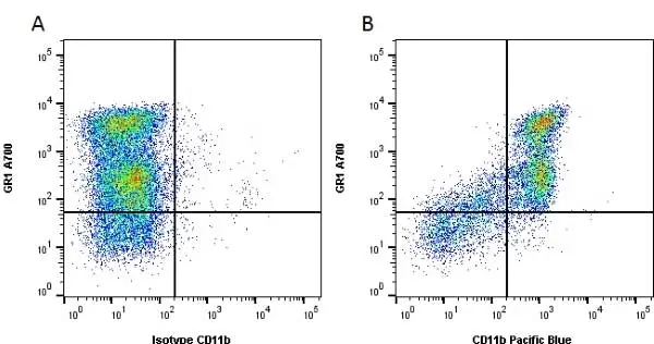
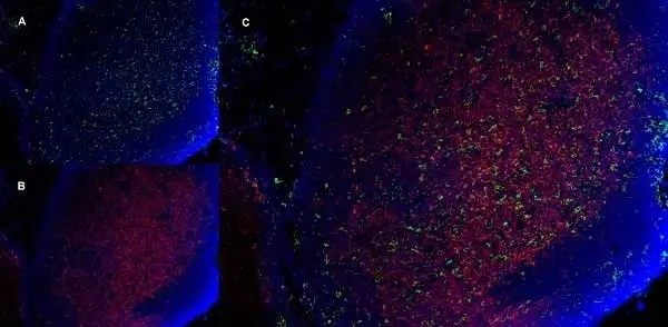
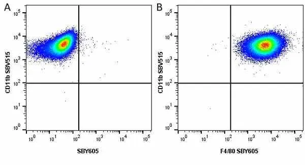
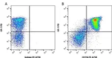
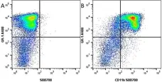
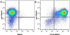
Figure B. Alexa Fluor® 700 conjugated Rat anti Mouse Gr-1 antibody, clone RB6-8C5 (MCA2387A700) and Pacific Blue® conjugated Rat anti Mouse CD11b antibody, clone 5C6 (MCA711PB). All experiments performed on red cell lysed mouse bone marrow gated on mononuclear cells in the presence of Mouse Seroblock (BUF041A).
Data acquired on the ZE5 Cell Analyzer.

Data acquired on the ZE5 Cell Analyzer.
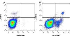
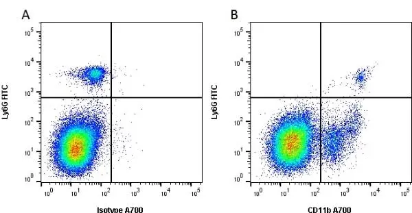
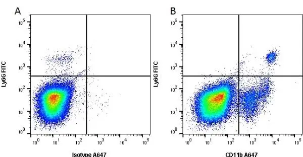
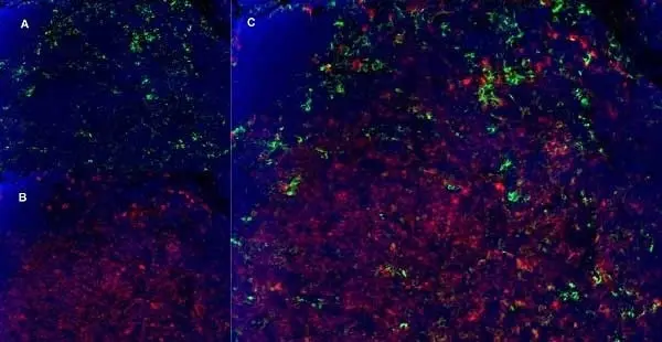
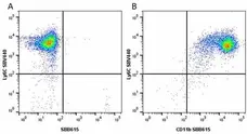
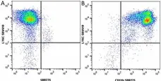
Figure B. FITC conjugated Rat anti Mouse Ly-6G antibody, clone 1A8 (MCA6077F) and Alexa Fluor® 647 conjugated Rat anti Mouse CD11b antibody, clone 5C6 (MCA711A647). All experiments performed on red cell lysed mouse blood in the presence of 10% mouse serum.
Data acquired on the ZE5 Cell Analyzer.

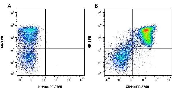
Figure B.Alexa Fluor®647 conjugated Rat anti Mouse Gr-1 antibody, clone RB6-8C5 (MCA2387A647) and RPE conjugated Rat anti Mouse CD11b antibody, clone 5C6 (MCA711PE).
All experiments performed on murine bone marrow in the presence of murine SeroBlock (BUF041A).
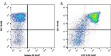
Figure B. Alexa Fluor 488 conjugated Rat anti Mouse GR-1 (MCA2387A488) and PE-Alexa Fluor 647 conjugated Rat anti Mouse CD11b (MCA711P647). All experiments performed on red cell lysed murine bone marrow blood gated live cells in the presence of 10% mouse serum. Data acquired on the ZE5 Cell Analyzer.
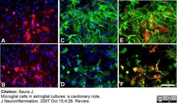
Rat anti mouse CD11b antibody, clone 5C6 (MCA711) used for the identification of microglia in mixed glial cultures by immunofluorescence.
Image caption:
Murine primary cortical mixed glial culture were treated with LPS (1 μg/ml) and IFNγ (0.5 ng/ml) for 24 hours and immunostained for NOS2 (A, B), GFAP (C) or CD11b (D). A and C show the same field and E is their merged image. B and D show the same field and F is their merged image. In control cultures NOS2-immunoreactive cells were not observed (data not shown). There is (LPS + IFNγ)-induced NOS2 expression in numerous cells (A, B). NOS2-positive cells were almost never GFAP immunoreactive (A, C, E) indicating a lack of NOS2 expression in most astrocytes. In contrast, virtually all NOS2-positive cells (>98%) were identified as microglia by their CD11b immunoreactivity. Nuclei are counterstained with Hoechst 33258 in A-D. NOS2-positive cells were identified with a rabbit anti-NOS2 antibody (1:500, BD Biosciences), GFAP-positive cells with a mouse anti-GFAP antibody (1:1000, Sigma) and CD11b-positive cells with 5C6 mouse anti-CD11b antibody (1:400, Serotec). Bar, 100 μm.
From: Saura J.
Microglial cells in astroglial cultures: a cautionary note.
J Neuroinflammation. 2007 Oct 15;4:26. Review.
This is from an open access article distributed under the terms of a Creative Commons Attribution License.

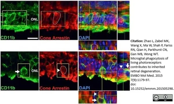
Rat anti Mouse CD11b antibody, clone 5C6 (MCA711) used to identify microglia in the retinal outer nuclear layer in a murine model of retinitis pigmentosa by immunofluorescence.
Image caption:
Infiltrating microglia phagocytose rods that are immunonegative for markers of early apoptosis and do not phagocytose cones during rod degeneration.
Photoreceptor cones are not phagocytosed by microglia during rod degeneration. (Upper panels) At P21–23, although infiltrating microglia in the ONL (CD11b, green) contain phagocytosed nuclei (DAPI, blue), none of these were found to be associated with cone arrestin immunopositivity (red), despite the close proximity of arrestin-positive cone somata (highlighted by circled area) to infiltrating microglia. (Lower panels) Example of a CD11b-positive microglial cell in the ONL juxtaposed closely to an arrestin-positive soma (highlighted by circled area). Analysis of orthogonal views of the confocal image stack demonstrates the absence of cone phagocytosis by microglia. Scale bar, 10 μm.
From: Zhao L, Zabel MK, Wang X, Ma W, Shah P, Fariss RN, Qian H, Parkhurst CN, Gan WB, Wong WT.
Microglial phagocytosis of living photoreceptors contributes to inherited retinal degeneration.
EMBO Mol Med. 2015 Jul 2;7(9):1179-97.
This is from an open access article distributed under the terms of a Creative Commons Attribution License.
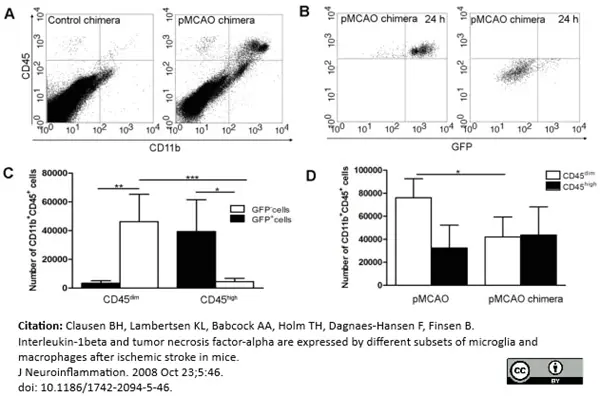
Rat anti Mouse CD11b antibody clone 5C6 (MCA711) used to identify macrophages in mixed populations by flow cytometry.
Image caption:
Infiltration of GFP+ BM-cells in infarct and peri-infarct regions. (A-B) Dot plots of viable macrophages/granulocytes (CD11b+CD45high, top right quadrants) and microglia (CD11b+CD45dim, bottom right quadrants) in cortex from BM-chimeric unmanipulated mice and mice exposed to pMCAO. (C) Bar graph showing mean numbers of CD11b+CD45dim microglia and CD11b+CD45high macrophages/granulocytes in BM-chimeric mice 24 hours after pMCAO, subdivided based on expression of GFP (n = 5). Approximately 92% of of the CD45high population were GFP+ . (D) Estimation and comparison of mean numbers of CD11b+CD45dim microglia in non-chimeric (n = 10) versus BM-chimeric mice (n = 5) 24 hours after of pMCAO shows significantly fewer CD11b+CD45dim microglial cells in irradiated mice. *P<0.05, **P<0.01, and ***P<0.001.
From: Clausen BH, Lambertsen KL, Babcock AA, Holm TH, Dagnaes-Hansen F, Finsen B.
Interleukin-1beta and tumor necrosis factor-alpha are expressed by different subsets of microglia and macrophages after ischemic stroke in mice.
J Neuroinflammation. 2008 Oct 23;5:46.
This is from an open access article distributed under the terms of a Creative Commons Attribution License.
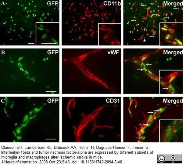
Rat anti Mouse CD11b antibody, clone 5C6 (MCA711) used to identify macrophages associated with vessels by immunofluorescence.
Image caption:
BM-derived GFP+ single cells and vessel-associated cells express CD11b. Fluorescence microscopy for GFP combined with immunofluorescence detection of (A) CD11b, (B) vWF and (C) CD31, 24 hours after pMCAO. (A) Fluorescence detection of GFP and CD11b showed that most GFP+ cells co-expressed CD11b (yellow cells, indicated by arrows), and intermingled with CD11b+ host cells. Note also that a few GFP+ cells did not co-express CD11b (arrow head). Insert shows high magnification of GFP+ cells, some of which co-express CD11b, aggregated around a vessel. (B, C) Fluorescence detection of GFP and the endothelial cell markers vWF (B) and CD31 (C). Inserts show higher magnification of sections of the same vessels. Although there are indications that single vWF+ cells co-express GFP (arrows in B), this could not be reproduced using staining for CD31, and the majority of vWF+ and CD31+ cells showed no co-expression of GFP. Instead, GFP remained confined to round and elongated cells located in the juxtavascular space (insert in C). CD11b+ cells were visualized using Alexa Fluor® 568-conjugated goat anti-rat IgG, vWF+ and CD31+ cells using Alexa Fluor® 546-conjugated goat anti-rabbit IgG and Alexa Fluor® 594-conjugated goat anti-rat IgG, respectively. Scale bars: 20 μm (A-C)..
From: Clausen BH, Lambertsen KL, Babcock AA, Holm TH, Dagnaes-Hansen F, Finsen B.
Interleukin-1beta and tumor necrosis factor-alpha are expressed by different subsets of microglia and macrophages after ischemic stroke in mice.
J Neuroinflammation. 2008 Oct 23;5:46.
doi: 10.1186/1742-2094-5-46.
This is from an open access article distributed under the terms of a Creative Commons Attribution License.
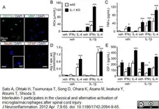
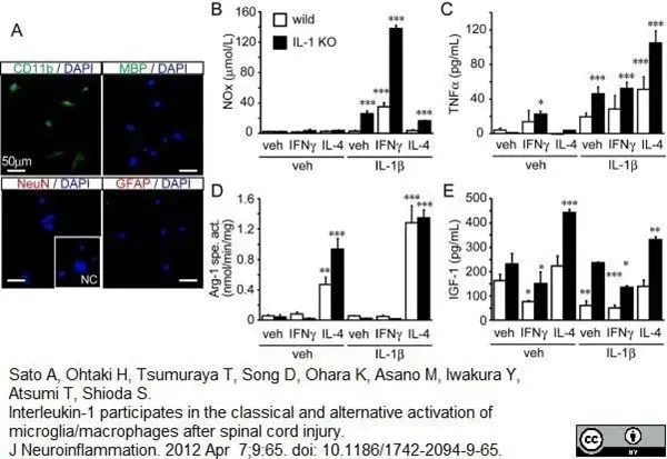
Rat anti mouse CD11b antibody, clone 5C6 (MCA711) used to identify microglia in mouse brain by immunofluorescence.
Image caption:
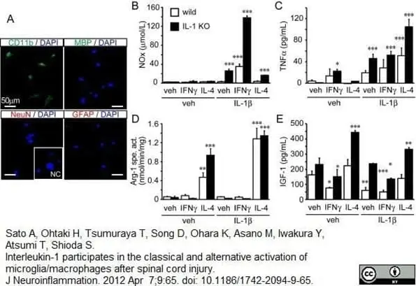
Activation of adult primary microglial cells in wild-type and IL-1 KO mice. (A) Primary microglial cells were obtained from young adult wild-type mice. The cells stain with the microglial marker CD11b, but not with the neuronal and astroglial markers, NeuN and GFAP, respectively. A few cells are stained with the oligodendroglial cell marker, MBP. NC (inset) is the primary antibody-free negative control. The microglial cells (n = 3 each group) were stimulated for 24 hours in the presence of the vehicle alone, or supplemented with IFN&gama; or IL-4 in the presence or absence of IL-1β. Total NO (NOx; B), TNFα (C), arginase specific activity (Arg-1 spe. act.; D) and IGF-1 (E) were determined from the media or cell suspensions. (B) NOx levels increase upon exposure of the cells to IL-1β and in a synergistic manner upon co-treatment of cells with IL-1β and IFNγ, but not when the cotreatment is with IL-4. (C) TNFα levels increase upon exposure of the cells to IFNγ, and further upon co-treatment with IL-1β. Surprisingly, the co-treatment of the cells with IL-4 and IL-1β induced the highest TNFα level among the experimental treatments used. (D) Arg1-specific activity increased significantly upon exposure to IL-4 and further increased when IL-4 and IL-1β were employed together. (E) IGF-1 levels decreased with exposure of the cells to IFNγ and increased in response to IL-4. The response was partially inhibited by cotreatment of the cells with IL-1β. Data are expressed as mean ± SD (n = 3). *: P<0.05, **: P<0.01, ***: P<0.001 compared with the vehicle-treated group in each genotype (one-way ANOVA followed by Dunnett post-hoc test). ANOVA, analysis of variance; IGF-1, insulin-like growth factor.
From: Sato A, Ohtaki H, Tsumuraya T, Song D, Ohara K, Asano M, Iwakura Y, Atsumi T, Shioda S.
Interleukin-1 participates in the classical and alternative activation of microglia/macrophages after spinal cord injury.J Neuroinflammation. 2012 Apr 7;9:65.
This is from an open access article distributed under the terms of a Creative Commons Attribution License.
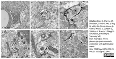
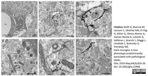
Rat anti Mouse CD11b antibody, clone 5C6 (MCA711) used for the evaluation of CD11b expression on microglia by immuno-electron microscopy.
Image caption:
Phenotypic characterization of the dark microglia, using immunoperoxidase staining in the CA1 lacunosum‐moleculare of stressed CX3CR1 knockout mice (A–C, F), or a nontransgenic control mouse (D, E). A: Focal staining for GFP in a dark microglial cell (dc) from a CX3CR1‐GFP mouse. In contrast, normal microglia display strong and diffuse immunoreactivity for IBA1 throughout their cytoplasm. B, C: Examples of dark microglia staining for the myeloid cell marker CD11b, which forms CR3 involved in phagocytosis, strongly expressed at the plasma membrane of their processes encircling synaptic elements. D, E: Dark microglia's staining for 4D4, a recently discovered marker of homeostatic microglia, at the extremity of their ramified processes. In contrast, the dark microglia do not stain for P2RY12 (F), another marker of homeostatic microglia that is abundant in microglial processes (m). a = astrocytic process, bl = basal lamina, bv = blood vessel, s = dendritic spine, and t = axon terminals. Asterisks show the extracellular space. Scale bars = 1 μm.
From: Bisht K, Sharma KP, Lecours C, Sánchez MG, El Hajj H, Milior G, Olmos-Alonso A, Gómez-Nicola D, Luheshi G, Vallières L, Branchi I, Maggi L, Limatola C, Butovsky O, Tremblay MÈ.
Dark microglia: A new phenotype predominantly associated with pathological states.
Glia. 2016 May;64(5):826-39.
This is from an open access article distributed under the terms of a Creative Commons Attribution License.
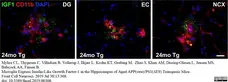
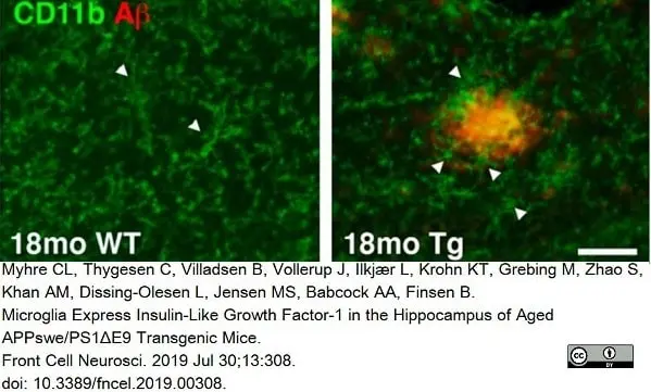
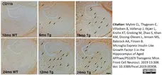
Rat anti Mouse CD11b antibody, clone 5C6 (MCA711) used for the demonstration of CD11b expressing cells in the brains of mice by immunofluorescence on formalin fixed cryosections.
Image caption:
IGF-1 is expressed in neurons and a subset of microglia in APPswe/PS1ΔE9 Tg mice.
(E) Double immunofluorescence for IGF-1 (green) and CD11b (red), and nuclear staining with DAPI (blue). Co-localization of IGF-1 to CD11b+ microglia is visualized by the punctuate yellow staining, whereas the IGF-1+ fiber-like structures in the plaques remain green. Scale-bar 20μm.
From: Myhre CL, Thygesen C, Villadsen B, Vollerup J, Ilkjær L, Krohn KT, Grebing M, Zhao S, Khan AM, Dissing-Olesen L, Jensen MS, Babcock AA, Finsen B.
Microglia Express Insulin-Like Growth Factor-1 in the Hippocampus of Aged APP(swe)/PS1(ΔE9) Transgenic Mice.
Front Cell Neurosci. 2019 Jul 30;13:308.
doi: 10.3389/fncel.2019.00308.
This image is from an open access article distributed under the terms of a Creative Commons Attribution License.
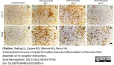
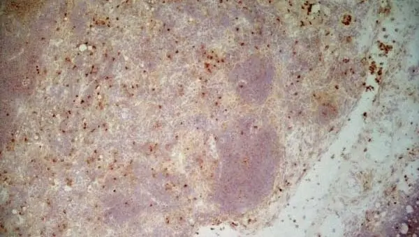
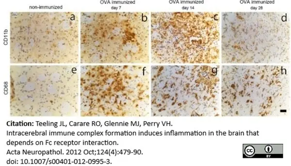
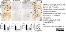
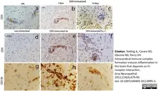
Rat anti Mouse CD11b antibody, clone 5C6 (MCA711) used to demonstrate microglial distribution in murine brain by immunohistohistochemistry on acetone fixed tissue cryosections.
Image caption:
Kinetics of macrophage and microglia activation in the brain after intracerebral injection of OVA. OVA-immunized mice or non-immunized mice received a unilateral injection of OVA into the striatum and brain tissue was assessed for presence of macrophage activation at day 7, day 14 and day 28. Images in the top panel shows CD11b immuno-reactivity in control non-immunized mice (a) and after 7 days (b), 14 days (c) or 28 days (d) in OVA-immunized mice: CD68 immunoreactivity in the lower panel in control (e) and after 7 days (f), 14 days (g) and 28 days (h) in immunized mice. Representative data of n = 3 per treatment and time point is shown. Scale bar 50 μm
From: Teeling JL, Carare RO, Glennie MJ, Perry VH.
Intracerebral immune complex formation induces inflammation in the brain that depends on Fc receptor interaction.
Acta Neuropathol. 2012 Oct;124(4):479-90.
doi: 10.1007/s00401-012-0995-3.
This image is from an open access article distributed under the terms of a Creative Commons Attribution License.
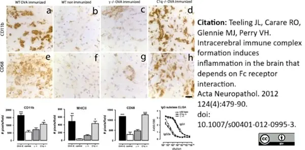
Rat anti Mouse CD11b antibody, clone 5C6 (MCA711) used to demonstrate microglial distribution in murine brain by immunohistohistochemistry on acetone fixed tissue cryosections.
Image caption:
Macrophage and microglia activation in the brain after intracerebral injection of OVA in wild type, C1q−/− mice and Fcγ−/−. Wild type, C1q−/− mice and Fcγ−/− mice on a BALB/c background were immunized against OVA followed by unilateral injection of OVA into the striatum. Macrophage and microglia activation was assessed by immunocytochemistry. Images in the top panel show CD11b immunoreactivity 3 days after intracerebral injection of OVA in OVA-immunized wild type (a), non-immunized wild type (b) or OVA-immunized Fcγ−/− (c) and OVA-immunized C1q−/− (d). Images in the middle panel shows CD68 immunoreactivity 3 days after intracerebral injection of OVA in OVA-immunized wild type (e), non-immunized wild type (f), OVA-immunized Fcγ−/− (g) and OVA-immunized C1q−/− (h). Scale bar 50 μm. In the bottom panel the number of DAB-positive pixels/field (cells and their processes) in the injected hemisphere was quantified as described in “Materials and methods”. Levels of circulating anti-OVA antibodies (IgG1 and IgG2a) were determined by ELISA. Data is expressed as A450/570 values. Closed circle and dotted line represent wild type, closed diamonds represents C1q−/− mice and closed squares represents Fcγ−/− mice. Statistical analysis: One-way ANOVA, Dunnett’s post-test. Data are expressed as mean of n = 3–6 per group. The experiment was performed once
From: Teeling JL, Carare RO, Glennie MJ, Perry VH.
Intracerebral immune complex formation induces inflammation in the brain that depends on Fc receptor interaction.
Acta Neuropathol. 2012 Oct;124(4):479-90.
doi: 10.1007/s00401-012-0995-3.
This image is from an open access article distributed under the terms of a Creative Commons Attribution License.
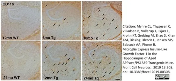
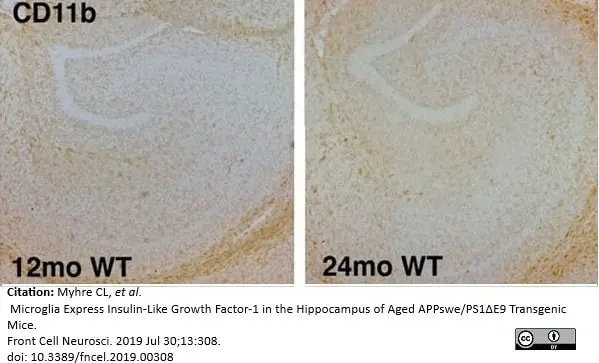
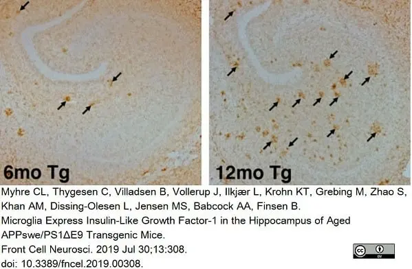
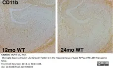
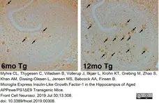
Rat anti Mouse CD11b antibody, clone 5C6 (MCA711) used for the demonstration of CD11b expressing cells in the brains of mice by immunohistochemistry on formalin fixed cryosections.
Image caption:
Altered microglial distribution in the hippocampus of APPswe/PS1ΔE9 Tg mice. (A) Low magnification panels show changes in CD11b immunoreactivity in the hippocampus of 6-, 12-, 18-, and 24-month-old Tg mice. The hippocampus from 12- and 24-month-old WT mice is shown for comparison.
From: Myhre CL, Thygesen C, Villadsen B, Vollerup J, Ilkjær L, Krohn KT, Grebing M, Zhao S, Khan AM, Dissing-Olesen L, Jensen MS, Babcock AA, Finsen B.
Microglia Express Insulin-Like Growth Factor-1 in the Hippocampus of Aged APP(swe)/PS1(ΔE9) Transgenic Mice.
Front Cell Neurosci. 2019 Jul 30;13:308.
doi: 10.3389/fncel.2019.00308.
This image is from an open access article distributed under the terms of a Creative Commons Attribution License.
Rat anti Mouse CD11b antibody, clone 5C6 (MCA711G) used to label microglia in mouse brain by immunohistochemistry on formalin fixed cryosections.
Image caption:
Altered microglial distribution in the hippocampus of APPswe/PS1ΔE9 Tg mice. (A) Low magnification panels show changes in CD11b immunoreactivity in the hippocampus of 6-, 12-, 18-, and 24-month-old Tg mice. The hippocampus from 12- and 24-month-old WT mice is shown for comparison.
From: Myhre CL, Thygesen C, Villadsen B, Vollerup J, Ilkjær L, Krohn KT, Grebing M, Zhao S, Khan AM, Dissing-Olesen L, Jensen MS, Babcock AA, Finsen B.
Microglia Express Insulin-Like Growth Factor-1 in the Hippocampus of Aged APPswe/PS1ΔE9 Transgenic Mice.
Front Cell Neurosci. 2019 Jul 30;13:308.
doi: 10.3389/fncel.2019.00308.
This image is from an open access article distributed under terms of a Creative Commons Attribution License.
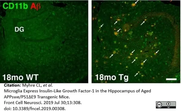
Rat anti Mouse CD11b antibody, clone 5C6 (MCA711G) used to label microglia in mouse brain by immunofluorescence on formalin fixed cryosections.
Image caption:
Combined immunofluorescence staining for CD11b and Aβ shows increased CD11b immunoreactivity near Aβ plaques in low magnification images from 18-month-old Tg mice (B). No plaques are observed in age-matched WT mice (B).
From: Myhre CL, Thygesen C, Villadsen B, Vollerup J, Ilkjær L, Krohn KT, Grebing M, Zhao S, Khan AM, Dissing-Olesen L, Jensen MS, Babcock AA, Finsen B.
Microglia Express Insulin-Like Growth Factor-1 in the Hippocampus of Aged APPswe/PS1ΔE9 Transgenic Mice.
Front Cell Neurosci. 2019 Jul 30;13:308.
doi: 10.3389/fncel.2019.00308.
This image is from an open access article distributed under terms of a Creative Commons Attribution License.
Rat anti Mouse CD11b antibody, clone 5C6 (MCA711G) used to label microglia in mouse brain by immunofluorescence on formalin fixed cryosections.
Image caption:
Arrowheads in (C) point to CD11b+ microglia. Microglial cells are clustered around Aβ plaques in Tg mice, but not in WT mice. The photomicrographs in (C) were both obtained in the dentate molecular layer. DG, dentate gyrus. Scale bar: 500 μm (C).
From: Myhre CL, Thygesen C, Villadsen B, Vollerup J, Ilkjær L, Krohn KT, Grebing M, Zhao S, Khan AM, Dissing-Olesen L, Jensen MS, Babcock AA, Finsen B.
Microglia Express Insulin-Like Growth Factor-1 in the Hippocampus of Aged APPswe/PS1ΔE9 Transgenic Mice.
Front Cell Neurosci. 2019 Jul 30;13:308.
doi: 10.3389/fncel.2019.00308.
This image is from an open access article distributed under terms of a Creative Commons Attribution License.
Data acquired on the ZE5 Cell Analyzer.
Data acquired on the ZE5 Cell Analyzer.
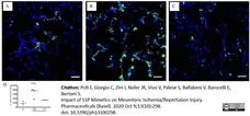
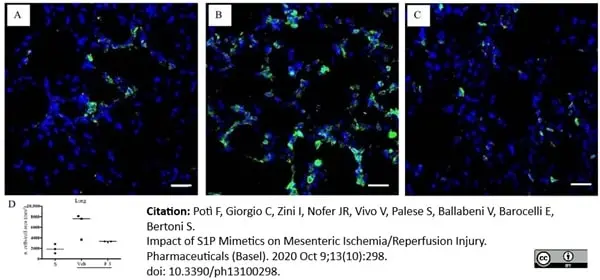
Rat anti Mouse CD11b antibody, clone 5C6 (MCA711) used to stain CD11+ cells in murine pulmonary tissue by immunofluorescence on cryostat sections.
Image caption:
Effects of FTY720 on I/R-induced CD11b+ cells recruitment in the lung. Representative immunofluorescence-stained sections visualizing CD11b+ cells (green fluorescence) in lung tissues excised from S mice (A) and from I/R mice administered with vehicle (B) or FTY720 3 mg/kg i.v. (C). Images were taken at 63× magnification through the oil immersion objective (scale bar: 25 μm). Sections were counterstained with DAPI for nuclear morphology (blue fluorescence). (D) Ratio of the number of CD11b+ cells divided by the cell total area in sections of lung tissues excised from vehicle-treated S mice (n = 3) and I/R mice administered with the vehicle (Veh) (n = 3) or with FTY720 3 mg/kg i.v. (F 3) (n = 3) (horizontal bar at the median value).
From:Potì F, Giorgio C, Zini I, Nofer JR, Vivo V, Palese S, Ballabeni V, Barocelli E, Bertoni S.
Impact of S1P Mimetics on Mesenteric Ischemia/Reperfusion Injury.
Pharmaceuticals (Basel). 2020 Oct 9;13(10):298.
doi: 10.3390/ph13100298.
This image is from an open access article distributed under terms of a Creative Commons Attribution License.
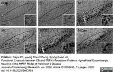
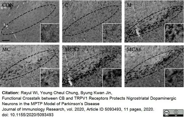
Rat anti Mouse CD11b antibody, clone 5C6 (MCA711) used to demonstrate microglia in mouse brain by immunohistochemistry on cryostat sections.
Image caption:
Crosstalk between CB and TRPV1 inhibits glial activation and expression of proinflammatory cytokines in the SN of MPTP-treated mice in vivo. Mice were intraperitoneally given an injection of PBS or MPTP. All mice intraperitoneally received vehicle as controls or cannabinoid (CB) antagonist (AM251 or AM630; 0.1 mg/kg/day) for 1 day or 3 days at 30 min before capsaicin (C) and 1 hour before MPTP and a single injection of capsaicin (0.5 mg/kg) at 30 min before MPTP.
Photomicrographs of CD11b+ microglia in the SN of MPTP-treated mice in vivo. Mice that received PBS as a control (CON); capsaicin (C); MPTP (M); MPTP and capsaicin (MC); MPTP, capsaicin, and AM251 (MCA2); or MPTP, capsaicin, and AM630 (MCA6) were sacrificed 3 days after the last MPTP injection. Brains were removed and coronal sections (30 μm) were cut using a sliding microtome. Every sixth serial section was selected and immunostained with CD11b antibody for microglia. Insets show higher magnification.
From: Rayul Wi, Young Cheul Chung, Byung Kwan Jin,
Functional Crosstalk between CB and TRPV1 Receptors Protects Nigrostriatal Dopaminergic Neurons in the MPTP Model of Parkinson’s Disease,
J Immunol Res, 2020, Article ID 5093493, 11 pages.
doi: 10.1155/2020/5093493.
This image is from an open access article distributed under terms of a Creative Commons Attribution License.

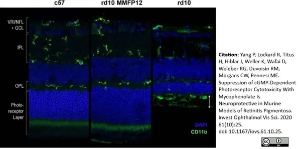
Rat anti Mouse CD11b antibody, clone 5C6 (MCA711) used to label microglia in murine retinal tissue by immunofluorescence.
Image caption:
Effect of MMF on microglia in rd10 mice. (A) Immunostaining of CD11b 1:1000 (green) showed that microglia at P22 in rd10 mice treated with MMF beginning at P12 (MMFP12) appeared to be similar to that of c57 mice; they were in an inactive highly-ramified state within the inner retinal layers, as opposed to naïve rd10 mice where infiltrating microglia/macrophages develop an amoeboid morphology and migrate into the outer retina and subretinal space (white arrows).
From: Yang P, Lockard R, Titus H, Hiblar J, Weller K, Wafai D, Weleber RG, Duvoisin RM, Morgans CW, Pennesi ME.
Suppression of cGMP-Dependent Photoreceptor Cytotoxicity With Mycophenolate Is Neuroprotective in Murine Models of Retinitis Pigmentosa.
Invest Ophthalmol Vis Sci. 2020 Aug 3;61(10):25.
doi: 10.1167/iovs.61.10.25.
This image is from an open access article distributed under terms of a Creative Commons Attribution License.
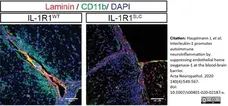
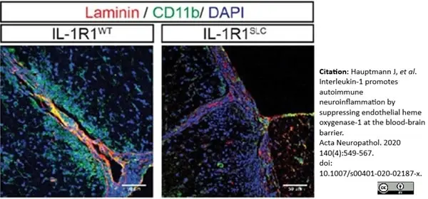
Rat anti Mouse CD11b antibody, clone 5C6 (MCA711) used to show CD11+ myeloid cells in CNS parenchyma by immunofluorescence.
Image caption:
BBB-EC IL-1 signaling promotes leukocyte migration.
Immunofluorescence analysis of spinal cord tissue at EAE onset showing CD11b+ myeloid cells infiltrating the CNS parenchyma. Spinal cord sections were stained for CD11b (green, in f, scale bar = 50 μm) and DAPI (blue) together with pan-laminin staining (red).
From: Hauptmann J, et al. Interleukin-1 promotes autoimmune neuroinflammation by suppressing endothelial heme oxygenase-1 at the blood-brain barrier.
Acta Neuropathol. 2020 Oct;140(4):549-67.
doi: 10.1007/s00401-020-02187-x.
This image is from an open access article distributed under terms of a Creative Commons Attribution License.
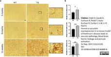
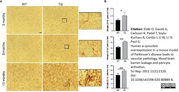
Rat anti Mouse CD11b antibody, clone 5C6 (MCA711) used to identify microglia in murine dorsal striatum by immunohistochemistry on formalin fixed cryosections.
Image caption:
CD11b+ microglia in the dorsolateral striatum of WT and TG mice. (a) Images showing CD11b+ microglia in WT and TG mice at 3, 8, and 13 months of age. Zoomed images illustrating CD11b+ microglia morphology. (b) Quantification of CD11b+ cell number per view field at 3 months (n= 4 WT, 4 TG); 8 months (n= 3 WT, 2 TG); and 13 months of age (n= 5 WT, 4 TG). Two-tailed student’s t-test: *p<0.05, p**<0.01. Scale bar: 20 μm.
From: Elabi O, Gaceb A, Carlsson R, Padel T, Soylu-Kucharz R, Cortijo I, Li W, Li JY, Paul G. Human α-synuclein overexpression in a mouse model of Parkinson's disease leads to vascular pathology, blood brain barrier leakage and pericyte activation.
Sci Rep. 2021 Jan 13;11(1):1120.
doi: 10.1038/s41598-020-80889-8
This image is from an open access article distributed under terms of a Creative Commons Attribution License.
Rat anti Mouse CD11b antibody, clone 5C6 (MCA711) used for the demonstration of microglia in murine brain by immunohistochemistry on acetone fixed cryosections.
Image caption:
T-cell recruitment after immune complex formation. OVA-immunized mice received a unilateral injection of OVA into the striatum and brain tissue was assessed for presence of CD3-positive T cells at 24 h (a), day 7 (b) and day 14 (c) after OVA injection. Non-immunized mice were used as control (d, g). OVA-immunized wild type (e, h) and Fcγ−/− (f, I) mice received a unilateral injection of OVA into the striatum and tissue was analysed for CD3 (d, e, f) or CD11b (g, h, I) immunoreactivity at 7 days. Representative data of n = 2–3 per time point are shown. Scale bar 100μm
From: Teeling JL, Carare RO, Glennie MJ, Perry VH.
Intracerebral immune complex formation induces inflammation in the brain that depends on Fc receptor interaction.
Acta Neuropathol. 2012 Oct;124(4):479-90.
doi: 10.1007/s00401-012-0995-3.
This image is from an open access article distributed under the terms of a Creative Commons Attribution License.
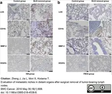
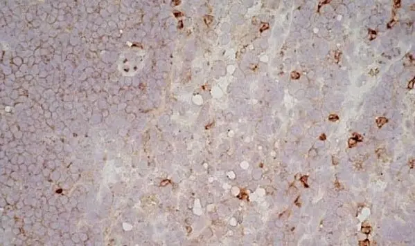
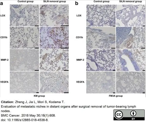
Rat anti Mouse CD11b antibody, clone 5C6 (MCA711G) used to identify CD11b expressing cells in murine lung by immunohistochemistry on formalin fixed, paraffin embedded tissue sections.
Image caption:
The changes of metastatic niche in lung samples. The expression of LOX, CD11b, MMP-2 and VEGFA were determined using IHC staining in the KM (a) and FM3A (b) groups. Bar: 50 μm
From: Zheng J, Jia L, Mori S, Kodama T.
Evaluation of metastatic niches in distant organs after surgical removal of tumor-bearing lymph nodes.
BMC Cancer. 2018 May 30;18(1):608.
doi: 10.1186/s12885-018-4538-8.
This image is from an open access article distributed under terms of a Creative Commons Attribution License.
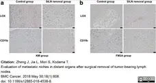
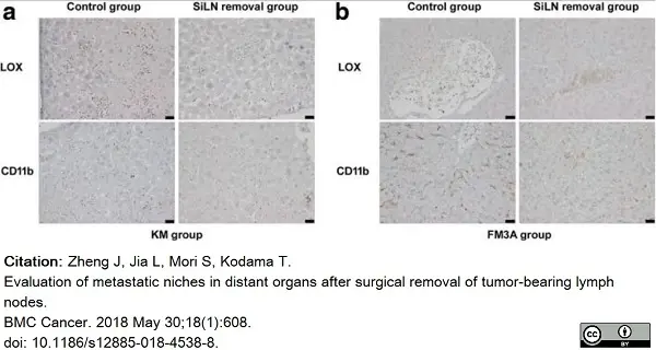
Rat anti Mouse CD11b antibody, clone 5C6 (MCA711G) used to identify CD11b expressing cells in murine liver by immunohistochemistry on formalin fixed, paraffin embedded tissue sections.
Image caption:
The changes of metastatic niche in liver samples. The expression of LOX and CD11b were observed using IHC staining in the KM (a) and FM3A (b) groups. Bar: 20 μm
From: Zheng J, Jia L, Mori S, Kodama T.
Evaluation of metastatic niches in distant organs after surgical removal of tumor-bearing lymph nodes.
BMC Cancer. 2018 May 30;18(1):608.
doi: 10.1186/s12885-018-4538-8.
This image is from an open access article distributed under terms of a Creative Commons Attribution License.
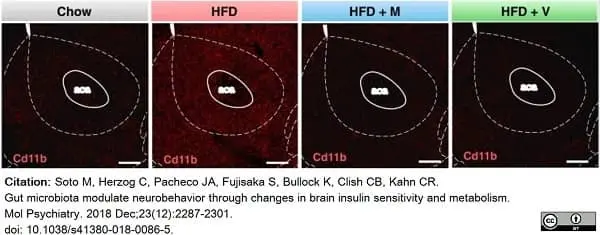
Rat anti Mouse CD11b antibody, clone 5C6 (MCA711G) used to inflammatory infiltrate in murine brain by immunofluorescence.
Image caption:
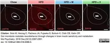
Representative images of the Nacc from chow and HFD-fed mice treated or not with antibiotics, stained with Cd11b antibody (red). Scale bars, 200 μm. C chow, H HFD, M HFD + metronidazole, V HFD + vancomycin.
From: Soto M, Herzog C, Pacheco JA, Fujisaka S, Bullock K, Clish CB, Kahn CR.
Gut microbiota modulate neurobehavior through changes in brain insulin sensitivity and metabolism.
Mol Psychiatry. 2018 Dec;23(12):2287-2301.
doi: 10.1038/s41380-018-0086-5.
This image is from an open access article distributed under terms of a Creative Commons Attribution License.
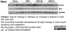
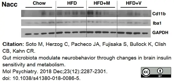
Rat anti Mouse CD11b antibody, clone 5C6 (MCA711G) used to inflammatory infiltrate in murine brain by western blotting.
Image caption:
Antibiotic treatment ameliorates HFD-induced inflammation in the brain. (a,b) Representative western blot of Cd11b and Iba1.
From: Soto M, Herzog C, Pacheco JA, Fujisaka S, Bullock K, Clish CB, Kahn CR.
Gut microbiota modulate neurobehavior through changes in brain insulin sensitivity and metabolism.
Mol Psychiatry. 2018 Dec;23(12):2287-2301.
doi: 10.1038/s41380-018-0086-5.
This image is from an open access article distributed under terms of a Creative Commons Attribution License.
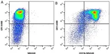
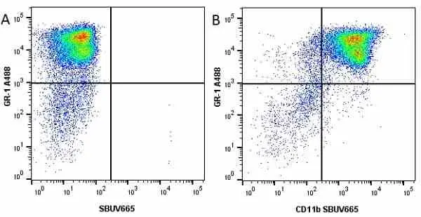
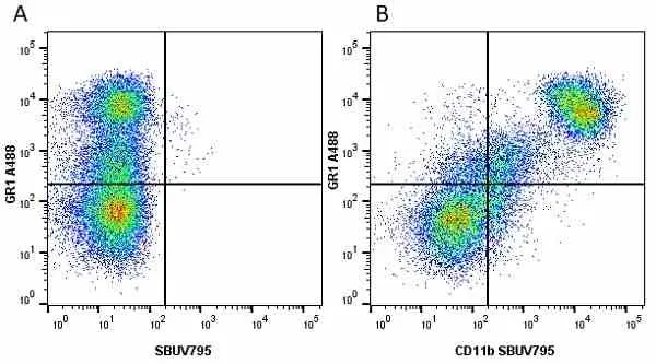
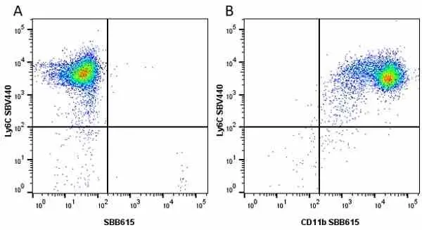
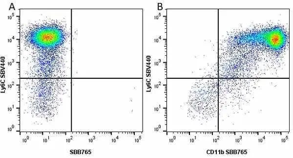
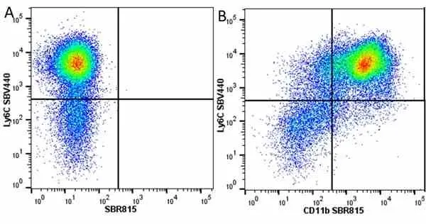
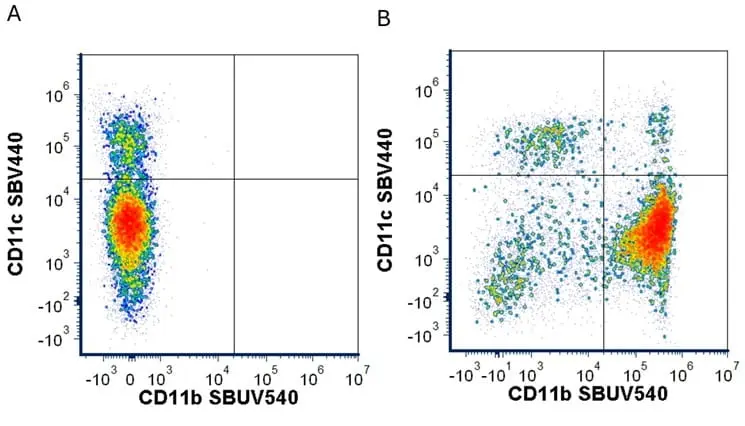
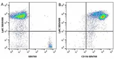
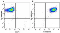
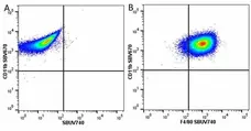
Figure B. Alexa Fluor® 488 conjugated Rat anti Mouse GR1 antibody, clone RB6-8C5 (MCA2387A488) and StarBright Violet 440 conjugated Rat anti Mouse CD11b antibody, clone 5C6 (MCA711SBV440). All experiments performed on mouse bone marrow gated on live single cells, in the presence of 10% mouse serum.
Data acquired on the ZE5 Cell Analyzer.
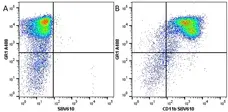
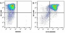
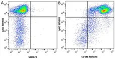
Figure B. Alexa Fluor® 488 conjugated Rat anti mouse GR1 antibody, clone RB6-8C5 (MCA2387A488) and StarBright Violet 610 conjugated Rat anti Mouse CD11b antibody, clone 5C6 (MCA711SBV610). All experiments performed on murine bone marrow gated on live single cells, in the presence of 10% mouse serum.
Data acquired on the ZE5 Cell Analyzer.
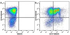
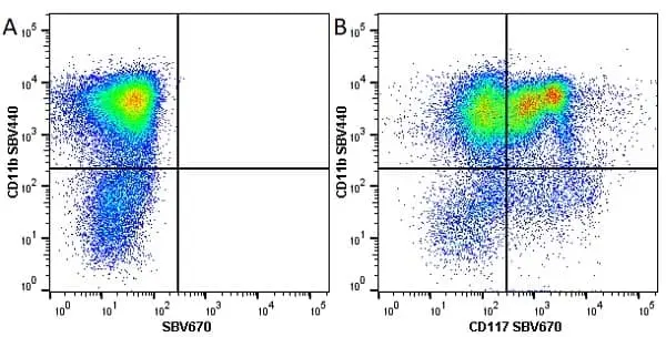
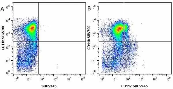
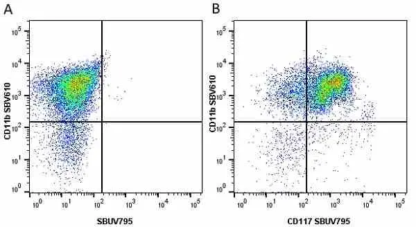
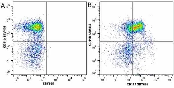
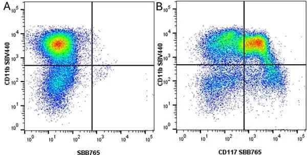
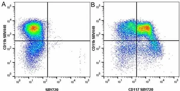
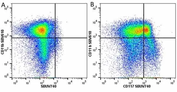
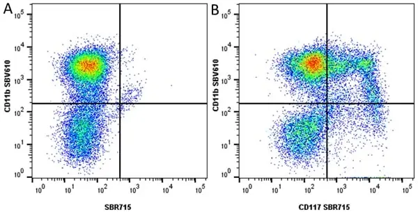
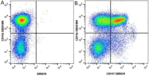
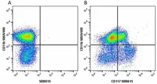
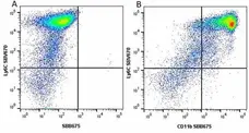
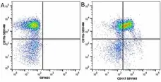
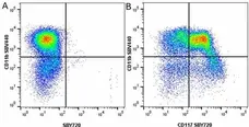
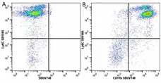
Figure B. StarBright Violet 440 conjugated Rat anti Mouse CD11b antibody, clone 5C6 (MCA711SBV440) and StarBright Violet 670 conjugated Rat anti Mouse CD117 antibody, clone 2B8 (MCA1365SBV670). All experiments performed on red cell lysed murine bone marrow gated on live single cells, in the presence of 10% mouse serum.
Data acquired on the ZE5 Cell Analyzer.
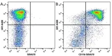
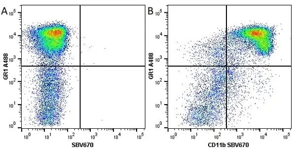
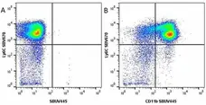
Figure B. Alexa Fluor® 488 conjugated Rat anti Mouse Gr-1 antibody, clone RB6-8C5 (MCA2387A488) and StarBright Violet 670 conjugated Rat anti Mouse CD11b antibody, clone 5C6 (MCA711SBV670). All experiments performed on red cell lysed murine bone marrow gated on live single cells, in the presence of 10% mouse serum.
Data acquired on the ZE5 Cell Analyzer

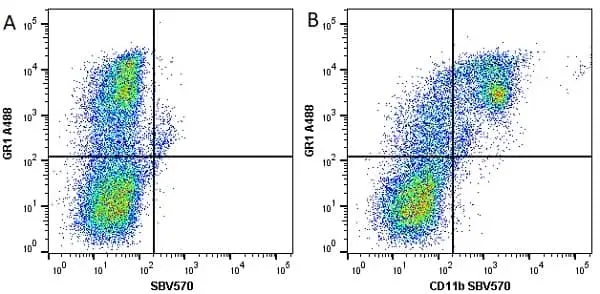
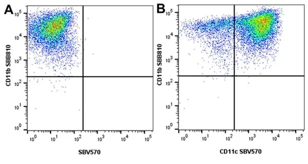
Figure B. Alexa Fluor® 488 conjugated Rat anti Mouse GR1 antibody, clone RB6-8C5 (MCA2387A488) and StarBright Violet 570 conjugated Rat anti mouse CD11b antibody, clone 5C6 (MCA711SBV570). All experiments performed on murine bone marrow cells gated on live single cells, in the presence of 10% mouse serum.
Data acquired on the ZE5 Cell Analyzer.
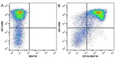
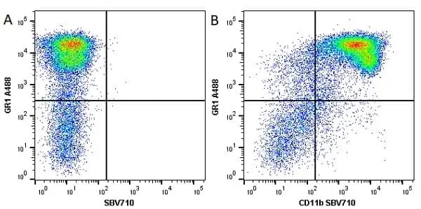
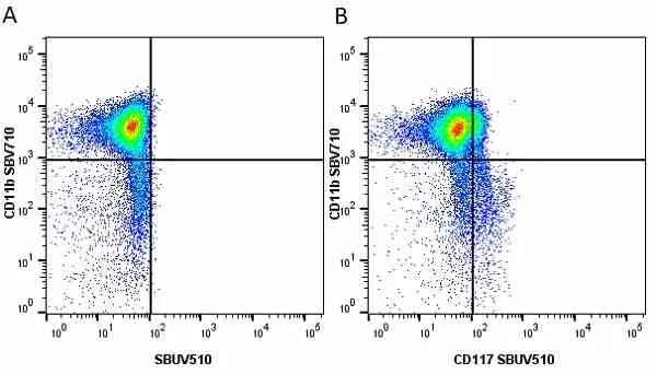
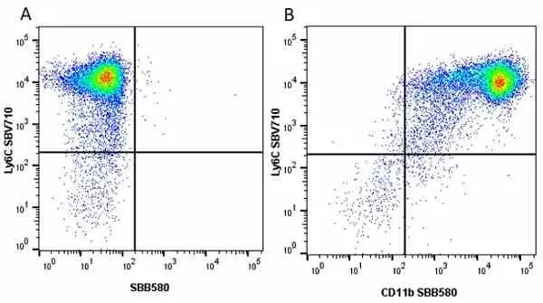
Figure B. Alexa Fluor® 488 conjugated Rat anti Mouse Gr-1 antibody, clone RB6-8C5 (MCA2387A488) and StarBright Violet 710 conjugated Rat anti Mouse CD11b antibody, clone 5C6 (MCA711SBV710). All experiments performed on murine bone marrow gated on live single cells, in the presence of 10% mouse serum.
Data acquired on the ZE5 Cell Analyzer.
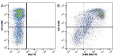
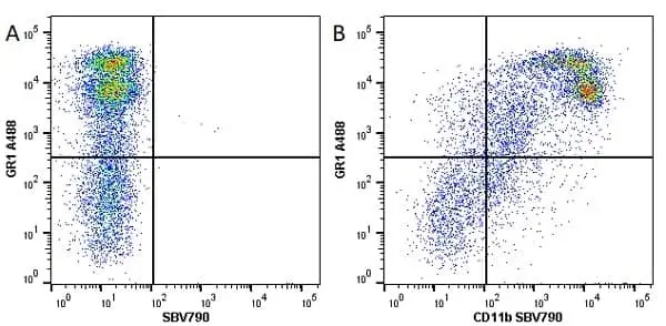
Figure B. Alexa Fluor® 488 conjugated Rat anti Mouse GR1 antibody, clone RB6-8C5 (MCA2387A488) and StarBright Violet 790 conjugated Rat anti Mouse CD11b antibody, clone 5C6 (MCA711SBV790). All experiments performed on murine bone marrow gated on live single cells, in the presence of 10% mouse serum. Data acquired on the ZE5 Cell Analyzer.
Data acquired on the ZE5 Cell Analyzer.
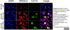
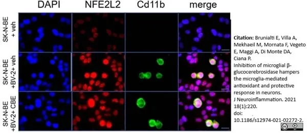
Rat anti Mouse CD11b antibody, clone 5C6 (MCA711) used to label BV-2 murine microglial cells by immunofluorescence.
Image caption:
Nuclear localization of NFE2L2 in neuronal-microglia culture treated with CBE. Representative immunocytochemistry analysis of SK-N-BE and BV-2 cell lines in monoculture and co-culture, treated with 200 μM CBE or vehicle for 48 hours; cells were co-stained with anti- NFE2L2 (red) and anti-CD11b antibodies (green), and with DAPI (blue).
From: Brunialti E, Villa A, Mekhaeil M, Mornata F, Vegeto E, Maggi A, Di Monte DA, Ciana P.
Inhibition of microglial β-glucocerebrosidase hampers the microglia-mediated antioxidant and protective response in neurons.
J Neuroinflammation. 2021 Sep 22;18(1):220.
doi: 10.1186/s12974-021-02272-2.
This image is from an open access article distributed under terms of a Creative Commons Attribution License.
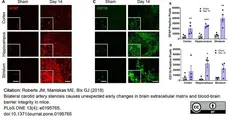
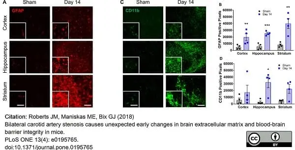
Rat anti Mouse CD11b antibody, clone 5C6 (MCA711) used to identify CD11b positive cells in mouse brain tissues by immunofluorescence.
Image caption:
Changes in glial cells are observed in multiple regions of the brain following BCAS.
A) Representative images of GFAP staining (red) within the cortex, hippocampus and striatum of sham or BCAS-treated (14 days) mice. Scale bar = 100μm. Inset images are a magnified portion to show detail. Scale bar = 300μm. B) Quantification of GFAP-positive pixels. N = 4 **p<0.01, ***p<0.001 C) Representative images of CD11b staining (green) within the cortex, hippocampus and striatum of sham or BCAS-treated (14 days) mice. Scale bar = 100μm. Inset images are a magnified portion to show detail. Scale bar = 300μm. D) Quantification of CD11b-positive pixels. N = 4 *p<0.05.
From: Citation: Roberts JM, Maniskas ME, Bix GJ (2018)
Bilateral carotid artery stenosis causes unexpected early changes in brain extracellular matrix and blood-brain barrier integrity in mice.
PLoS ONE 13(4): e0195765.
doi: 10.1371/journal.pone.0195765
This image is from an open access article distributed under terms of a Creative Commons Attribution License.
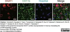
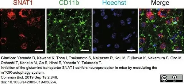
Rat anti Mouse CD11b antibody, clone 5C6 (MCA711) used to label microglia in mouse brain by immunbofluorescence.
Image caption:
Analysis of Slc38a1 expression in mouse tissues.
Identification of SNAT1-expressing cells in the cerebral cortex. Double-immunohistochemical staining using antibodies against SNAT1 and CD11b (microglial marker). Nuclei were counterstained with Hoechst 33342. Scale bars = 20 μm.
From: Yamada D, Kawabe K, Tosa I, Tsukamoto S, Nakazato R, Kou M, Fujikawa K, Nakamura S, Ono M, Oohashi T, Kaneko M, Go S, Hinoi E, Yoneda Y, Takarada T.
Inhibition of the glutamine transporter SNAT1 confers neuroprotection in mice by modulating the mTOR-autophagy system.
Commun Biol. 2019 Sep 18;2:346.
doi: 10.1038/s42003-019-0582-4.
This image is from an open access article distributed under terms of a Creative Commons Attribution License.
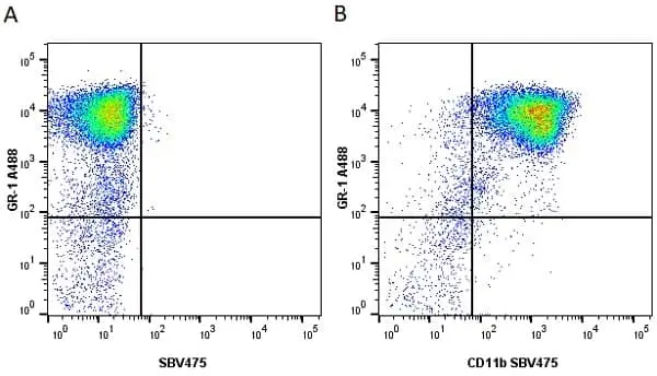
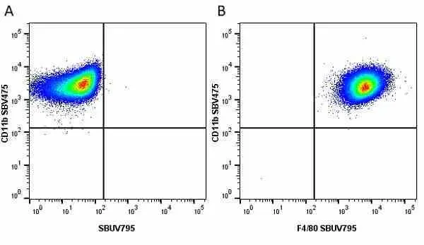
Figure B. Alexa Fluor® 488 conjugated Rat anti Mouse Gr-1 antibody, clone RB6-8C5 (MCA2387A488) and StarBright Violet 475 conjugated Rat anti Mouse CD11b antibody, clone 5C6 (MCA711SBV475). All experiments performed on red cell lysed murine bone marrow cells gated on live single cells, in the presence of 10% mouse serum.
Data acquired on the ZE5 Cell Analyzer.
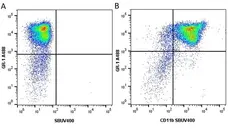
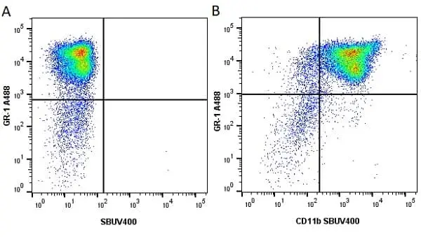
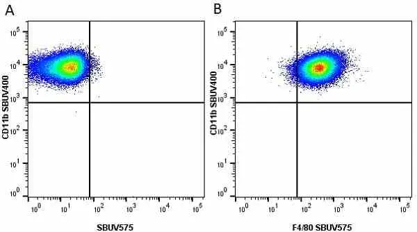
Figure B. Alexa Fluor® 488 conjugated Rat anti Mouse Gr-1 antibody, clone RB6-8C5 (MCA2387A488) and StarBright UltraViolet 400 conjugated Rat anti Mouse CD11b (MCA711SBUV400). . All experiments performed on red cell lysed murine bone marrow cells gated on live single cells, in the presence of 10% mouse serum.
Data acquired on the ZE5 Cell Analyzer.
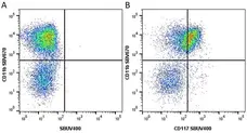
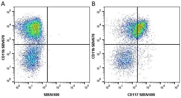
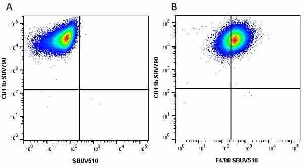
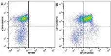
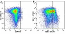
Figure B. StarBright Violet 670 conjugated Rat anti Mouse CD11b antibody, clone 5C6 (MCA711SBV670) and StarBright UltraViolet 400 conjugated Rat anti Human CD117 antibody, clone 2B8 (MCA1365SBUV400). All experiments performed on murine bone marrow gated on live single cells, in the presence of 10% mouse serum.
Data acquired on the ZE5 Cell Analyzer.
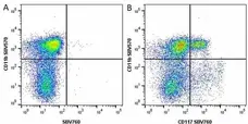
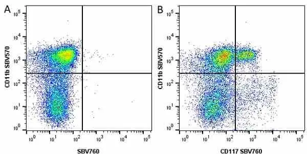
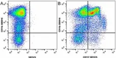
Figure B. StarBright Violet 570 conjugated Rat anti Mouse CD11b antibody, clone 5C6 (MCA711SBV670) and StarBright UltraViolet 400 conjugated Rat anti Human CD117 antibody, clone 2B8 (MCA1365SBV760). All experiments performed on murine bone marrow gated on live single cells, in the presence of 10% mouse serum.
Data acquired on the ZE5 Cell Analyzer.
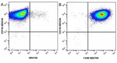
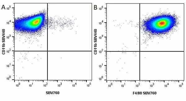
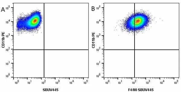
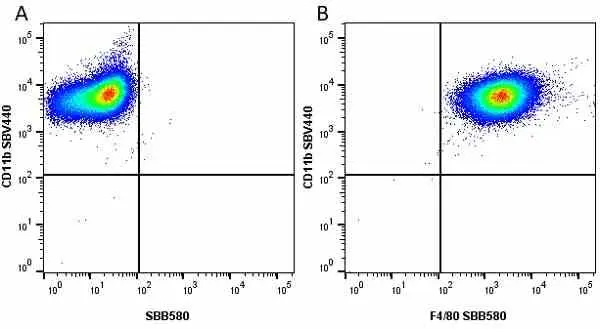
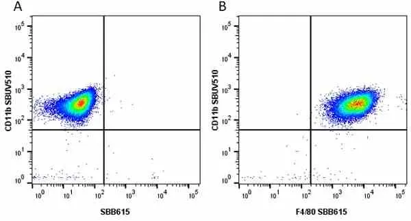
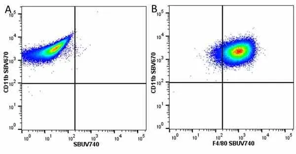
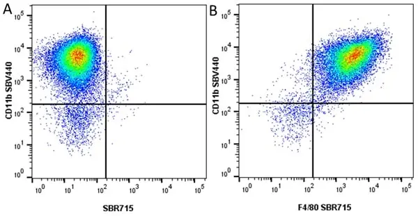
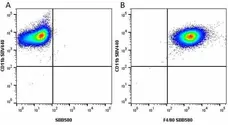
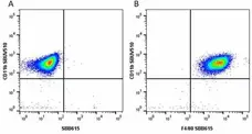
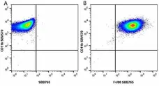
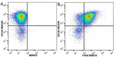
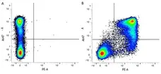
Figure B. StarBright Violet 440 conjugated Rat anti Mouse CD11b antibody, clone 5C6 (MCA711SBV440) and StarBright Violet 760 conjugated Rat anti Mouse F4/80 (MCA497SBV760). All experiments performed on J774.2 cell gated on live single cells, in the presence of 10% mouse serum.
Data acquired on the ZE5 Cell Analyzer.


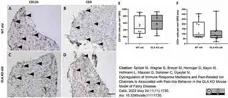
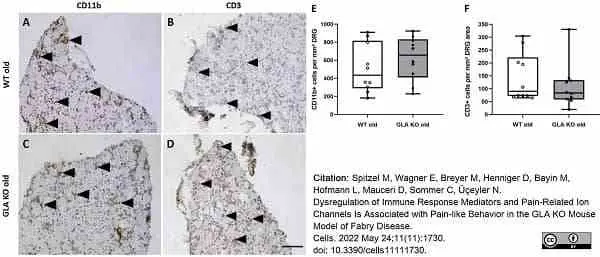
Rat anti Mouse CD11b antibody, clone 5C6 (MCA711) used to label imfiltrating macrophages in the dorsal root ganglia of mice by immunohistochemistry in cryosections.
Image caption:
Macrophage and T-cell infiltration in DRG of old WT and GLA KO mice. (A–D) Representative photomicrographs of CD11b+ macrophages and CD3+ T-cells (arrowheads) in DRG of old WT ((A,B), respectively) and old GLA KO mice ((C,D), respectively). (E) Quantification of CD11b+ macrophages per mm2 DRG area in old WT (◯, n = 10) and old GLA KO mice (●, n = 10). (F) Quantification of CD3+ T-cells per mm² DRG area in old WT ◯, n = 10) and old GLA KO mice (●, n = 10). Abbreviations: CD = Cluster of Differentiation; DRG = dorsal root ganglia; GLA KO = alpha-galactosidase A knockout; WT = wildtype. Scale bar: 100 μm.
From: Spitzel M, Wagner E, Breyer M, Henniger D, Bayin M, Hofmann L, Mauceri D, Sommer C, Üçeyler N.
Dysregulation of Immune Response Mediators and Pain-Related Ion Channels Is Associated with Pain-like Behavior in the GLA KO Mouse Model of Fabry Disease.
Cells. 2022 May 24;11(11):1730.
doi: 10.3390/cells11111730.
This image is from an open access article distributed under terms of a Creative Commons Attribution License.
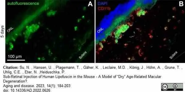
Rat anti Mouse CD11b antibody, clone 5C6 (MCA711) used to label microglia in murine retina by immunofluorescence.
Image caption:
Microglial cells have phagocytosed LF. (A) Autofluorescence of a retinal cryosection five days after subretinal injection of LF. Aggregates of autofluorescent material in the subretinal space are clearly visible. (B) The same site of the sample, after immunostaining against CD11b using a red fluorescent dye. Co-localisation of LF autofluorescence and CD11b can be clearly seen.
From: Nan Su , Uwe Hansen , Tanja Plagemann , Karin Gäher , M. Dominik Leclaire , Jeannette König , Annika Höhn , Tilman Grune , Constantin E. Uhlig , Nicole Eter , Peter Heiduschka.
Sub-Retinal Injection of Human Lipofuscin in the Mouse - A Model of “Dry” Age-Related Macular Degeneration?.
Aging and disease. 2023, 14(1): 184-203.
doi: 10.14336/AD.2022.0626
This image is from an open access article distributed under terms of a Creative Commons Attribution License.


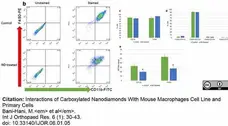
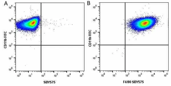
Fitc conjugated Rat anti Mouse CD11b antibody, clone 5C6 (MCA711F) used to label macrophages and evaluate CD11b expression by flow cytometry.
Image caption:
Effects of ND on macrophages differentiation. (b) Representative graphs from FACS analysis showing the percent of cells expressing macrophage surface markers, FITC-CD11b and PE-F4/80. (c) Percent of cells expressing CD11b or F4/80. (d) Percent of each cell population based on their surface markers. (e) Effects of ND on the number of surface markers expressed per cell. Results represent fold change in MFI as compared to unstained control from four independent experiments. * p<0.05.
From: Interactions of Carboxylated Nanodiamonds With Mouse Macrophages Cell Line and Primary Cells
Bani-Hani, M. et al.
Int J Orthopaed Res. 6 (1): 30-43.
Doi: 10.33140/IJOR.06.01.05
This image is from an open access article distributed under terms of a Creative Commons Attribution License.
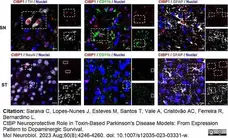
Rat anti Mouse CD11b antibody, clone 5C6 (MCA711G) used to stain microglia in mouse brain by immunofluorescence.
Image caption:
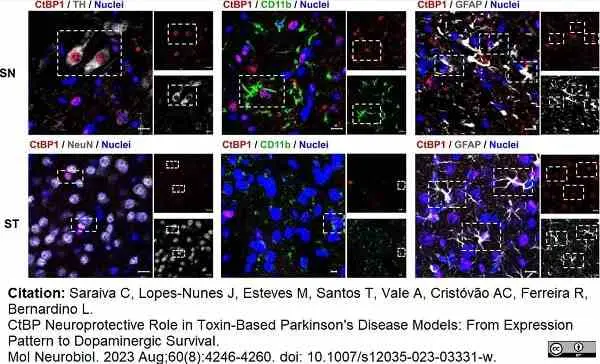
CtBP1 cellular and subcellular localization in the substantia nigra and striatum of young adult mice in vivo. Representative images of the cellular and subcellular localization of CtBP1 in the substantia nigra (SN) and striatum (ST) of young adult mice. CtBP1 (red) is expressed in tyrosine hydroxylase (TH) neurons (dopaminergic marker, top left panel – SN, gray), CD11b cells (microglial marker, middle panels, green), GFAP cells (astrocytic marker, right panels, gray), and mature neurons (NeuN, lower left panel – ST, gray) in both the SN and ST. Nuclei are stained in blue. Dashed rectangles highlight double-positive cells. Scale bar 20 μm
From: Saraiva C, Lopes-Nunes J, Esteves M, Santos T, Vale A, Cristóvão AC, Ferreira R, Bernardino L.
CtBP Neuroprotective Role in Toxin-Based Parkinson's Disease Models: From Expression Pattern to Dopaminergic Survival.
Mol Neurobiol. 2023 Aug;60(8):4246-4260.
doi: 10.1007/s12035-023-03331-w..
This image is from an open access article distributed under terms of a Creative Commons Attribution License.
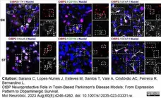
Rat anti Mouse CD11b antibody, clone 5C6 (MCA711G) used to stain microglia in mouse brain by immunofluorescence.
Image caption:
CtBP2 cellular and subcellular localization in the substantia nigra and striatum of young adult mice in vivo. Representative images of the cellular and subcellular localization of CtBP2 in the substantia nigra (SN) and striatum (ST) of young adult mice. CtBP2 (red) is expressed in tyrosine hydroxylase (TH) neurons (dopaminergic marker, top left panel – SN, gray), CD11b cells (microglial marker, middle panels, green), GFAP cells (astrocytic marker, right panels, gray), and mature neurons (NeuN, lower left panel – ST, gray) in both the SN and ST. Nuclei are stained in blue. Dashed rectangles highlight double-positive cells. Scale bar 20 μm
From: Saraiva C, Lopes-Nunes J, Esteves M, Santos T, Vale A, Cristóvão AC, Ferreira R, Bernardino L.
CtBP Neuroprotective Role in Toxin-Based Parkinson's Disease Models: From Expression Pattern to Dopaminergic Survival.
Mol Neurobiol. 2023 Aug;60(8):4246-4260.
doi: 10.1007/s12035-023-03331-w..
This image is from an open access article distributed under terms of a Creative Commons Attribution License.
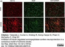
Rat anti Mouse CD11b antibody, clone 5C6 (MCA711) used to label cells in mouse brain by immunofluorescence.
Image caption:
Upregulation of IRAP expression in CD11b and GFAP positive cells following stroke. Representative immunofluorescent images demonstrating (A) upregulation of IRAP in activated CD11b positive cells within the stroked cortical core.
From: Telianidis J, Hunter A, Widdop R, Kemp-Harper B, Pham V, McCarthy C, Chai SY.
Inhibition of insulin-regulated aminopeptidase confers neuroprotection in a conscious model of ischemic stroke.
Sci Rep. 2023 Nov 13;13(1):19722.
doi: 10.1038/s41598-023-46072-5.
This image is from an open access article distributed under terms of a Creative Commons Attribution License.
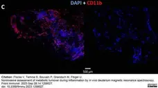
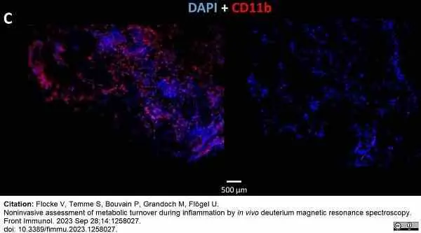
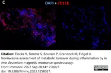
Rat anti Mouse CD11b antibody, clone 5C6 (MCA711) used to label inflammatory cells from matrigel puugs implante in the necks of mice.
Image caption:
In vivo and ex vivo validation of the experimental inflammatory focus. (C) Immunohistochemistry of excised plugs confirmed the in vivo findings in (B).
From: Flocke V, Temme S, Bouvain P, Grandoch M, Flögel U.
Noninvasive assessment of metabolic turnover during inflammation by in vivo deuterium magnetic resonance spectroscopy.
Front Immunol. 2023 Sep 28;14:1258027.
doi: 10.3389/fimmu.2023.1258027.
This image is from an open access article distributed under terms of a Creative Commons Attribution License.

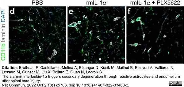
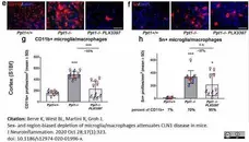
Rat anti Mouse CD11b antibody, clone 5C6 (MCA711) used to label microglia, macrophages and neutrophils in murine spinal cord by immunofluorescence.
Image caption:
Microglia alleviate IL-1α-mediated neuroinflammation and oligodendrocyte loss independently of their expression of IL-1R1.
d–f Confocal images showing the presence of CD11b+ cells (green cells in d–f; CD11b stains microglia, macrophages and neutrophils) in the spinal cord of C57BL/6 mice injected with either PBS (d), rmIL-1α (e) or rmIL-1α + PLX5622 (f) at 24 h post-injection.
From: Bretheau F, Castellanos-Molina A, Bélanger D, Kusik M, Mailhot B, Boisvert A, Vallières N, Lessard M, Gunzer M, Liu X, Boilard É, Quan N, Lacroix S.
The alarmin interleukin-1α triggers secondary degeneration through reactive astrocytes and endothelium after spinal cord injury.
Nat Commun. 2022 Oct 2;13 (1): 5786.
doi: 10.1038/s41467-022-33463-x..
This image is from an open access article distributed under terms of a Creative Commons Attribution License.
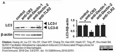
Rat anti Mouse CD11b antibody, clone 5C6 (MCA711EL) used to treat mouse macrophages prior to western blotting analysis.
Image caption:
Dectin-1-mediated recognition is crucial for H. capsulatum-induced LC3-II formation.
Macrophages from WT mice were treated with blocking antibodies against CR3, Dectin-1, Dectin-2, or TLR2 (10 μg/ml each) (A) or laminarin at indicated concentrations (F) for 1 h prior to stimulation with H. capsulatum (MOI = 5) for 1 h. Cell lysates were extracted and analyzed by Western blotting. (B) Macrophages from WT, Clec7a−/−, Clec4n−/− and Itgam−/− mice were stimulated with or without (0 min) H. capsulatum (MOI = 5) for 30 and 60 min. Cell lysates were subjected to Western blotting. Data shown in the right hand panel are relative intensity of LC3-II normalized against the corresponding β-actin, mean ± SEM are shown
From: Huang JH, Liu CY, Wu SY, Chen WY, Chang TH, Kan HW, Hsieh ST, Ting JP, Wu-Hsieh BA.
NLRX1 Facilitates Histoplasma capsulatum-Induced LC3-Associated Phagocytosis for Cytokine Production in Macrophages.
Front Immunol. 2018 Dec 3;9:2761.
doi: 10.3389/fimmu.2018.02761.
This image is from an open access article distributed under terms of a Creative Commons Attribution License.

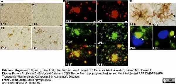
Rat anti Mouse CD11b antibody, clone 5C6 (MCA711) used to label CD11b expressing cells in murine brain by immunohistochemistry on cryosections and by immunofluorescence.
Image caption:
CD11b+ cells clustering around Aβ plaques display Aβ-containing vacuole-like structures. (A) IHC staining showing CD11b+ cells in the neocortex of PBS- and LPS-injected Tg and Wt mice. Cells with altered morphology and with some forming clusters are more abundant in Tg mice compared to Wt mice. (B) Double immunofluorescence staining showing co-localization (yellow) of 6E10+ Aβ plaques (red) and CD11b+ cells (green) in both PBS- and LPS-injected Tg mice. (C) CD11b+ cells with vacuole-like structures are observed in both PBS- and LPS-injected Tg mice, and double immunofluorescence shows some vacuoles to be Aβ+ (yellow). 25 μm (A), 20 μm (B), 50 μm (C, top), and 10 μm (bottom).
From:Thygesen C, Ilkjær L, Kempf SJ, Hemdrup AL, von Linstow CU, Babcock AA, Darvesh S, Larsen MR, Finsen B.
Diverse Protein Profiles in CNS Myeloid Cells and CNS Tissue From Lipopolysaccharide- and Vehicle-Injected APPSWE/PS1ΔE9 Transgenic Mice Implicate Cathepsin Z in Alzheimer's Disease.
Front Cell Neurosci. 2018 Nov 6;12:397.
doi: 10.3389/fncel.2018.00397..
This image is from an open access article distributed under terms of a Creative Commons Attribution License.
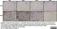
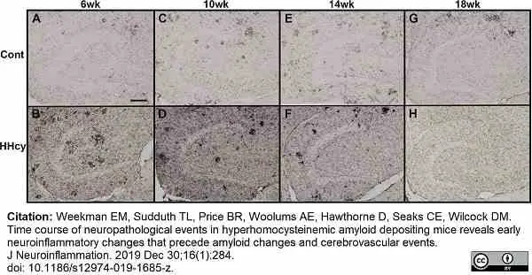
Rat anti Mouse CD11b antibody, clone 5C6 (MCA711) used to label CD11b expressing cells in murine dentate gyrus by immunohistochemistry on cryosections.
Image caption:
Neuroinflammatory changes begin at 6 weeks on the HHcy diet in the APP/PS1 mice. a–h Representative images of CD11b staining in the dentate gyrus of APP/PS1 mice on control diet or HHcy diet for 6, 10, 14, or 18 weeks are shown. Scale bar in A = 100 μm.
From:Weekman EM, Sudduth TL, Price BR, Woolums AE, Hawthorne D, Seaks CE, Wilcock DM. Time course of neuropathological events in hyperhomocysteinemic amyloid depositing mice reveals early neuroinflammatory changes that precede amyloid changes and cerebrovascular events.
J Neuroinflammation. 2019 Dec 30;16(1):284.
doi: 10.1186/s12974-019-1685-z..
This image is from an open access article distributed under terms of a Creative Commons Attribution License.
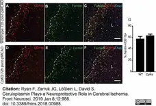
Rat anti Mouse CD11b antibody, clone 5C6 (MCA711) used to label microglia in mouse brain by immunofluorescence.
Image caption:
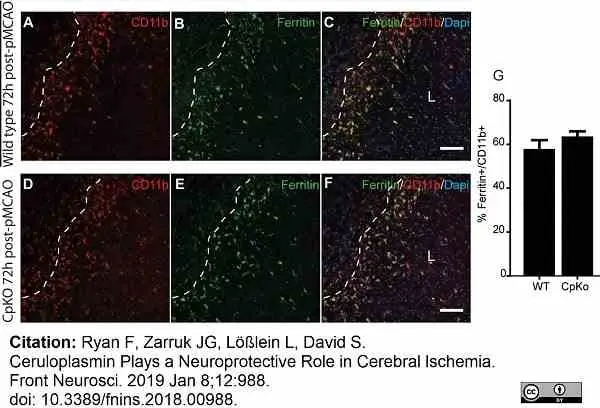
Immunofluorescence labeling for ferritin in macrophage/microglia. Double immunofluorescence labeling show that ferritin+/CD11b+ macrophage/microglia are located mainly in the lesion (L) along the lesion border in both wildtype (A–C) and CpKO (D–F) mice 72 h after pMCAO. Dotted line demarcates the lesion boundary. The merged images (C,F) show CD11b, ferritin and DAPI nuclear staining. Note that the number of CD11b+/ferritin+ doubled labeled cells are not significantly different in the two genotypes (G). Scale bar = 100 μm.
From: Ryan F, Zarruk JG, Lößlein L, David S.
Ceruloplasmin Plays a Neuroprotective Role in Cerebral Ischemia.
Front Neurosci. 2019 Jan 8;12:988.
doi: 10.3389/fnins.2018.00988.
This image is from an open access article distributed under terms of a Creative Commons Attribution License.

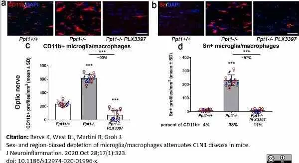
Rat anti Mouse CD11b antibody, clone 5C6 (MCA711G) used to label CD11b expressing microglial cells in mouse optic nerve by immunofluorescence.
Image caption:
Region-biased depletion of CD11b+ and Sn+ cells in the CNS of CLN1 mice upon treatment with PLX3397. a, b Representative fluorescent microscopic images of immunohistochemically labeled CD11b+ or activated Sn+ microglia/macrophages in longitudinal optic nerve sections of 6-month-old Ppt1+/+, Ppt1−/−, and PLX3397-treated Ppt1−/− mice after 5 months of treatment. Scale bar: 50 μm. c, d Quantification of CD11b+ or Sn+ cells showed a significant reduction in number in optic nerves after PLX3397 treatment. Percentages of Sn+ cells related to CD11b+ cells are indicated below the corresponding groups. n = 5 male and 5 female mice per group. One-way ANOVA and Tukey’s post hoc tests. *P <0.05; ***P <0.001
From: Berve K, West BL, Martini R, Groh J.
Sex- and region-biased depletion of microglia/macrophages attenuates CLN1 disease in mice.
J Neuroinflammation. 2020 Oct 28;17(1):323.
doi: 10.1186/s12974-020-01996-x.
This image is from an open access article distributed under terms of a Creative Commons Attribution License.
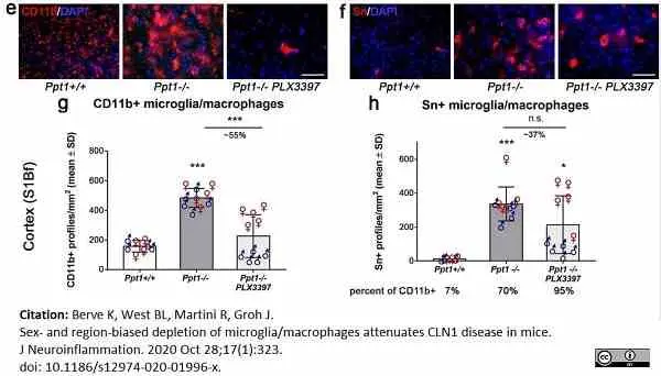
Rat anti Mouse CD11b antibody, clone 5C6 (MCA711G) used to label CD11b expressing microglial cells in mouse somatosensory barrel field (S1Bf) cortex by immunofluorescence.
Image caption:
Region-biased depletion of CD11b+ and Sn+ cells in the CNS of CLN1 mice upon treatment with PLX3397. e, f Representative fluorescent microscopic images of immunohistochemically labeled CD11b+ or activated Sn+ microglia/macrophages in S1Bf cortex region of 6-month-old Ppt1+/+, Ppt1−/− mice, and PLX3397-treated Ppt1−/− mice after 5 months of treatment. Scale bar: 50 μm. g, h Quantification of CD11b+ or Sn+ cells showed a significant or non-significant tendential reduction in number in the S1Bf cortex region after PLX3397 treatment, respectively. Percentages of Sn+ cells related to CD11b+ cells are indicated below the corresponding groups. n = 5 male and 5 female mice per group. One-way ANOVA and Tukey’s post hoc tests. *P <0.05; ***P <0.001
From: Berve K, West BL, Martini R, Groh J.
Sex- and region-biased depletion of microglia/macrophages attenuates CLN1 disease in mice.
J Neuroinflammation. 2020 Oct 28;17(1):323.
doi: 10.1186/s12974-020-01996-x.
This image is from an open access article distributed under terms of a Creative Commons Attribution License.
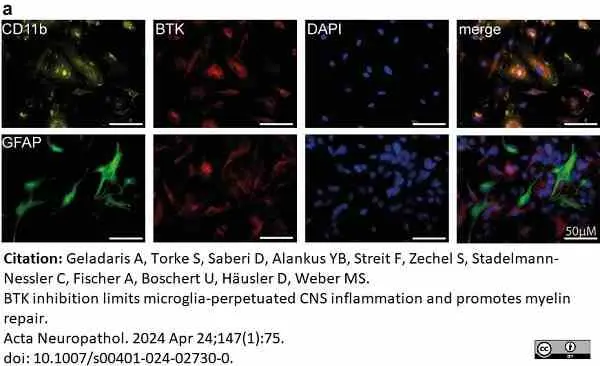
Mouse anti myelin proteolipid protein antibody, clone plpc1 (MCA839G) used to stain myelin in mouse brain by immunofluorescence.
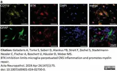
Image caption:
BTK is expressed in microglia and upregulated in CNS inflammation. a Primary mixed glia culture isolated from C57BL/6 J were fixed and stained for BTK expression.
From: Geladaris A, Torke S, Saberi D, Alankus YB, Streit F, Zechel S, Stadelmann-Nessler C, Fischer A, Boschert U, Häusler D, Weber MS.
BTK inhibition limits microglia-perpetuated CNS inflammation and promotes myelin repair.
Acta Neuropathol. 2024 Apr 24;147(1):75.
doi: 10.1007/s00401-024-02730-0.
This image is from an open access article distributed under terms of a Creative Commons Attribution License.
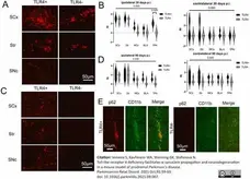
Rat anti Mouse CD68 antibody, clone FA-11 (MCA1957) used to label microglia in mouse brain by immunofluorescence.
Image caption:
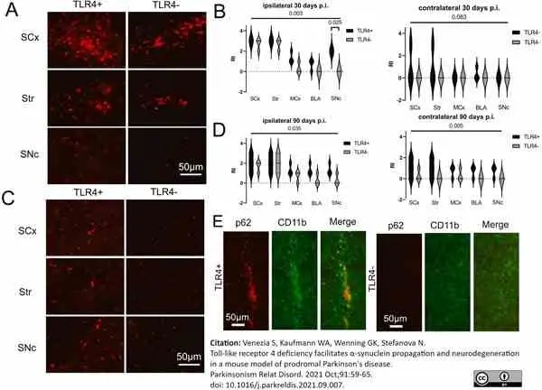
CD68-positive microglia in the brains of mice receiving hu-αS PFFs striatal inoculation. (A) Representative images of immunofluorescence for CD68 in wild type (TLR+) and TLR4-deficient (TLR4-) mice 30 days after the hu-αS PFFs striatal inoculation, ipsilaterally. (B) Violin plots with a comparison of the distribution of the CD68-positive signal in TLR4+ and TLR4- mice 30 days after the hu-αS PFFs striatal inoculation; non-parametric Friedman's two-way ANOVA by rank and Kruskal-Wallis multiple comparisons test. (C) Representative images of immunofluorescence for CD68 in wild type (TLR+) and TLR4-deficient (TLR4-) mice 90 days after the hu-αS PFFs striatal inoculation, contralaterally (D) Comparison of the intensity of CD68-positive signal in TLR4+ and TLR4- mice 90 days after the hu-αS PFFs striatal inoculation; non-parametric Friedman's two-way ANOVA by rank and Kruskal-Wallis multiple comparisons test. Abbreviations: STR, striatum; SCx, sensory cortex; MCx, motor cortex; BLA, basolateral amygdala; SNc, substantia nigra pars compacta; p. i., post inoculation. (E) Double immunofluorescence for p62 (red) and CD11b (green) in TLR+ and TLR4- brains, 90 days after hu-αS PFFs striatal inoculation.
From: Venezia S, Kaufmann WA, Wenning GK, Stefanova N.
Toll-like receptor 4 deficiency facilitates α-synuclein propagation and neurodegeneration in a mouse model of prodromal Parkinson's disease.
Parkinsonism Relat Disord. 2021 Oct;91:59-65.
doi: 10.1016/j.parkreldis.2021.09.007.
This image is from an open access article distributed under terms of a Creative Commons Attribution License.
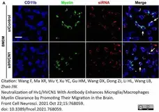
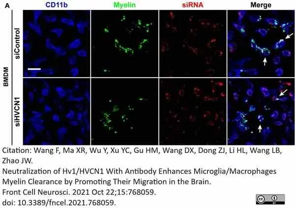
Rat anti Mouse CD11b antibody, clone 5C6 (MCA711G) used to identify bone marrow derived macrophages by immunofluorescence.
Image caption:
Knockdown of HVCN1 does not affect myelin phagocytosis and lysosome acidification in vitro. (A) Representative immunofluorescence images show that Hvcn1 knockdown by Cy3 labeled siRNA (red) does not affect myelin phagocytosis (green) in CD11b+ BMDM (blue). Scale bar = 100 μm. Arrows indicate two example BMDM cells that phagocytized myelin.
From: Wang F, Ma XR, Wu Y, Xu YC, Gu HM, Wang DX, Dong ZJ, Li HL, Wang LB, Zhao JW.
Neutralization of Hv1/HVCN1 With Antibody Enhances Microglia/Macrophages Myelin Clearance by Promoting Their Migration in the Brain.
Front Cell Neurosci. 2021 Oct 22;15:768059.
doi: 10.3389/fncel.2021.768059.
This image is from an open access article distributed under terms of a Creative Commons Attribution License.


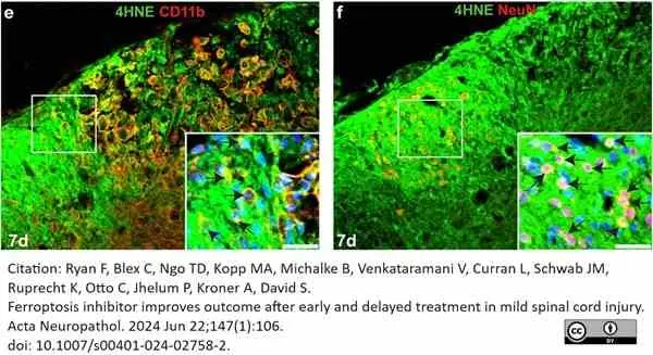
Rat anti Mouse CD11b antibody, clone 5C6 (MCA711) used to label macrophages in spinal cord of mice by immunofluorescence.
Image caption:
Changes in levels of 4-HNE in the injured mouse spinal cord.
Double immunofluorescence labeling of the dorsal horn region labeled for 4HNE/CD11b (e) and 4HNE/NeuN (f). Note the double labeled CD11b + macrophages and NeuN + neurons in the insets in e and f, respectively; nuclei in the insets stained with DAPI.
From: Ryan F, Blex C, Ngo TD, Kopp MA, Michalke B, Venkataramani V, Curran L, Schwab JM, Ruprecht K, Otto C, Jhelum P, Kroner A, David S.
Ferroptosis inhibitor improves outcome after early and delayed treatment in mild spinal cord injury.
Acta Neuropathol. 2024 Jun 22;147(1):106.
doi: 10.1007/s00401-024-02758-2.
This image is from an open access article distributed under terms of a Creative Commons Attribution License.
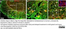
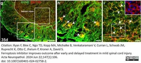
Rat anti Mouse CD11b antibody, clone 5C6 (MCA711) used to label macrophages in spinal cord of mice by immunofluorescence.
Image caption:
Changes in levels of 4-HNE in the injured mouse spinal cord.
g Double immunofluorescence labeling of 4HNE/CD11b at 35 days post-SCI. Note the strong 4HNE labeling in CD11b+ macrophages in the white matter and in the lesion core. Regions outlined within the white squares in panel g are shown at higher magnification in panels h (white matter), and i (lesion core). The double labeled cells appear yellow. The large-rounded cells within the lesion core are macrophages as indicated by the single labeling channel for CD11b (red; inset in panel i). The strong 4-HNE labeling in and around the core of the lesion provides evidence of widespread lipid peroxidation that can contribute to oxidative damage even at later time periods (5 weeks) after SCI. Scale bars = 100 μm; inset = 25 μm
From: Ryan F, Blex C, Ngo TD, Kopp MA, Michalke B, Venkataramani V, Curran L, Schwab JM, Ruprecht K, Otto C, Jhelum P, Kroner A, David S.
Ferroptosis inhibitor improves outcome after early and delayed treatment in mild spinal cord injury.
Acta Neuropathol. 2024 Jun 22;147(1):106.
doi: 10.1007/s00401-024-02758-2.
This image is from an open access article distributed under terms of a Creative Commons Attribution License.
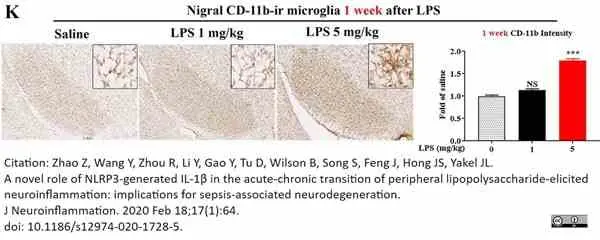
Rat anti Mouse CD11b antibody, clone 5C6 (MCA711G) used to label microglia by immunohistochemistry of free floating coronal brain slices.
Image caption:
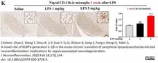
Peripheral LPS dose-dependently increases brain mature IL-1β production and causes sustained nigral microglial activation.
(k): At 1 week after injection of LPS (1 or 5 mg/kg, ip) or saline vehicle in C57BL/6 J mice (n = 3/group for each timepoint), nigral microglial CD-11b (k) immunostaining was performed. Representative images were shown. Scale bar = 300 μm. The histograms showed the density CD-11b (k) quantified by with ImageJ. **p <0.01 and ***p <0.001 compared with saline vehicle group, and NS between LPS 1 and 5 mg/kg groups. One-way ANOVA followed by Bonferroni post hoc multiple comparison test.
From: Zhao Z, Wang Y, Zhou R, Li Y, Gao Y, Tu D, Wilson B, Song S, Feng J, Hong JS, Yakel JL.
A novel role of NLRP3-generated IL-1β in the acute-chronic transition of peripheral lipopolysaccharide-elicited neuroinflammation: implications for sepsis-associated neurodegeneration.
J Neuroinflammation. 2020 Feb 18;17(1):64.
doi: 10.1186/s12974-020-1728-5.
This image is from an open access article distributed under terms of a Creative Commons Attribution License.
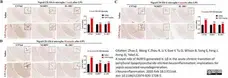
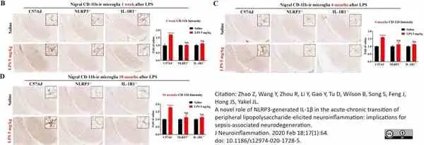
Rat anti Mouse CD11b antibody, clone 5C6 (MCA711G) used to label microglia by immunohistochemistry of free floating coronal brain slices.
Image caption:
High dose LPS injection produces long-lasting microglial activation in WT mice, but not in NLRP3−/− or IL-1R1−/− mice. Following a single injection of LPS (5 mg/kg; ip) or saline vehicle, mice were perfused1 week, 4 and 10 months thereafter for nigral microglial CD-11b immunostaining (n = 3/group for each time point). Representative images at each time point were shown (b–d). Scale bar = 300 μm. Results are expressed as folds of time-matched vehicle control. Histograms represent degree of microglial activation quantified by measuring the density of CD-11b staining with ImageJ. ***p <0.001, ****p <0.0001, and NS compared to respective saline vehicle group. Two-way ANOVA followed by Bonferroni post hoc multiple comparison test.
From: Zhao Z, Wang Y, Zhou R, Li Y, Gao Y, Tu D, Wilson B, Song S, Feng J, Hong JS, Yakel JL.
A novel role of NLRP3-generated IL-1β in the acute-chronic transition of peripheral lipopolysaccharide-elicited neuroinflammation: implications for sepsis-associated neurodegeneration.
J Neuroinflammation. 2020 Feb 18;17(1):64.
doi: 10.1186/s12974-020-1728-5.
This image is from an open access article distributed under terms of a Creative Commons Attribution License.
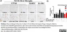
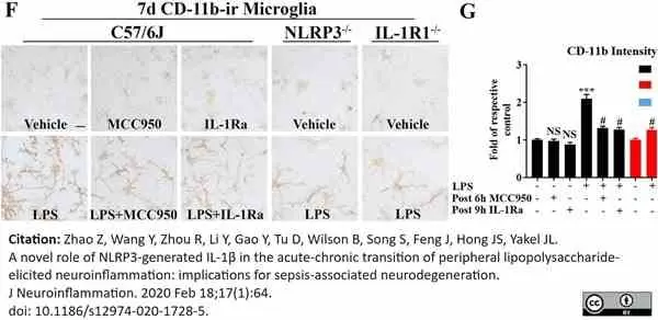
Rat anti Mouse CD11b antibody, clone 5C6 (MCA711G) used to label microglia by immunohistochemistry of free floating coronal brain slices.
Image caption:
Genetic or pharmacological inhibition of NLRP3 or IL-1R1 represses LPS-elicited production of chronic inflammatory mediators in neuron-glial cultures. Neuron-glial cultures prepared from WT, NLRP3−/−, and IL-1R1−/− mice were treated with LPS (20 ng/ml) or vehicle medium. At 7 days after LPS and post-treatment of MCC950 or IL-1Ra, representative images of microglial CD-11b (the α-chain of Mac1, indicating Mac1 expression here) immunostaining (f) and the intensity of CD-11b (g) measured by ImageJ were shown. Bar = 50 μm. g NS, #p <0.001 compared with WT vehicle group, and ***p <0.001 compared with WT LPS group. Two-way ANOVA followed by Bonferroni post hoc multiple comparison test.
From: Zhao Z, Wang Y, Zhou R, Li Y, Gao Y, Tu D, Wilson B, Song S, Feng J, Hong JS, Yakel JL.
A novel role of NLRP3-generated IL-1β in the acute-chronic transition of peripheral lipopolysaccharide-elicited neuroinflammation: implications for sepsis-associated neurodegeneration.
J Neuroinflammation. 2020 Feb 18;17(1):64.
doi: 10.1186/s12974-020-1728-5.
This image is from an open access article distributed under terms of a Creative Commons Attribution License.


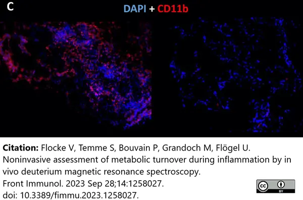
Rat anti Mouse CD11b antibody, clone 5C6 (MCA711) used to label inflammatory myeloid cells by immunofluorescence.
Image caption:
Ex vivo validation of the experimental inflammatory focus.
(C) Immunohistochemistry of excised plugs confirmed the in vivo findings.
From: Flocke V, Temme S, Bouvain P, Grandoch M, Flögel U.
Noninvasive assessment of metabolic turnover during inflammation by in vivo deuterium magnetic resonance spectroscopy.
Front Immunol. 2023 Sep 28;14:1258027.
doi: 10.3389/fimmu.2023.1258027.
This image is from an open access article distributed under terms of a
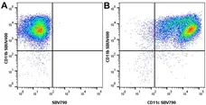
Figure B. StarBright UltraViolet 400 conjugated Rat anti Mouse CD11b antibody, clone 5C6 (MCA711SBUV400) and StarBright Violet 790 conjugated Hamster anti Mouse CD11c antibody, clone N418 (MCA1369SBV790). All experiments performed on bone marrow derived dendritic cells in the presence of 10% mouse serum.
Data acquired on the ZE5 Cell Analyzer.
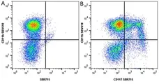
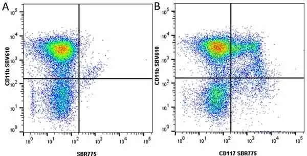
Figure B. StarBright Violet 610 conjugated Rat anti Mouse CD11b antibody, clone 5C6 (MCA711SBV610) and StarBright Red 715 conjugated Rat anti Mouse CD117 antibody, clone 2B8 (MCA1365SBR715). All experiments performed on red cell lysed mouse blood gated on live single cell lymphocytes, in the presence of 10% mouse serum.
Data acquired on the ZE5 Cell Analyzer.
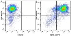
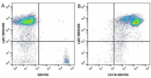
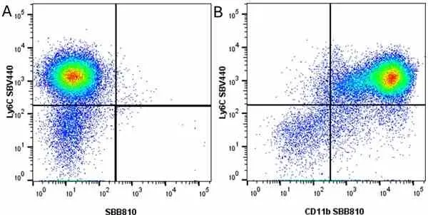
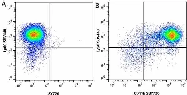
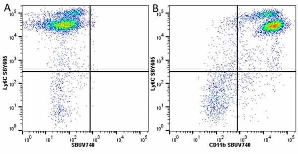
Figure B. StarBright Violet 610 conjugated Rat anti Mouse Ly-6C antibody, clone ER-MP20 (MCA2389SBV610) and StarBright Red 715 conjugated Rat anti Mouse CD11b antibody, clone 5C6 (MCA711SBR715). All experiments performed on red cell lysed murine blood gated live cells in the presence of 10% mouse serum.
Data acquired on the ZE5 Cell Analyzer.
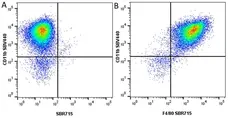
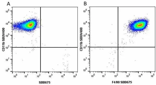
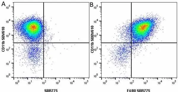
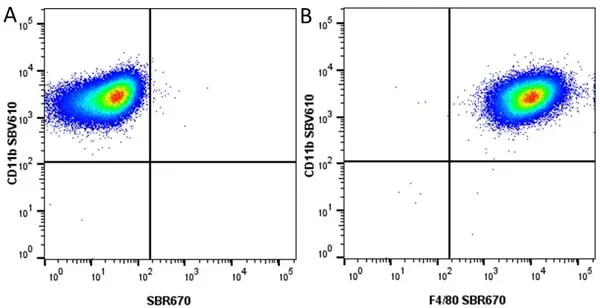
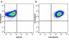
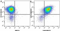
Figure B. StarBright Violet 610 conjugated Rat anti Mouse CD11b antibody, clone 5C6 (MCA711SBV610) and StarBright Red 670 conjugated Rat anti Mouse F4/80 antibody, clone A3-1(MCA497SBR670). All experiments performed on J774.2 cells gated on live single cells, in the presence of 10% mouse serum.
Data acquired on the ZE5 Cell Analyzer.

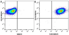
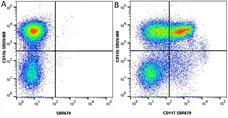
Figure B. Alexa Fluor® 647 conjugated Rat anti Mouse Gr-1 antibody, clone RB^-8C5 (MCA2387A647) and RPE conjugated Rat anti Mouse CD11b antibosy, clone 5C6 (MCA711PE). All experiments performed on murine bone marrow in the presence of Murine SeroBlock (BUF041A).
Rat anti mouse CD11b antibody, clone 5C6 (MCA711) used to identify microglia in mouse brain by immunofluorescence.
Image caption:
Activation of adult primary microglial cells in wild-type and IL-1 KO mice. (A) Primary microglial cells were obtained from young adult wild-type mice. The cells stain with the microglial marker CD11b, but not with the neuronal and astroglial markers, NeuN and GFAP, respectively. A few cells are stained with the oligodendroglial cell marker, MBP. NC (inset) is the primary antibody-free negative control. The microglial cells (n = 3 each group) were stimulated for 24 hours in the presence of the vehicle alone, or supplemented with IFN&gama; or IL-4 in the presence or absence of IL-1β. Total NO (NOx; B), TNFα (C), arginase specific activity (Arg-1 spe. act.; D) and IGF-1 (E) were determined from the media or cell suspensions. (B) NOx levels increase upon exposure of the cells to IL-1β and in a synergistic manner upon co-treatment of cells with IL-1β and IFNγ, but not when the cotreatment is with IL-4. (C) TNFα levels increase upon exposure of the cells to IFNγ, and further upon co-treatment with IL-1β. Surprisingly, the co-treatment of the cells with IL-4 and IL-1β induced the highest TNFα level among the experimental treatments used. (D) Arg1-specific activity increased significantly upon exposure to IL-4 and further increased when IL-4 and IL-1β were employed together. (E) IGF-1 levels decreased with exposure of the cells to IFNγ and increased in response to IL-4. The response was partially inhibited by cotreatment of the cells with IL-1β. Data are expressed as mean ± SD (n = 3). *: P<0.05, **: P<0.01, ***: P<0.001 compared with the vehicle-treated group in each genotype (one-way ANOVA followed by Dunnett post-hoc test). ANOVA, analysis of variance; IGF-1, insulin-like growth factor.
From: Sato A, Ohtaki H, Tsumuraya T, Song D, Ohara K, Asano M, Iwakura Y, Atsumi T, Shioda S.
Interleukin-1 participates in the classical and alternative activation of microglia/macrophages after spinal cord injury.J Neuroinflammation. 2012 Apr 7;9:65..
doi: 10.1186/1742-2094-9-65.
This is from an open access article distributed under the terms of a Creative Commons Attribution License.
Rat anti Mouse CD11b antibody, clone 5C6 (MCA711) used to identify macrophages associated with vessels by immunofluorescence.
Image caption:
BM-derived GFP+ single cells and vessel-associated cells express CD11b. Fluorescence microscopy for GFP combined with immunofluorescence detection of (A) CD11b, (B) vWF and (C) CD31, 24 hours after pMCAO. (A) Fluorescence detection of GFP and CD11b showed that most GFP+ cells co-expressed CD11b (yellow cells, indicated by arrows), and intermingled with CD11b+ host cells. Note also that a few GFP+ cells did not co-express CD11b (arrow head). Insert shows high magnification of GFP+ cells, some of which co-express CD11b, aggregated around a vessel. (B, C) Fluorescence detection of GFP and the endothelial cell markers vWF (B) and CD31 (C). Inserts show higher magnification of sections of the same vessels. Although there are indications that single vWF+ cells co-express GFP (arrows in B), this could not be reproduced using staining for CD31, and the majority of vWF+ and CD31+ cells showed no co-expression of GFP. Instead, GFP remained confined to round and elongated cells located in the juxtavascular space (insert in C). CD11b+ cells were visualized using Alexa Fluor® 568-conjugated goat anti-rat IgG, vWF+ and CD31+ cells using Alexa Fluor® 546-conjugated goat anti-rabbit IgG and Alexa Fluor® 594-conjugated goat anti-rat IgG, respectively. Scale bars: 20 μm (A-C)..
From: Clausen BH, Lambertsen KL, Babcock AA, Holm TH, Dagnaes-Hansen F, Finsen B.
Interleukin-1beta and tumor necrosis factor-alpha are expressed by different subsets of microglia and macrophages after ischemic stroke in mice.
J Neuroinflammation. 2008 Oct 23;5:46.
doi: 10.1186/1742-2094-5-46.
This is from an open access article distributed under the terms of a Creative Commons Attribution License.
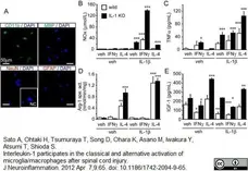
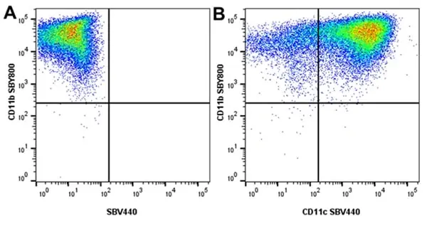
Figure B. StarBright Yellow 800 conjugated Rat anti Mouse CD11b antibody, clone 5C6 (MCA711SBY800) and StarBright Violet 440 conjugated Hamster anti Mouse CD11c antibody, clone N418 (MCA1369SBV440).
All experiments performed on bone marrow derived dendritic cells (BMDC), in the presence of 10% mouse serum.Data acquired on the ZE5 Cell analyser.
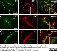
Figure B. StarBright Blue 810 conjugated Rat anti Mouse CD11b antibody, clone 5C6 (MCA711SBB810) and StarBright Violet 570 conjugated Hamster anti Mouse CD11c antibody, clone N418 (MCA1369SBV570).
All experiments performed on bone marrow derived dendritic cells (BMDC), in the presence of 10% mouse serum. Data acquired on the ZE5 Cell analyser.

All experiments performed on bone marrow derived dendritic cells (BMDC), in the presence of 10% mouse serum. Data acquired on the ZE5 Cell analyser.

Figure B. StarBright Violet 515 conjugated Rat anti Mouse CD11b antibody, clone 5C6 (MCA711SBV515) and StarBright Blue 700 conjugated Hamster anti Mouse CD11c antibody, clone N418 (MCA1369SBB700).
All experiments performed on bone marrow derived dendritic cells (BMDC), in the presence of 10% mouse serum.Data acquired on the ZE5 Cell analyser.
Filter by Application:
F IF C EM P WB BA VI Reset| Rat anti Mouse CD11b antibody, clone 5C6 recognizes CD11b, also known as the integrin alpha M chain. CD11b is implicated in various adhesive interactions of monocytes, macrophages and granulocytes as well as in mediating the uptake of complement-coated particles. Rat anti Mouse CD11b antibody, clone 5C6 immunoprecipitates a heterodimer of ~165 and ~95 kDa. This clone also exhibits various functional properties, reportedly inhibiting adhesion in vitro and inflammatory recruitment in vivo. Rat anti Mouse CD11b antibody, clone 5C6 also inhibits delayed hypersensitivity, potentiates bacterial infections and inhibits type 1 diabetes. |

Our CD11b (5C6) Antibody has been referenced in >161 publications* *Based on June 2020 data from CiteAb's antibody search engine. |
- Target Species
- Mouse
- Species Cross-Reactivity
-
Target Species Cross Reactivity Human - N.B. Antibody reactivity and working conditions may vary between species.
- Product Form
- Purified IgG - liquid
- Preparation
- Purified IgG prepared by affinity chromatography on Protein G from tissue culture supernatant
- Buffer Solution
- Phosphate buffered saline
- Preservative Stabilisers
0.09% Sodium Azide - Carrier Free
- Yes
- Immunogen
- Thioglycollate-elicited peritoneal macrophages (TPM)
- Approx. Protein Concentrations
- IgG concentration 1 mg/ml
- Fusion Partners
- Spleen cells from AO rats were fused with cells of the Y3 rat myeloma cell line
- Regulatory
- For research purposes only
- Guarantee
- 12 months from date of despatch
This product is shipped at ambient temperature. It is recommended to aliquot and store at -20°C on receipt. When thawed, aliquot the sample as needed. Keep aliquots at 2-8°C for short term use (up to 4 weeks) and store the remaining aliquots at -20°C.
Avoid repeated freezing and thawing as this may denature the antibody. Storage in frost-free freezers is not recommended.
Avoid repeated freezing and thawing as this may denature the antibody. Storage in frost-free freezers is not recommended.
This product has been reported to work in the following applications. This information is derived from testing within our laboratories, peer-reviewed publications or personal communications from the originators. Please refer to references indicated for further information. For general protocol recommendations, please visit the antibody protocols page.
| Application Name | Verified | Min Dilution | Max Dilution |
|---|---|---|---|
| Flow Cytometry |  |
1/100 | |
| Immunofluorescence | |
||
| Immunohistology - Frozen | |
||
| Immunoprecipitation | |
Where this antibody has not been tested for use in a particular technique this does not necessarily exclude its use in such procedures. Suggested working dilutions are given as a guide only. It is recommended that the user titrates the antibody for use in their own system using appropriate negative/positive controls.
- Flow Cytometry
- Use 10ul of the suggested working dilution to label 106 cells in 100ul
- Histology Positive Control Tissue
- Spleen
References for CD11b antibody
-
Rosen, H. and Gordon, S. (1987) Monoclonal antibody to the murine type 3 complement receptor inhibits adhesion of myelomonocytic cells in vitro and inflammatory cell recruitment in vivo.
J Exp Med. 166: 1685-701. -
Rosen, H. et al. (1989) Antibody to the murine type 3 complement receptor inhibits T lymphocyte-dependent recruitment of myelomonocytic cells in vivo.
J Exp Med. 169: 535-48. -
Serafini, B. et al. (2000) Intracerebral recruitment and maturation of dendritic cells in the onset and progression of experimental autoimmune encephalomyelitis.
Am J Pathol. 157: 1991-2002. -
Platt, N. et al. (2000) Apoptotic thymocyte clearance in scavenger receptor class A-deficient mice is apparently normal.
J Immunol. 164: 4861-7. -
Carenini, S. et al. (2001) The role of macrophages in demyelinating peripheral nervous system of mice heterozygously deficient in p0.
J Cell Biol. 152: 301-8. -
Engwerda, C.R. et al. (2002) Locally up-regulated lymphotoxin alpha, not systemic tumor necrosis factor alpha, is the principle mediator of murine cerebral malaria.
J Exp Med. 195: 1371-7. -
Mittal, A. et al. (2003) CD11b+ cells are the major source of oxidative stress in UV radiation-irradiated skin: possible role in photoaging and photocarcinogenesis.
Photochem Photobiol. 77 (3): 259-64. -
Lesnik, P. et al. (2003) Decreased atherosclerosis in CX3CR1-/- mice reveals a role for fractalkine in atherogenesis.
J Clin Invest. 111: 333-40.
View The Latest Product References
-
Di Filippo, C. et al. (2005) Cannabinoid CB2 receptor activation reduces mouse myocardial ischemia-reperfusion injury: involvement of cytokine/chemokines and PMN.
J Leukoc Biol. 75 (3): 453-9. -
Lin, H.H. et al. (2005) The macrophage F4/80 receptor is required for the induction of antigen-specific efferent regulatory T cells in peripheral tolerance.
J Exp Med. 201: 1615-25. -
Mennini, T. et al. (2006) Nonhematopoietic erythropoietin derivatives prevent motoneuron degeneration in vitro and in vivo.
Mol Med. 12: 153-60. -
Saura, J. (2007) Microglial cells in astroglial cultures: a cautionary note.
J Neuroinflammation. 4: 26. -
Kondo, Y. et al. (2007) Osteopetrotic (op/op) mice have reduced microglia, no Abeta deposition, and no changes in dopaminergic neurons.
J Neuroinflammation. 4: 31. -
Halle, A. et al. (2008) The NALP3 inflammasome is involved in the innate immune response to amyloid-beta.
Nat Immunol. 9: 857-65. -
Devey, L. et al. (2008) Tissue-resident Macrophages protect the Liver From Ischemia Reperfusion Injury via a Heme Oxygenase-1-Dependent mechanism.
Mol Ther. 1: 65-72. -
Hickman, S.E. et al. (2008) Microglial dysfunction and defective beta-amyloid clearance pathways in aging Alzheimer's disease mice.
J Neurosci. 28 (33): 8354-60. -
Khorooshi, R. et al. (2008) NF-kappaB-driven STAT2 and CCL2 expression in astrocytes in response to brain injury.
J Immunol.181: 7284-91. -
Basso, A.S. et al. (2008) Reversal of axonal loss and disability in a mouse model of progressive multiple sclerosis.
J Clin Invest. 118: 1532-43. -
Weberpals, M. et al. (2009) NOS2 gene deficiency protects from sepsis-induced long-term cognitive deficits.
J Neurosci. 29: 14177-84. -
Valerio, A. et al. (2009) Leptin is induced in the ischemic cerebral cortex and exerts neuroprotection through NF-kappaB/c-Rel-dependent transcription.
Stroke. 40: 610-7. -
Traka, .M. et al (2010) A genetic mouse model of adult-onset, pervasive central nervous system demyelination with robust remyelination.
Brain. 133: 3017-29. -
Kim, D. et al. (2010) NADPH oxidase 2-derived reactive oxygen species in spinal cord microglia contribute to peripheral nerve injury-induced neuropathic pain.
Proc Natl Acad Sci U S A. 107: 14851-6. -
Lu, J. et al. (2010) Ursolic acid attenuates D-galactose-induced inflammatory response in mouse prefrontal cortex through inhibiting AGEs/RAGE/NF-κB pathway activation.
Cereb Cortex. 20: 2540-8. -
Heneka, M.T. et al. (2010) Locus ceruleus controls Alzheimer's disease pathology by modulating microglial functions through norepinephrine.
Proc Natl Acad Sci U S A. 107: 6058-63. -
Dohi, K. et al. (2010) Gp91phox (NOX2) in classically activated microglia exacerbates traumatic brain injury.
J Neuroinflammation. 7: 41. -
Cui, Y.F. et al. (2010) Embryonic stem cell-derived L1 overexpressing neural aggregates enhance recovery in Parkinsonian mice.
Brain. 133: 189-204. -
Wu, T. et al. (2011) Expression and cellular localization of cyclooxygenases and prostaglandin E synthases in the hemorrhagic brain.
J Neuroinflammation. 8:22. -
Tysseling, V.M.et al. (2011) SDF1 in the dorsal corticospinal tract promotes CXCR4+ cell migration after spinal cord injury.
J Neuroinflammation. 8:16. -
McDonald, J.U. et al. (2011) In vivo functional analysis and genetic modification of in vitro-derived mouse neutrophils.
FASEB J. 25: 1972-82. -
Terwel, D. et al. (2011) Critical Role of Astroglial Apolipoprotein E and Liver X Receptor-{alpha} Expression for Microglial A{beta} Phagocytosis.
J Neurosci. 31: 7049-59. -
Sato, A. et al. (2012) Interleukin-1 participates in the classical and alternative activation of microglia/macrophages after spinal cord injury.
J Neuroinflammation. 9: 65. -
Heydenreich, N. et al. (2012) C1-inhibitor protects from brain ischemia-reperfusion injury by combined antiinflammatory and antithrombotic mechanisms.
Stroke. 43 (9): 2457-67. -
Yamanaka M et al. (2012) PPARγ/RXRα-induced and CD36-mediated microglial amyloid-β phagocytosis results in cognitive improvement in amyloid precursor protein/presenilin 1 mice.
J Neurosci. 32 (48): 17321-31. -
Mencl, S. et al. (2014) FTY720 does not protect from traumatic brain injury in mice despite reducing posttraumatic inflammation.
J Neuroimmunol. 274 (1-2): 125-31. -
Natrajan, M.S. et al. (2015) Retinoid X receptor activation reverses age-related deficiencies in myelin debris phagocytosis and remyelination.
Brain. 138 (Pt 12): 3581-97. -
Kan, M.J. et al. (2015) Arginine deprivation and immune suppression in a mouse model of Alzheimer's disease.
J Neurosci. 35 (15): 5969-82. -
Babcock, A.A. et al. (2015) Cytokine-producing microglia have an altered beta-amyloid load in aged APP/PS1 Tg mice.
Brain Behav Immun. 48: 86-101. -
Kaindlstorfer, C. et al. (2015) Failure of Neuroprotection Despite Microglial Suppression by Delayed-Start Myeloperoxidase Inhibition in a Model of Advanced Multiple System Atrophy: Clinical Implications.
Neurotox Res. 28 (3): 185-94. -
Liu, Z. et al. (2016) Transforming growth factor-β1 acts via TβR-I on microglia to protect against MPP(+)-induced dopaminergic neuronal loss.
Brain Behav Immun. 51: 131-43. -
Garcia-Mesa, Y. et al. (2017) Immortalization of primary microglia: a new platform to study HIV regulation in the central nervous system.
J Neurovirol. 23 (1): 47-66. -
Lu, Y. et al. (2016) Annexin A10 is involved in the development and maintenance of neuropathic pain in mice.
Neurosci Lett. 631: 1-6. -
Hristova M et al. (2016) Inhibition of Signal Transducer and Activator of Transcription 3 (STAT3) reduces neonatal hypoxic-ischaemic brain damage.
J Neurochem. 136 (5): 981-994. -
Ye, M. et al. (2016) Neuroprotective effects of bee venom phospholipase A2 in the 3xTg AD mouse model of Alzheimer's disease.
J Neuroinflammation. 13 (1): 10. -
Nagai, J. et al. (2016) Inhibition of CRMP2 phosphorylation repairs CNS by regulating neurotrophic and inhibitory responses.
Exp Neurol. 277: 283-95. -
Tachibana, M. et al. (2016) Rescuing effects of RXR agonist bexarotene on aging-related synapse loss depend on neuronal LRP1.
Exp Neurol. 277: 1-9. -
Sun, H. et al. (2016) Aquaporin-4 mediates communication between astrocyte and microglia: Implications of neuroinflammation in experimental Parkinson's disease.
Neuroscience. 317: 65-75. -
Shinohara, M. et al. (2016) APOE2 eases cognitive decline during Aging: Clinical and preclinical evaluations.
Ann Neurol. 79 (5): 758-74. -
Kami, K. et al. (2016) Histone Acetylation in Microglia Contributes to Exercise-Induced Hypoalgesia in Neuropathic Pain Model Mice.
J Pain. 17 (5): 588-99. -
Bisht, K. et al. (2016) Dark microglia: A new phenotype predominantly associated with pathological states.
Glia. 64 (5): 826-39. -
Hilla, A.M. et al. (2017) Microglia Are Irrelevant for Neuronal Degeneration and Axon Regeneration after Acute Injury.
J Neurosci. 37 (25): 6113-24. -
Paizs, M. et al. (2017) Axotomy Leads to Reduced Calcium Increase and Earlier Termination of CCL2 Release in Spinal Motoneurons with Upregulated Parvalbumin Followed by Decreased Neighboring Microglial Activation.
CNS Neurol Disord Drug Targets. 16 (3): 356-67. -
Pulido-Salgado, M. et al. (2017) Myeloid C/EBPβ deficiency reshapes microglial gene expression and is protective in experimental autoimmune encephalomyelitis.
J Neuroinflammation. 14 (1): 54. -
Thomsen, M.S. et al. (2017) Synthesis and deposition of basement membrane proteins by primary brain capillary endothelial cells in a murine model of the blood-brain barrier.
J Neurochem. 140 (5): 741-754. -
Laurent, C. et al. (2017) Hippocampal T cell infiltration promotes neuroinflammation and cognitive decline in a mouse model of tauopathy.
Brain. 140 (Pt 1): 184-200. -
Schuhmann, M.K. et al. (2017) Blocking of platelet glycoprotein receptor Ib reduces "thrombo-inflammation" in mice with acute ischemic stroke.
J Neuroinflammation. 14 (1): 18. -
Rabl, R. et al. (2017) Early start of progressive motor deficits in Line 61 α-synuclein transgenic mice.
BMC Neurosci. 18 (1): 22. -
Crépeaux, G. et al. (2017) Non-linear dose-response of aluminium hydroxide adjuvant particles: Selective low dose neurotoxicity.
Toxicology. 375: 48-57. -
Da Ros, F. et al. (2017) Targeting Interleukin-1β Protects from Aortic Aneurysms Induced by Disrupted Transforming Growth Factor β Signaling.
Immunity. 47 (5): 959-973.e9. -
Hou, L. et al. (2018) Taurine protects noradrenergic locus coeruleus neurons in a mouse Parkinson's disease model by inhibiting microglial M1 polarization.
Amino Acids. 50 (5): 547-556. -
Zheng, J. et al. (2018) Evaluation of metastatic niches in distant organs after surgical removal of tumor-bearing lymph nodes.
BMC Cancer. 18 (1): 608. -
Soto, M. et al. (2018) Gut microbiota modulate neurobehavior through changes in brain insulin sensitivity and metabolism.
Mol Psychiatry. 23 (12): 2287-301. -
Mouton-Liger, F. et al. (2018) Parkin deficiency modulates NLRP3 inflammasome activation by attenuating an A20-dependent negative feedback loop.
Glia. 66 (8): 1736-51. -
Roberts, J.M. et al. (2018) Bilateral carotid artery stenosis causes unexpected early changes in brain extracellular matrix and blood-brain barrier integrity in mice.
PLoS One. 13 (4): e0195765. -
Cope, E.C. et al. (2018) Microglia Play an Active Role in Obesity-Associated Cognitive Decline.
J Neurosci. 38 (41): 8889-904. -
Song, S. et al. (2019) Noradrenergic dysfunction accelerates LPS-elicited inflammation-related ascending sequential neurodegeneration and deficits in non-motor/motor functions.
Brain Behav Immun. 81: 374-87. -
Liu, Z. et al. (2019) IL-17A exacerbates neuroinflammation and neurodegeneration by activating microglia in rodent models of Parkinson's disease.
Brain Behav Immun. 81: 630-45. -
Gomez-Nicola, D. et al. (2019) Measuring Microglial Turnover in the Adult Brain.
Methods Mol Biol. 2034: 207-15. -
Di Benedetto, G. et al. (2019) Beneficial effects of curtailing immune susceptibility in an Alzheimer's disease model.
J Neuroinflammation. 16 (1): 166. -
Bernier, L.P. et al. (2019) Nanoscale Surveillance of the Brain by Microglia via cAMP-Regulated Filopodia.
Cell Rep. 27 (10): 2895-2908.e4. -
Tunesi, M. et al. (2019) Hydrogel-based delivery of Tat-fused protein Hsp70 protects dopaminergic cells in vitro and in a mouse model of Parkinson’s disease
NPG Asia Materials. 11: 28. -
Myhre, C.L. et al. (2019) Microglia Express Insulin-Like Growth Factor-1 in the Hippocampus of Aged APPswe/PS1ΔE9 Transgenic Mice.
Front Cell Neurosci. 13: 308. -
Hauptmann, J. et al. (2020) Interleukin-1 promotes autoimmune neuroinflammation by suppressing endothelial heme oxygenase-1 at the blood-brain barrier.
Acta Neuropathol. 140 (4): 549-67. -
Yang, P. et al. (2020) Suppression of cGMP-Dependent Photoreceptor Cytotoxicity With Mycophenolate Is Neuroprotective in Murine Models of Retinitis Pigmentosa.
Invest Ophthalmol Vis Sci. 61 (10): 25. -
Potì, F. et al. (2020) Impact of S1P Mimetics on Mesenteric Ischemia/Reperfusion Injury.
Pharmaceuticals (Basel). 13 (10) 298. -
Wi, R. et al. (2020) Functional Crosstalk between CB and TRPV1 Receptors Protects Nigrostriatal Dopaminergic Neurons in the MPTP Model of Parkinson's Disease.
J Immunol Res. 2020: 5093493. -
Madore, C. et al. (2020) Essential omega-3 fatty acids tune microglial phagocytosis of synaptic elements in the mouse developing brain.
Nat Commun. 11 (1): 6133. -
Ellman, D.G. et al. (2020) Conditional Ablation of Myeloid TNF Improves Functional Outcome and Decreases Lesion Size after Spinal Cord Injury in Mice.
Cells. 9 (11): 2407. -
Elabi, O. et al. (2021) Human α-synuclein overexpression in a mouse model of Parkinson's disease leads to vascular pathology, blood brain barrier leakage and pericyte activation.
Sci Rep. 11 (1): 1120. -
Yoshizaki, S. et al. (2021) Microglial inflammation after chronic spinal cord injury is enhanced by reactive astrocytes via the fibronectin/β1 integrin pathway.
J Neuroinflammation. 18 (1): 12. -
Mañucat-Tan, N. et al. (2021) Hypochlorite-induced aggregation of fibrinogen underlies a novel antioxidant role in blood plasma.
Redox Biol. 40: 101847. -
Brunialti, E. et al. (2021) Inhibition of microglial β-glucocerebrosidase hampers the microglia-mediated antioxidant and protective response in neurons.
J Neuroinflammation. 18 (1): 220. -
Spitzel, M. et al. (2022) Dysregulation of Immune Response Mediators and Pain-Related Ion Channels Is Associated with Pain-like Behavior in the GLA KO Mouse Model of Fabry Disease.
Cells. 11 (11): 1730. -
Ji, N. et al. (2022) VSIG4 Attenuates NLRP3 and Ameliorates Neuroinflammation via JAK2-STAT3-A20 Pathway after Intracerebral Hemorrhage in Mice.
Neurotox Res. 40 (1): 78-88. -
Shanaki-Bavarsad, M. et al. (2022) Astrocyte-targeted Overproduction of IL-10 Reduces Neurodegeneration after TBI.
Exp Neurobiol. 31 (3): 173-95. -
Bretheau, F. et al. (2022) The alarmin interleukin-1α triggers secondary degeneration through reactive astrocytes and endothelium after spinal cord injury.
Nat Commun. 13 (1): 5786. -
Bani-Hani, M. et al. (2023) Interactions of Carboxylated Nanodiamonds With Mouse Macrophages Cell Line and Primary Cells
Int J Orthopaed Res. 6 (1): 30-43. -
Saraiva, C. et al. (2023) CtBP Neuroprotective Role in Toxin-Based Parkinson's Disease Models: From Expression Pattern to Dopaminergic Survival.
Mol Neurobiol. 60 (8): 4246-60. -
Nelke, C. et al. (2023) K(2P)2.1 is a regulator of inflammatory cell responses in idiopathic inflammatory myopathies.
J Autoimmun. 142: 103136. -
Telianidis, J. et al. (2023) Inhibition of insulin-regulated aminopeptidase confers neuroprotection in a conscious model of ischemic stroke.
Sci Rep. 13 (1): 19722. -
Flocke, V. et al. (2023) Noninvasive assessment of metabolic turnover during inflammation by in vivo deuterium magnetic resonance spectroscopy.
Front Immunol. 14: 1258027. -
Heiduschka, P. et al. (2023) Sub-Retinal Injection of Human Lipofuscin in the Mouse - A Model of “Dry” Age-Related Macular Degeneration?
Aging and disease. 14 (1): 184. -
Lim, J. et al. (2018) Characterizing the Mechanisms of Nonopsonic Uptake of Cryptococci by Macrophages.
J Immunol. 200 (10): 3539-46. -
Huang, J.H. et al. (2018) NLRX1 Facilitates Histoplasma capsulatum-Induced LC3-Associated Phagocytosis for Cytokine Production in Macrophages.
Front Immunol. 9: 2761. -
Frey, O. et al. (2018) Induction of chronic destructive arthritis in SCID mice by arthritogenic fibroblast-like synoviocytes derived from mice with antigen-induced arthritis.
Arthritis Res Ther. 20 (1): 261. -
Friker, L.L. et al. (2020) β-Amyloid Clustering around ASC Fibrils Boosts Its Toxicity in Microglia.
Cell Rep. 30 (11): 3743-54.e6. -
Hickman, S.E. et al. (2019) Heterozygous CX3CR1 Deficiency in Microglia Restores Neuronal β-Amyloid Clearance Pathways and Slows Progression of Alzheimer's Like-Disease in PS1-APP Mice.
Front Immunol. 10: 2780. -
Kim, D.H. et al. (2018) Effect of growth differentiation factor-15 secreted by human umbilical cord blood-derived mesenchymal stem cells on amyloid beta levels in in vitro and in vivo models of Alzheimer's disease.
Biochem Biophys Res Commun. 504 (4): 933-40. -
Maezawa, I. et al. (2018) Kv1.3 inhibition as a potential microglia-targeted therapy for Alzheimer's disease: preclinical proof of concept.
Brain. 141 (2): 596-612. -
Price, B.R. et al. (2020) Therapeutic Trem2 activation ameliorates amyloid-beta deposition and improves cognition in the 5XFAD model of amyloid deposition.
J Neuroinflammation. 17 (1): 238. -
Thygesen, C. et al. (2018) Diverse Protein Profiles in CNS Myeloid Cells and CNS Tissue From Lipopolysaccharide- and Vehicle-Injected APPSWE/PS1ΔE9 Transgenic Mice Implicate Cathepsin Z in Alzheimer's Disease.
Front Cell Neurosci. 12: 397. -
Weekman, E.M. et al. (2019) Time course of neuropathological events in hyperhomocysteinemic amyloid depositing mice reveals early neuroinflammatory changes that precede amyloid changes and cerebrovascular events.
J Neuroinflammation. 16 (1): 284. -
Thei, L. et al. (2018) Extracellular signal-regulated kinase 2 has duality in function between neuronal and astrocyte expression following neonatal hypoxic-ischaemic cerebral injury.
J Physiol. 596 (23): 6043-62. -
Ryan, F. et al. (2018) Ceruloplasmin Plays a Neuroprotective Role in Cerebral Ischemia.
Front Neurosci. 12: 988. -
Rizzi, C. et al. (2018) NGF steers microglia toward a neuroprotective phenotype.
Glia. 66 (7): 1395-416. -
Haight, E.S. et al. (2020) Of mice, microglia, and (wo)men: a case series and mechanistic investigation of hydroxychloroquine for complex regional pain syndrome.
Pain Rep. 5 (5): e841. -
Berve, K. et al. (2020) Sex- and region-biased depletion of microglia/macrophages attenuates CLN1 disease in mice.
J Neuroinflammation. 17 (1): 323. -
Su, N. et al. (2023) Sub-Retinal Injection of Human Lipofuscin in the Mouse - A Model of "Dry" Age-Related Macular Degeneration?
Aging Dis. 14 (1): 184-203. -
Geladaris, A. et al. (2024) BTK inhibition limits microglia-perpetuated CNS inflammation and promotes myelin repair.
Acta Neuropathol. 147 (1): 75. -
Venezia, S. et al. (2021) Toll-like receptor 4 deficiency facilitates α-synuclein propagation and neurodegeneration in a mouse model of prodromal Parkinson's disease.
Parkinsonism Relat Disord. 91: 59-65. -
Klawonn, A.M. et al. (2021) Microglial activation elicits a negative affective state through prostaglandin-mediated modulation of striatal neurons.
Immunity. 54 (2): 225-234.e6. -
Wang, F. et al. (2021) Neutralization of Hv1/HVCN1 With Antibody Enhances Microglia/Macrophages Myelin Clearance by Promoting Their Migration in the Brain.
Front Cell Neurosci. 15: 768059. -
Ryan, F. et al. (2024) Ferroptosis inhibitor improves outcome after early and delayed treatment in mild spinal cord injury.
Acta Neuropathol. 147 (1): 106. -
Zhao, Z. et al. (2020) A novel role of NLRP3-generated IL-1β in the acute-chronic transition of peripheral lipopolysaccharide-elicited neuroinflammation: implications for sepsis-associated neurodegeneration.
J Neuroinflammation. 17 (1): 64. -
Schuhmann, M.K. et al. (2020) CD84 Links T Cell and Platelet Activity in Cerebral Thrombo-Inflammation in Acute Stroke.
Circ Res. 127 (8): 1023-1035. -
Flocke, V. et al. (2023) Noninvasive assessment of metabolic turnover during inflammation by in vivo deuterium magnetic resonance spectroscopy.
Front Immunol. 14: 1258027. -
Jang, G. et al. (2024) Visceral adiposity is associated with iron deposition and myelin loss in the brains of aged mice.
Neurochem Int. 179: 105833. -
Ilse, V. et al. (2026) Reconstituting central nervous system niche cues partially restores homeostatic-like features in cultured murine primary microglia.
Sci Rep. 16 (1): 20998
- Synonyms
- CD11
- Integrin Alpha M Chain
- MAC-1
- RRID
- AB_321292
- UniProt
- P05555
- Entrez Gene
- Itgam
- GO Terms
- GO:0001846 opsonin binding
- GO:0004872 receptor activity
- GO:0008305 integrin complex
- GO:0005634 nucleus
- GO:0007159 leukocyte cell-cell adhesion
- GO:0007229 integrin-mediated signaling pathway
- GO:0008201 heparin binding
- GO:0009897 external side of plasma membrane
- GO:0030593 neutrophil chemotaxis
- View More GO Terms
- GO:0043395 heparan sulfate proteoglycan binding
- GO:0045123 cellular extravasation
- GO:0050798 activated T cell proliferation
View more products with CD11b specificity
Please Note: All Products are "FOR RESEARCH PURPOSES ONLY"
View all Anti-Mouse ProductsAlways be the first to know.
When we launch new products and resources to help you achieve more in the lab.
Yes, sign me up