StarBright Red Dyes

- On This Page
- StarBright Red Dye Range
- SBR670
- SBR715
- SBR775
- SBR815
- SBR Multicolor Panel Data
- Associated reading
Excitable by the 640 nm laser, StarBright™ Red (SBR) Dyes, are the perfect dyes to expand your capabilities in both conventional and full spectrum flow cytometry.
Designed to be bright, used in any buffer and with outstanding stability, StarBright Red Dyes should be your first choice when building panels using the 640 nm laser.
StarBright Red Dye Range
StarBright Dye |
Max Ex, nm |
Max Em, nm |
Brightness (1-5) |
ZE5 Cell Analyzer Optimal Filter |
Comparison Dyes |
Launch Date |
|---|---|---|---|---|---|---|
|
653 |
666 |
4 |
670/30 |
A647, APC |
Available now |
|
|
638 |
712 |
4 |
720/50 |
A700,APC-R700 |
Available now |
|
|
653 |
778 |
3 |
775/50 |
APC-Cy7, APC-H7, APC-Fire750 |
Available now |
|
|
654 |
811 |
3 |
800LP |
APC-Fire810 |
Available now |
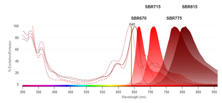
Fig. 1. Excitation and emission spectra of StarBright Red Dyes.
StarBright Red Dyes are perfect replacements for APC and APC tandems in your flow cytometry experiments. They are bright, with unique spectra, not photosensitive, and can be fixed in both formaldehyde and alcohol-based fixatives with minimal changes in performance and spectra, making these dyes an excellent choice for both conventional and full spectrum flow cytometry.
StarBright Red Dyes can be used either with/without a special buffer depending on the requirements of the fluorophores that the StarBright Red Dyes are multiplexed with, offering you flexibility when building panels.
StarBright Red 670 (SBR670) Dye
SBR670 is the first of our new StarBright Red Dye series. It is a bright 640 nm laser excitable dye emitting at 666 nm with a narrow excitation and emission profile (Figure 2).
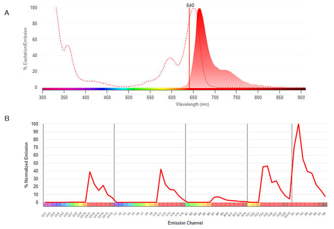
Fig. 2. SBR670. A, Conventional spectra. Excitation by the red laser (dotted line) with emission shown in the shaded histogram. B, Normalized dye signature. Full emission spectrum showing the emission at all wavelengths. Data were collected on a 5-L Cytek Aurora Flow Cytometry System using SpectroFlo Software.
SBR670 has been found to be brighter than Alexa Fluor 647 (A647) (Figure 3) and allophycocyanin (APC) (not shown). It can be used with any common staining buffer and can be fixed in PFA or alcohol-based buffers with minimal loss of signal.
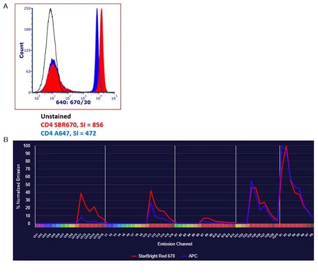
Fig. 3. Comparison of SBR670 with APC and A647. A, Human peripheral blood was stained with CD4 SBR670 (MCA1267SBR670) (red), or CD4 A647 (MCA1267A647) (blue) and analyzed on the ZE5 Cell Analyzer detected using the 670/30 filter. All antibodies were titrated prior to use to determine the optimal concentration. B, Full normalized emission spectrum showing the emission at all wavelengths of APC and SBR670. Data were collected on a 5-L Cytek Aurora Flow Cytometry System using SpectroFlo Software.
Furthermore, as it has a unique spectrum, a similarity score of 0.94, and has a maximal emission in a different channel, StarBright Red 670 Dye can be easily used with APC in full spectrum flow cytometry multicolor panels (Figure 3).
StarBright Red 715 (SBR715) Dye
SBR715 is a very bright 640 nm laser excitable dye emitting at 712 nm with a narrow excitation and emission profile. It has a unique spectrum, with a similarity score of 0.83 compared to A700 (Figure 4).
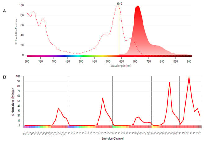
Fig. 4. SBR715. A, Conventional spectra. Excitation by the red laser (dotted line) with emission shown in the shaded histogram. B, Normalized dye signature. Full emission spectrum showing the emission at all wavelengths. Data were collected on a 5-L Cytek Aurora Flow Cytometry System using SpectroFlo Software.
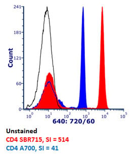
Fig. 5. Brightness comparison. Human peripheral blood was stained with CD4 SBR715 (MCA1267SBR715) (red), or CD4 A700 (MCA1267A700) (blue) and analyzed on the ZE5 Cell Analyzer detected using the 720/60 filter. All antibodies were titrated prior to use to determine the optimal concentration.
There may be some excitation by the 561 nm laser, so there may be spreading when used with dyes such as PE-Cy5.5 or SBY720, but careful panel design will help.
On the ZE5 Cell Analyzer SBR715 has been found to be over 10x brighter than Alexa Fluor 700 (A700) (Figure 5). It can be used with any common staining buffer and can be fixed in PFA or alcohol-based buffers with minimal loss of signal.
StarBright Red 775 (SBR775) Dye
SBR775 is a bright 640 nm laser excitable dye emitting at 778 nm with a unique excitation and emission profile compared to other dyes.
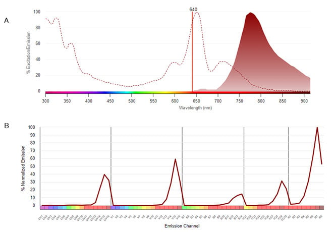
Fig. 6. SBR775. A, Conventional spectra. Excitation by the red laser (dotted line) with emission shown in the shaded histogram. B, Normalized dye signature. Full emission spectrum showing the emission at all wavelengths. Data were collected on a 5-L Cytek Aurora Flow Cytometry System using SpectroFlo Software.
The additional signal from the 355 nm and 405 nm lasers are responsible for the uniqueness with a similarity score of 0.88 compared to APC-Cy7 (Figure 6). SBR775 is as bright as APC-Cy7 with the added benefit of being able to be stably fixed in PFA and alcohol-based fixatives with no loss of signal, which can be a problem for APC-Cy7 (Figure 7).
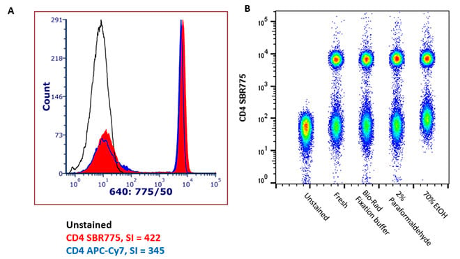
Fig. 7. Brightness comparison and stability after fixation. A, Human peripheral blood was stained with CD4 SBR775 (MCA1267SBR775) (red), or CD4 APC-Cy7 (blue) and analyzed on the ZE5 Cell Analyzer detected using the 775/50 filter. All antibodies were titrated prior to use to determine the optimal concentration. B, No loss of performance was observed when samples were fixed with either PFA or alcohol-based fixatives prior to acquisition. All samples were collected on the ZE5 Cell Analyzer. EtOH, ethanol; PFA, paraformaldehyde.
In addition, SBR775 does not bind to monocytes in a nonspecific manner as has been reported for cyanine-containing dyes (data not shown).
StarBright Red 815 (SBR815) Dye
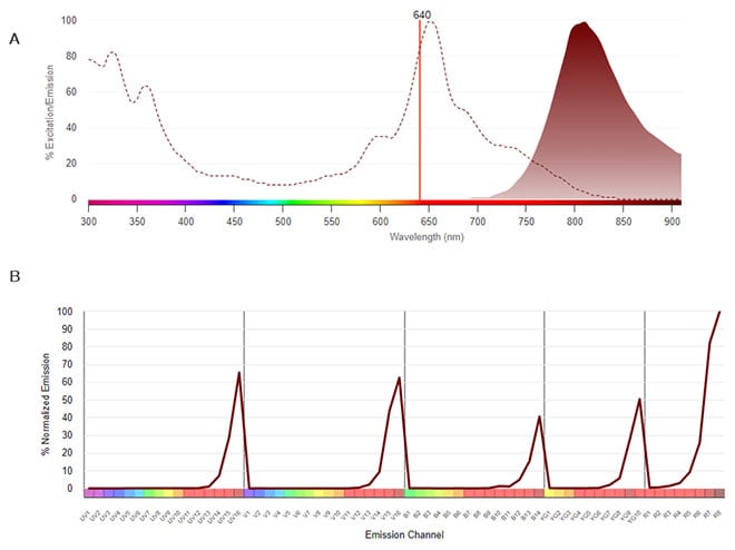
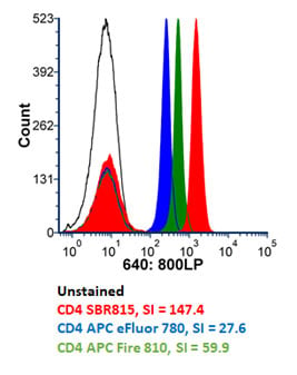
Fig. 9. Brightness comparison. Human peripheral blood was stained with CD4 SBR815 (MCA1267SBR815) (red), CD4 APC-Fire 810 (green) and CD4 APC-eFluor 780 (blue) and analyzed on the ZE5 Cell Analyzer detected using the 800LP filter. All antibodies were titrated prior to use to determine the optimal concentration.
Fig. 8. SBR815. A, Conventional spectra. Excitation by the red laser (dotted line) with emission shown in the shaded histogram. B, Normalized dye signature. Full emission spectrum showing the emission at all wavelengths. Data were collected on a 5-L Cytek Aurora Flow Cytometry System using SpectroFlo Software.
SBR815 is the fourth of our 640 nm excitable StarBright Dyes. A bright 640 nm laser excitable dye emitting at 811 nm with a unique excitation and emission profile compared to other dyes (Figure 8).
SBR815 is brighter than both APC-Fire 810 and APC-eFluor 780 on the ZE5 Cell Analyzer (Figure 9) with the added benefit of being able to be stably fixed in PFA and alcohol-based fixatives with no loss of signal (data not shown).
The additional signal from other lasers is responsible for the uniqueness with a similarity score of 0.81 compared to APC-Fire 810 and 0.79 compared to APC-eFluor 780. SBR815 has been successfully used together in a panel with APC-Fire 810, although significant spread means mutually exclusive markers are preferred (Figure 10).
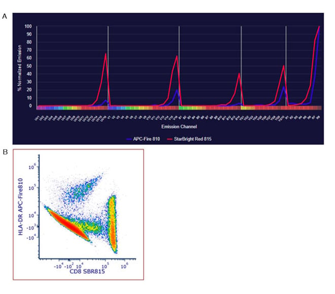
Fig. 10. SBR815 in full spectrum flow cytometry. A, the full emission spectrum of SBR815 and APC-Fire 810 showing the emission at all wavelengths. B, human peripheral blood cells were stained for HLA-DR APC-Fire 810, and CD8 SBR815 (MCA1226SBR815). Data were collected on the Cytek Aurora Flow Cytometry System using SpectroFlo Software.
StarBright Red Multicolor Panel Data
StarBright Red Dyes can be used successfully in combination with other StarBright Dyes and traditional fluorophores in conventional and spectral panels and can be found in posters we have presented at conferences.
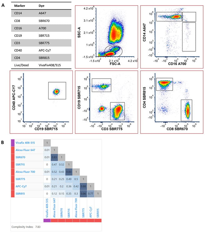
Fig. 11. Eight-color full spectrum flow cytometry panel containing seven 640 nm excitable dyes. A, Human peripheral blood was stained for CD14A647 (MCA1568A647), CD8SBR670 (MCA1226SBR670), CD16A700 (MCA2537A700), CD19SBR715 (MCA1940SBR715), CD3SBR775 (MCA463SBR775), CD40APC-Cy7, CD4SBR815 (MCA1267SBR815), and a 405 nm excitable viability dye VivaFix408/512 (1351113). Data generated on the Cytek Aurora Spectral Analyzer. B, Similarity matrix and complexity score for the eight-color panel.
In addition, we have successfully combined multiple StarBright Red Dyes with other 640 nm excitable dyes to create a stand-alone eight-color panel that contains seven 640 nm excitable dyes in full spectrum flow cytometry, expanding panel building possibilities (Figure 11).
StarBright Red Antibodies
| Description | Target | Format | Clone | Applications | Citations | Code |
|---|
Associated Reading
For more information on how these products can improve the effectiveness of your flow cytometry experiments, please take a look at our StarBright Dyes page.
Useful information about fluorescent dyes and immunophenotyping can be found in our flow cytometry explained knowledge hub. Alternatively, feel free to explore the associated resource links below, for more in-depth information about each cited topic. If you are interested in learning the fundamentals of this application, please view our flow cytometry basics guide.
