
TrailBlazer™ Tag and TrailBlazer StarBright™ Dye Label Kits in Veterinary Studies
 TrailBlazer™ Tag and TrailBlazer StarBright™ Dye Label Kits in Veterinary Studies
Flow cytometry is increasingly used for immunophenotyping in veterinary diagnostics, disease monitoring, and animal model research. Unlike in human and mouse research, veterinary flow cytometry panels are often constrained by the limited availability of fluorophore-conjugated antibodies.
Researchers working with feline, porcine, bovine, canine, and other veterinary species may need to split markers across multiple smaller panels, increasing sample requirements and making comprehensive immune profiling more challenging.
TrailBlazer Tag and StarBright Dye Label Kits help overcome these limitations by enabling purified antibodies to be conjugated to one of 32 premium StarBright Dyes, providing greater flexibility to build larger, better-optimized veterinary immunophenotyping panels and identify more cell populations within a single assay.
Why Use TrailBlazer Kits in Veterinary Research?
- Overcome fluorophore limitations—access to 32 StarBright Dye options for conjugating purified antibodies
- Optimize panel design—choose the optimal fluorophore combinations that help reduce spreading and improve population resolution
- Eliminate secondary antibodies—simplify staining and avoid secondary antibody binding to multiple primary antibodies of the same isotype
- Identify more cell populations in a single panel—expand smaller panels into higher plex panels for deeper immune profiling
- Save time and sample—combine multiple panels into one larger panel to reduce processing time, sample required, and for quicker analysis
TrailBlazer Kit Benefits
- Label a wide range of antibodies—use purified antibodies across different isotypes and storage buffers, including buffers containing BSA
- Generate stable labeled antibodies—one-year stability of kit conjugates means they can be treated like a purchased directly conjugated antibody
The examples below show how TrailBlazer Kits can be used to build larger flow cytometry panels by combining directly conjugated, cross-reactive, and TrailBlazer Kit-labeled antibodies to reveal more detailed information about the immune system.
Flow Cytometry Panels Using TrailBlazer Kits
Feline Panels
Expanding Panels with Cross-Reactive and TrailBlazer Kit-Labeled Antibodies
Although Bio-Rad has the largest range of feline antibodies, present fluorophore options restrict feline panel sizes, limiting the number of cell populations that can be identified. One of several possible 4-color panels is shown in this example (Table 1 and Figure 1), where CD4+ T cells (T helper cells) and their activation status can be identified in feline blood.
Table 1. Reagents used in the 4-color feline panel.
Target |
Fluorophore |
Catalog Number |
|---|---|---|
|
CD4 |
FITC |
|
|
CD5 |
PE |
|
|
Viability Dye |
VivaFix 583/603 |
|
|
CD134 |
A647 |
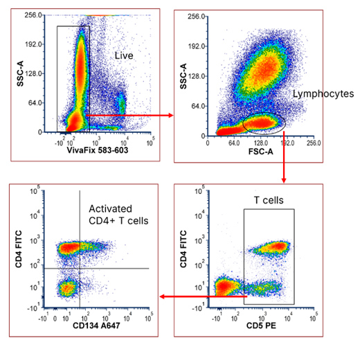
Fig. 1. 4-color feline panel. Red cell lysed feline blood was stained with a 4-color panel (Table 1) and acquired on a ZE5 Cell Analyzer. Single cell gate not shown.
To enable identification of B cells and monocytes, validated cross-reactive antibodies against CD3, CD14, and CD45RA were introduced, expanding the panel to 7-colors (Table 2 and Figure 2).
Table 2. Reagents used in the 7-color feline panel, including cross-reactive antibodies.
Target |
Fluorophore |
Catalog Number |
|---|---|---|
|
CD14 |
SBUV540 |
|
|
CD3 |
Pacific Blue |
|
|
CD4 |
FITC |
|
|
CD5 |
PE |
|
|
Viability Dye |
VivaFix 583/603 |
|
|
CD134 |
A647 |
|
|
CD45R |
SBR815 |
* Cross-reactive antibodies.
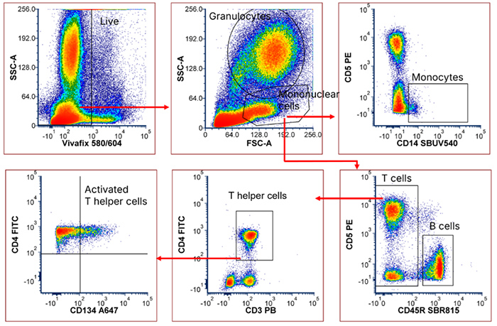
Fig. 2. 7-color feline panel including cross-reactive antibodies. Red cell lysed feline blood was stained with the panel shown in Table 2 and acquired on a ZE5 Cell Analyzer. Single cell gate not shown.
To enable identification of all major blood immune cell populations in this panel, including T helper cells, cytotoxic T cells, granulocytes, monocytes, and B cells, the panel was further expanded with TrailBlazer StarBright Dye Kit-labeled antibodies against CD8, CD18, and CD21, creating a larger 10-color panel (Table 3 and Figure 3).
Table 3. Reagents used in the 10-color feline panel. StarBright Dye TrailBlazer Kit-labeled antibodies were generated by using a TrailBlazer Tag Kit (12020038) and the indicated TrailBlazer StarBright Dye Label Kit.
Target |
Fluorophore |
Catalog Number |
TrailBlazer Label Kit Catalog Number |
|---|---|---|---|
|
CD8 |
SBUV400 |
||
|
CD14 |
SBUV540 |
||
|
CD3 |
Pacific Blue |
||
|
CD21 |
SBV760 |
MCA1781R * |
|
|
CD4 |
FITC |
||
|
CD5 |
PE |
||
|
Viability Dye |
VivaFix 583/603 |
||
|
CD18 |
SBY720 |
||
|
CD134 |
A647 |
||
|
CD45R |
SBR815 |
* Cross-reactive antibodies.
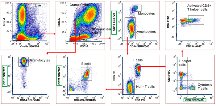
Fig. 3. 10-color feline panel incorporating cross-reactive antibodies and StarBright Dye TrailBlazer Kit-labeled antibodies. Red cell lysed feline blood was stained with a panel (Table 3) and acquired on a ZE5 Cell Analyzer. Single cell gate not shown. TrailBlazer Kit-labeled antibodies are highlighted in green.
Porcine
Identifying a Low Antigen Density T Cell Marker with TrailBlazer Kit-Labeled Antibodies
TrailBlazer Kits can be used to label antibodies against a wide range of targets, including low-density markers and markers expressed on small cell populations. In pigs, CD27 is an important marker for assessing T cell memory status, but limited fluorophore availability and low antigen density—which require a bright fluorophore—can make it challenging to include in larger panels. This challenge was addressed by conjugating the CD27 antibody to the bright StarBright Violet 670 fluorophore (brightness 5/5). TrailBlazer Kits also enabled CD44, a pan T cell marker, to be added to the panel using the slightly less bright but spectrally narrow StarBright Violet 515 dye (brightness 4/5).
The data (Figure 4) were consistent with published literature, and clearly identified distinct naive, central memory (CM), and effector memory (EM) T cell populations. The small increase in expression of CD11a as cells differentiated from naive to EM cells within the CD8 memory populations confirmed the validity of these populations (Álvarez et al., 2025).
Table 4. Reagents used in the porcine 9-color T cell panel. StarBright Dye TrailBlazer Kit-labeled antibodies were generated by using a TrailBlazer Tag Kit (12020038) and the indicated TrailBlazer StarBright Dye Label Kit.
Target |
Fluorophore |
Catalog Number |
TrailBlazer Label Kit Catalog Number |
|---|---|---|---|
|
CD3 |
SBUV400 |
||
|
Viability Dye |
DAPI |
||
|
CD8 |
SBUV740 |
||
|
CD45 |
PB |
||
|
CD44 |
SBV515 |
||
|
CD27 |
SBV670 |
||
|
CD11a |
FITC |
||
|
CD45RA |
PE |
||
|
CD4 |
A647 |
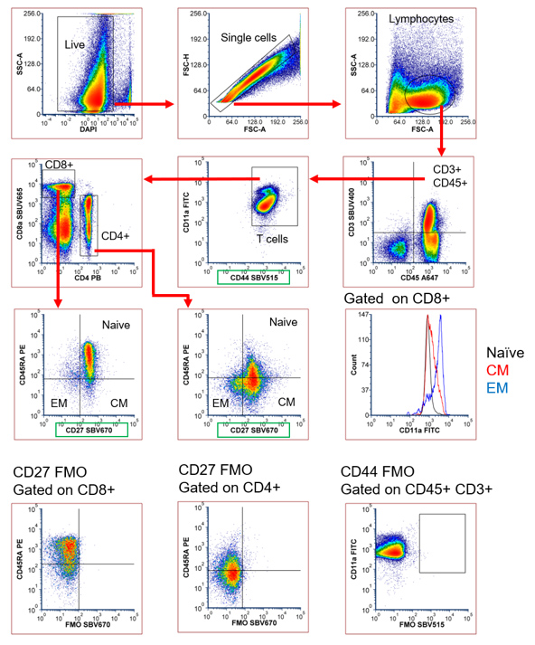
Fig. 4. Porcine 9-color T cell panel. Red cell lysed porcine blood was stained with an antibody panel shown in Table 4 and acquired on a ZE5 Cell Analyzer. TrailBlazer Kit-labeled antibodies are highlighted in green.
Large Immunophenotyping Panel—Multiplexing TrailBlazer Kit Conjugates
Beyond the cell type-specific panel, TrailBlazer Kits enabled a previously published 10-color general immunophenotyping panel to be expanded into a larger 19-color panel for high-dimensional immunophenotyping. This expanded panel enables deeper characterization of naive, central memory (CM), and effector memory (EM) T cells, regulatory T cells (T regs), B cell populations, and γδ T cells. Populations that previously required three separate panels to identify (Clark et al., 2019) can now be assessed in a single panel.
Table 5. Reagents used in the expanded 19-color porcine panel. StarBright Dye TrailBlazer Kit-labeled antibodies were generated by using a TrailBlazer Tag Kit (12020038) and the indicated TrailBlazer StarBright Dye Label Kit.
Target |
Fluorophore |
Catalog Number |
TrailBlazer Label Kit Catalog Number |
|---|---|---|---|
|
CD3 |
SBUV400 |
||
|
CD16 |
SBUV510 |
||
|
CD9 |
SBUV575 |
||
|
CD45 |
SBUV665 |
||
|
SWC5 |
SBUV740 |
||
|
CD8a |
SBUV795 |
||
|
FOXP3 |
BV421 |
404-5773-82 (Thermo) |
|
|
Viability Dye |
Vivafix 410/450 |
||
|
CD4 |
SBV515 |
||
|
SLA Class II |
SBV610 |
||
|
CD25 |
SBV710 |
||
|
CD45RA |
SBV790 |
||
|
CD21 |
FITC |
4530-02 (Southern Biotech) |
|
|
Granulocytes |
SBB700 |
||
|
γδTCR |
PE |
561486 (Waters) |
|
|
CD11R3 |
SBY605 |
||
|
CD2 |
PE-Cy5 |
300210 (BioLegend) |
|
|
CD27 |
A647 |
||
|
CD14 |
SBR775 |
* Cross-reactive antibody. Antibodies from Bio-Rad unless indicated.
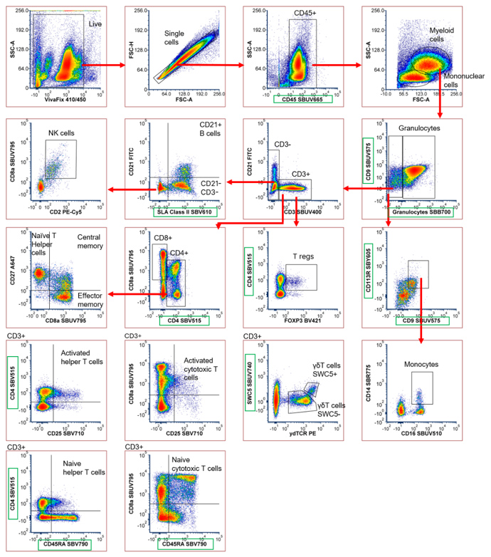
Fig. 5. Expanded 19-color porcine panel incorporating cross-reactive antibodies and StarBright Dye TrailBlazer Kit-labeled antibodies. Red cell lysed porcine blood was stained with an antibody panel shown in Table 5 and acquired on a ZE5 Cell Analyzer. TrailBlazer Kit-labeled antibodies are highlighted in green.
TrailBlazer Kits enable a step change in panel size, allowing more immune cell populations to be detected in a single assay. Whether working with feline, porcine, or other non-human species where reagent options are limited, these kits provide greater choice and flexibility to support more detailed immunophenotyping for research and diagnosis.
Discover the full range of TrailBlazer Tag and TrailBlazer StarBright Dye Label Kits.
View Range NowReferences
- Álvarez B et al. (2025). Phenotypes of porcine blood CD8β T cells and their capacity for IFN gamma production in the context of PRV vaccination. Dev Comp Immunol 165, 105347
- Clark D et al. Immunophenotyping Pig Immune Cells by Flow Cytometry: A Powerful Tool for Pharmacology and Toxicology Studies. Poster
Frequently Asked Questions
How can I increase the number of markers in a veterinary flow cytometry panel?
TrailBlazer Tag and StarBright Dye Label Kits allow purified antibodies to be conjugated to one of 32 StarBright Dyes, increasing fluorophore choice and enabling larger immunophenotyping panels.
Can cross-reactive antibodies be used in veterinary flow cytometry?
Yes. Cross-reactive antibodies are commonly used when species-specific reagents are unavailable. Combining these antibodies with TrailBlazer-conjugated antibodies can expand panel size substantially.
What species can TrailBlazer Kits support?
Any species where purified antibodies are available, including feline, porcine, canine, bovine, and other veterinary research models.
Why use directly conjugated antibodies instead of secondary antibodies?
Directly conjugated antibodies reduce workflow complexity and eliminate unwanted binding caused by secondary antibodies.
Useful TrailBlazer Tag and TrailBlazer StarBright Dye Label Kit Information
- TrailBlazer Tag and Label Kits — Frequently Asked QuestionsFurther information regarding antibody compatibility, technology, and how to select the right kit for your experiment.
- TrailBlazer Tag and Label Kits ProtocolFind out how to use these kits to get the best results. The protocols cover general kit instructions for use (both Tag and Label Kits).
- TrailBlazer Tag and Label Kits — Inquiry FormIf you have any questions about the TrailBlazer Kits, simply complete and submit a short form and a member of our team will respond to your inquiry.