PUREBLU™ DAPI


PUREBLU™ DAPI
- Product Type
- Accessory Reagent
- Specificity
- PUREBLU™ DAPI
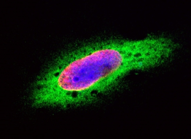
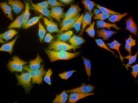
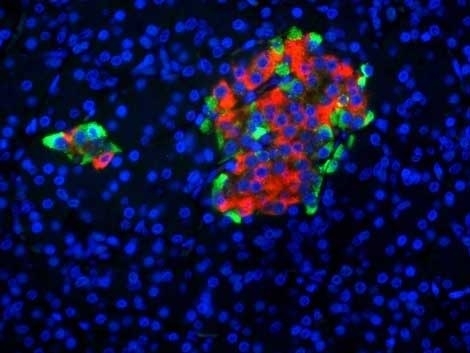
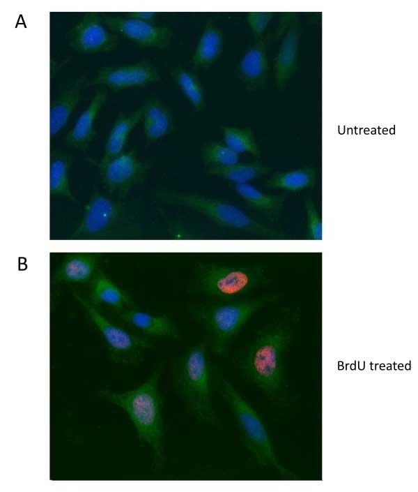
| DAPI is a cell permeable fluorescent compound (MW 350.3) that is able to stain the DNA of eukaryotic and prokaryotic cells by binding with high affinity to the minor groove of AT-rich DNA sequences. When DAPI is bound to DNA and excited by an ultraviolet light source, blue fluorescent emission can be detected with maximum emission at 461 nm. PureBlu DAPI has a characteristic Stokes shift of approximately 100 nm, which makes this dye an optimal choice when good spectral separation is desired. PureBlu DAPI is compatible with fixed and unfixed cells. |
- Reconstitution
- Reconstitute one vial of lyophilized PureBlu DAPI Dye with 500 μl of de-ionized water, then vortex briefly to make a 100x stock solution.
- Reagents In The Kit
- 5 vials, 50 μg each, DAPI nuclear staining dye powder
- Regulatory
- For research purposes only
- Guarantee
- Guaranteed until date of expiry. Please see product label.
- Acknowledgements
- Triton is a trademark of Dow Chemical Company
After reconstitution store at +4°C or at -20°C if preferred
This product is photosensitive and should be protected from light. PureBlu DAPI Dye is stable for 12 months from date of reconstitution if stored at - 20°C or 6 months at 2-8°C.
| Application Name | Verified | Min Dilution | Max Dilution |
|---|---|---|---|
| Flow Cytometry | 1/100 | ||
| Immunofluorescence | 1/100 |
- Instructions For Use
- Note: The optimal concentration for different cell types should be determined empirically.
Staining Procedure - Flow Cytometry Cell Viability
1. Grow cells of interest under conditions specific for the cell type. Harvest cells and obtain a single cell suspension.
2. Check cell numbers and viability using trypan blue.
3. Resuspend cells at 2 x 10⁶ cells/ml in 0.5ml Staining Buffer (BUF073) or 1x PBS.
4. Add 5μl of 100x PureBlu DAPI Dye stock solution to 0.5ml cells.
5. Stain at room temperature for 15 min in the dark.
6. Optional: Rinse cells with Staining Buffer (BUF073) or 1x PBS.
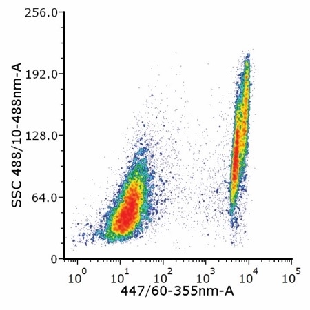
7. Proceed with analysis by flow cytometry. When bound to double-stranded DNA (dsDNA) PureBlu DAPI Dye can be excited with either a 355 nm or a 405 nm laser, with optimum emission at 461nm.
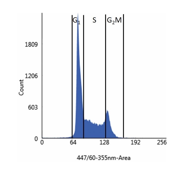
Staining Procedure - Flow Cytometry Cell DNA Content
1. Grow cells of interest under conditions specific for the cell type. Harvest cells and obtain a single cell suspension.
2. Check cell numbers and viability using trypan blue.
3. Wash cells in Staining Buffer (BUF073) or 1x PBS.
4. Fix in ice cold 70% ethanol for 2 hr at 4°C.
5. Wash cells in Staining Buffer (BUF073) or 1x PBS.
6. Stain cells with 5μl of PureBlu DAPI Dye stock solution to 0.5 ml cells at a concentration of 2 x 10⁶ cells/ml in cell staining buffer containing 0.1% Triton X-100.
Note: Optimal concentrations of cells and PureBlu DAPI may vary depending on cell type and should be determined through careful titration prior to final experimental analysis.
7. Stain at room temperature for 30 min in the dark.
8. Optional: Rinse cells with Staining Buffer (BUF073) or 1x PBS.
9. Proceed with analysis by flow cytometry. When bound to double-stranded DNA (dsDNA) PureBlu DAPI Dye can be excited with either a 355 nm or a 405 nm laser, with optimum emission at 461nm.
Staining Procedure - Cell Nuclear Visualization in Microscopy and Cell Imaging
Preparation of the 1x staining solution: Dilute the 100x stock solution 1/100 with PBS for a final concentration of 1 μg/ml.
1. Grow cells of interest under conditions specific for the cell type.
2. Rinse cells with 1x PBS.
3. Optional: Rinse cells with 1x PBS and permeabilize them with 1x PBST (0.1% Triton X-100 in 1x PBS) at room temperature for 5 min.
5. Rinse cells with 1x PBS.
6. Stain with 1x staining solution (diluted with PBS) at room temperature for 15 min.
7. Rinse cells with 1x PBS.
8. Optional: Remove PBS and mount cells in antifade mounting media.
9. Image cells.
| Description | Product Code | Applications | Pack Size | List Price | Your Price | Quantity | |
|---|---|---|---|---|---|---|---|
| Staining Buffer | BUF073 | F | 500 ml | Log in | |||
| List Price | Your Price | ||||||
| Log in | |||||||
| Description | Staining Buffer | ||||||
References for PUREBLU™ DAPI
-
She, D.T. et al. (2018) SIRT2 Inhibition Confers Neuroprotection by Downregulation of FOXO3a and MAPK Signaling Pathways in Ischemic Stroke.
Mol Neurobiol. 55 (12): 9188-203. -
Carter-Timofte, M.E. et al. (2021) Antiviral Potential of the Antimicrobial Drug Atovaquone against SARS-CoV-2 and Emerging Variants of Concern.
ACS Infect Dis. acsinfecdis.1c00278. -
Earnest, K.G. et al. (2021) Development and characterization of a DNA aptamer for MLL-AF9 expressing acute myeloid leukemia cells using whole cell-SELEX.
Sci Rep. 11 (1): 19174. -
Paterno, R. et al. (2021) Hippocampal gamma and sharp-wave ripple oscillations are altered in a Cntnap2 mouse model of autism spectrum disorder.
Cell Rep. 37 (6): 109970. -
Roux-Biejat, P. et al. (2021) Acid Sphingomyelinase Controls Early Phases of Skeletal Muscle Regeneration by Shaping the Macrophage Phenotype.
Cells. 10 (11): 3028. -
Nogueira-Rodrigues, J. et al. (2021) Rewired glycosylation activity promotes scarless regeneration and functional recovery in spiny mice after complete spinal cord transection.
Dev Cell. 57 (4): 440-450.e7. -
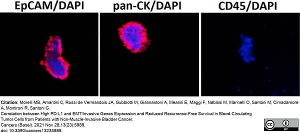
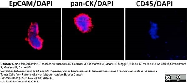
Morelli, M.B. et al. (2021) Correlation between High PD-L1 and EMT/Invasive Genes Expression and Reduced Recurrence-Free Survival in Blood-Circulating Tumor Cells from Patients with Non-Muscle-Invasive Bladder Cancer.
Cancers (Basel). 13 (23): 5989. -
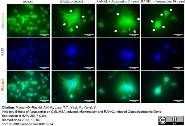
Mamun-Or-Rashid, A.N.M. et al. (2021) Inhibitory Effects of Astaxanthin on CML-HSA-Induced Inflammatory and RANKL-Induced Osteoclastogenic Gene Expression in RAW 264.7 Cells
Biomedicines. 10 (1): 54.
View The Latest Product References
-
Saito, N. et al. (2022) Impaired dental implant osseointegration in rat with streptozotocin-induced diabetes.
J Periodontal Res. 57 (2): 412-24. -
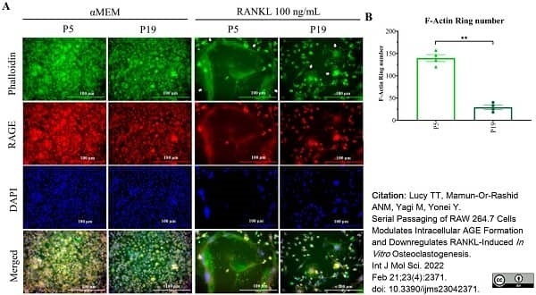
Lucy, T.T. et al. (2022) Serial Passaging of RAW 264.7 Cells Modulates Intracellular AGE Formation and Downregulates RANKL-Induced In Vitro. Osteoclastogenesis.
Int J Mol Sci. 23 (4): 2371. -
Foo, S.L. et al. (2022) Breast cancer metastasis to brain results in recruitment and activation of microglia through annexin-A1/formyl peptide receptor signaling.
Breast Cancer Res. 24 (1): 25. -
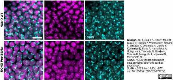
Itai, T. et al. (2023) A novel NONO variant that causes developmental delay and cardiac phenotypes
Sci Rep. 13 (1): 975. -
Alaryan, M.M. et al. (2023) Brassica Cover Crops and Natural Spongospora subterranea Infestation of Peat-Based Potting Mix May Increase Powdery Scab Risk on Potato.
Plant Dis. 107 (9): 2769-77. -
van Schalkwyk, M.C.I. et al. (2021) Development and Validation of a Good Manufacturing Process for IL-4-Driven Expansion of Chimeric Cytokine Receptor-Expressing CAR T-Cells.
Cells. 10 (7): 1797. -
Watanabe, T. et al. (2024) Senotherapeutic effect of Agrimona pilosa Ledeb. In targeting senescent cells in naturally aged mice
Food Bioscience. : 103903.
Please Note: All Products are "FOR RESEARCH PURPOSES ONLY"
Always be the first to know.
When we launch new products and resources to help you achieve more in the lab.
Yes, sign me up