CD45R antibody | RA3-6B2
Rat anti Mouse CD45R
- Product Type
- Monoclonal Antibody
- Clone
- RA3-6B2
- Isotype
- IgG2a
- Specificity
- CD45R

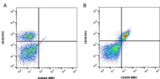
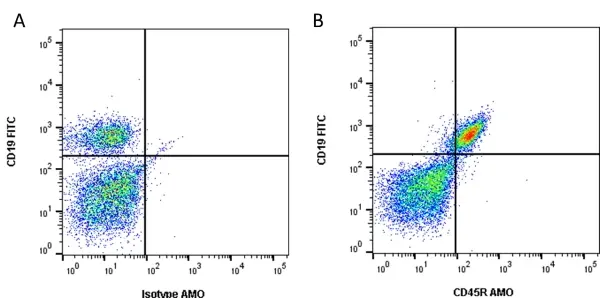
Figure B. FITC conjugated Rat anti Mouse CD19 antibody, clone 6D5 (MCA1439F) and Amethyst Orange conjugated Rat anti Mouse CD45R antibody, clone RA3-6B2 (MCA1258AMO). All experiments performed on mouse blood gated on live single cells, in the presence of 10% mouse serum.
Data acquired on the ZE5 Cell analyser.
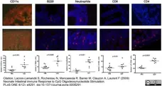
Rat anti Mouse CD45R antibody, clone RA3-6B2 (MCA1258) used for the enumeration of B cells in the intestinal mucosa by immunofluorescence.
Image caption:
Recruitment of inflammatory cells in the intestine of neonates after CpG-ODN treatment. Eight-day-old neonatal mice received 20 μg/g of CpG-ODN orally. Twenty-four hours after treatment, ilea were removed to analyze the recruitment of inflammatory cells by immunohistochemistry. Six to ten neonates from different litters (5 sections for each animal) per group were analyzed. Data were analyzed by non-parametric Mann-Whitney test.
From: Lacroix-Lamandé S, Rochereau N, Mancassola R, Barrier M, Clauzon A, Laurent F (2009)
Neonate Intestinal Immune Response to CpG Oligodeoxynucleotide Stimulation.
PLoS ONE 4(12): e8291.
doi: 10.1371/journal.pone.0008291
This image is from an open access article distributed under the terms of a Creative Commons Attribution License.
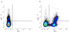
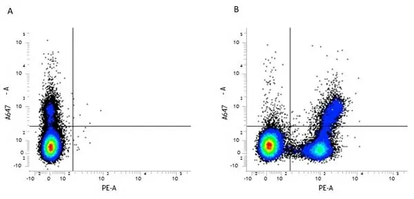
Figure B. Alexa Fluor® 647 conjugated Hamster anti Mouse CD79b antibody, clone HM79-11 (MCA1821A647) and RPE conjugated Rat anti Mouse CD45R antibody, clone RA3-6B2 (MCA1258PE). All experiments performed on murine bone marrow in the presence of murine SeroBlock (BUF041A).
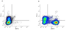
Figure B. RPE conjugated Mouse anti Mouse CD28 antibody, clone E18 (MCA2473PE) and Alexa Fluor® 647 conjugated Rat anti Mouse CD45 antibody, clone RA3-6B2 (MCA1258A647). All experiments performed on murine bone marrow in the presence of murine SeroBlock (BUF041A).
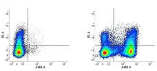
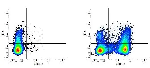
Figure B. RPE conjugated Rat anti Mouse CD36 antibody, clone MF3 (MCA2748PE) and Alexa Fluor® 488 conjugated Rat anti Mouse CD45R antibody, clone RA3-6B2 (MCA1258A488). All experiments performed on murine bone marrow in the presence of murine SeroBlock (BUF041A).
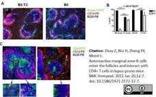
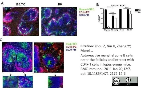
Rat anti Mouse CD45R antibody, clone RA3-6B2 (MCA1258G) used for the identification of marginal zone B cells in mouse spleen by immunofluorescence on cryostat sections.
Image caption:
Intrafollicular location of MZB cells in B6.TC spleens. A. Representative spleen sections from 3 mo old B6.TC (left) and B6 (right) mice stained with Moma-1-FITC, CD1d-PE and B220-PB. The B220+ CD1d+ MZB cells show as bright pink while the other B cells show as blue. The ring of Moma-1+ metallophillic macrophages delineates the MZ inner edge. B. Percentage of CD1d+ B220+ B cells relative to total B220+ B cells outside (MZ) and inside (FO) the Moma-1+ ring in 3 mo and 10 mo old B6 and B6.TC mice. The data show means and standard errors of the mean (SEM) calculated of 4 MZ and FO areas for each mouse. ***: p < 0.001 for t tests. C. Representative spleen sections from 10 mo old B6.TC (left) and B6 (right) mice stained with CD4-FITC, CD1d-PE and B220-PB. Boxed areas show multiple contacts between green B6.TC CD4 T cells and pink MZB cells, but not in the B6 spleen. Original magnification: 200X.
From: Zhou Z, Niu H, Zheng YY, Morel L.
Autoreactive marginal zone B cells enter the follicles and interact with CD4+ T cells in lupus-prone mice.
BMC Immunol. 2011 Jan 20;12:7.
doi: 10.1186/1471-2172-12-7
This image is from an open access article distributed under the terms of Creative Commons Attribution License.

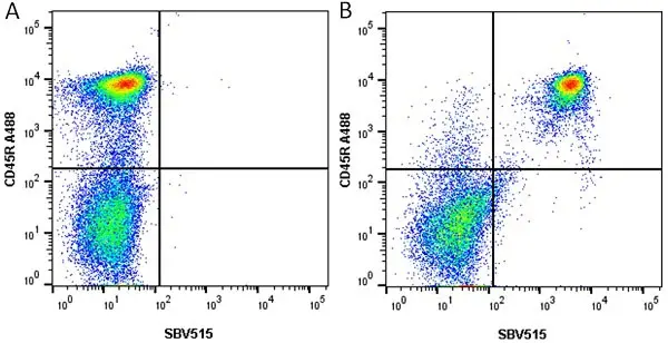
Figure B. Alexa Fluor® 488 conjugated Rat anti Mouse CD45R antibody, clone RA3-6B2 (MCA1258A488) and SBV515 conjugated Rat anti Mouse CD19 antibody, clone 6D5 (MCA1439SBV515). All experiments performed on red cell lysed mouse blood gated on live single cell lymphocytes, in the presence of 10% mouse serum.
Data acquired on the ZE5 Cell analyser.
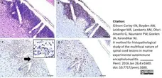
Rat anti Mouse CD45R antibody, clone RA3-6B2 (MCA1258G) used for the demonstration of B cells in murine spinal column by immunohistochemistry on formalin fixed, paraffin embedded tissue sections.
Image caption:
Representative images of key immunohistochemical markers run on a longitudinally sectioned, decalcified spinal column from an EAE mouse with a clinical EAE score of 4.
(A) HE, (B) B220 immunohistochemistry for (B) cells, (C) F4/80 immunohistochemistry for macrophages (arrow indicates area highlighted in inset, inset bar = 20 μm), (D) CD3 immunohistochemistry for T cells. Bars = 200 μm.
From: Gibson-Corley KN, Boyden AW, Leidinger MR, Lambertz AM, Ofori-Amanfo G, Naumann PW, Goeken JA, Karandikar NJ.
A method for histopathological study of the multifocal nature of spinal cord lesions in murine experimental autoimmune encephalomyelitis.
PeerJ. 2016 Jan 26; 4: e1600.
doi: 10.7717/peerj.1600.
This image is from an open access article distributed under the terms of a Creative Commons Attribution License.
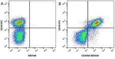
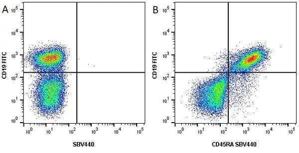
Figure B. FITC conjugated Rat anti Mouse CD19 antibody, clone 6D5 (MCA1439F) and SBV440 conjugated Rat anti Mouse CD45R antibody, clone RA3-6B2 (MCA1258SBV440). All experiments performed on mouse splenocytes in the presence of 10% mouse serum.
Data acquired on the ZE5 Cell analyser.

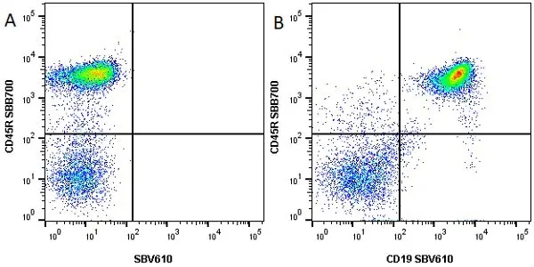
Figure B. StarBright Blue 700 conjugated Rat anti Mouse CD45R antibody, clone RA3-6B2 (MCA1258SBB700) and StarBright Violet 610 conjugated Rat anti Mouse CD19 antibody, clone 6D5 (MCA1439SBV610). All experiments performed on red blood lysed mouse blood gated on live single cell lymphocytes, in the presence of 10% mouse serum.
Data acquired on the ZE5 Cell analyser.

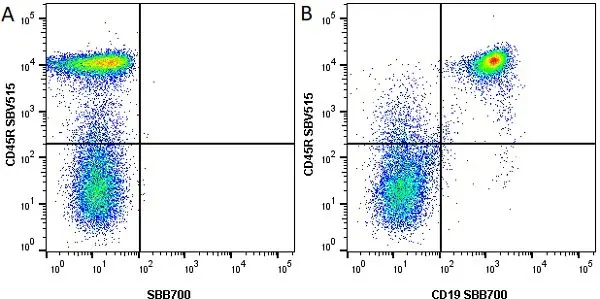
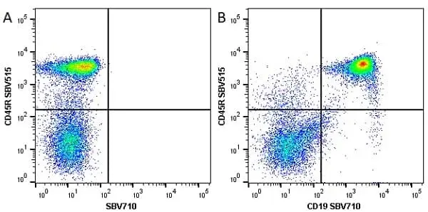
Figure B. StarBright Violet 515 conjugated Rat anti Mouse CD45R antibody, clone RA3-6B2 (MCA1258SBV515) and StarBright Blue 700 conjugated Rat anti Mouse CD19 antibody, clone 6D5 (MCA1439SBB700). All experiments performed on red cell lysed mouse blood gated on live single cell lymphocytes, in the presence of 10% mouse serum.
Data acquired on the ZE5 Cell analyser.
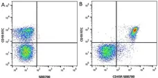
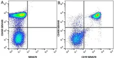
Figure B. StarBright Violet 440 conjugated Rat anti Mouse CD45R antibody, clone RA3-6B2 (MCA1258SBV440) and StarBright Violet 670 conjugated Rat anti Mouse CD19 antibody, clone 6D5 (MCA1439SBV670). All experiments performed on red cell lysed mouse blood gated on live lymphocytes, in the presence of 10% mouse serum.
Data acquired on the ZE5 Cell analyser.
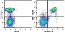
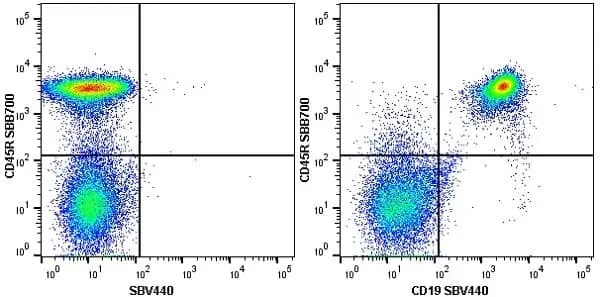
Figure B. StarBright Blue 700 conjugated Rat anti Mouse CD45R antibody, clone RA3-6B2 (MCA1258SBB700) and StarBright Violet 440 conjugated Rat anti Mouse CD19 antibody, clone 6D5 (MCA1439SBV440). All experiments performed on red blood lysed mouse blood gated on live single cell lymphocytes, in the presence of 10% mouse serum.
Data acquired on the ZE5 Cell analyser.

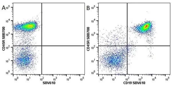
Figure B. StarBright Blue 700 conjugated Rat anti Mouse CD45R antibody, clone RA3-6B2 (MCA1258SBB700) and StarBright Violet 610 conjugated Rat anti Mouse CD19 antibody, clone 6D5 (MCA1439SBV610). All experiments performed on red blood lysed mouse blood gated on live single cell lymphocytes, in the presence of 10% mouse serum.
Data acquired on the ZE5 Cell analyser.
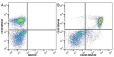
Figure B. StarBright Violet 440 conjugated Rat anti Mouse CD19 antibody, clone 6D5 (MCA1439SBV440) and StarBright Violet 610 conjugated Rat anti Mouse CD45R antibody, clone RA3-6B2 (MCA1258SBV610). All experiments performed on red blood lysed mouse blood gated on live single cell lymphocytes, in the presence of 10% mouse serum.
Data acquired on the ZE5 Cell analyser.
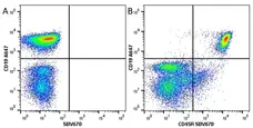
Figure B. Alexa Fluor® 647 conjugated Rat anti Mouse CD19 antibody, clone 6D5 (MCA1439A647) and StarBright Violet 670 conjugated Rat anti Human Mouse CD45R antibody, clone RA3-6B2 (MCA1258SBV670). All experiments performed on red cell lysed mouse blood gated on live single cell lymphocytes, in the presence of 10% mouse serum.
Data acquired on the ZE5 Cell analyser.

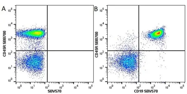
Figure B. StarBright Blue 700 conjugated Rat anti Mouse CD45R antibody, clone RA3-6B2 (MCA1258SBB700) and StarBright Violet 570 conjugated Rat anti Mouse CD19 antibody, clone 6D5 (MCA1439SBV570). All experiments performed on red cell lysed mouse blood gated on live single cell lymphocytes, in the presence of 10% mouse serum.
Data acquired on the ZE5 Cell analyser.
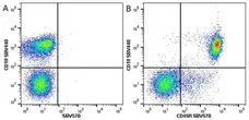
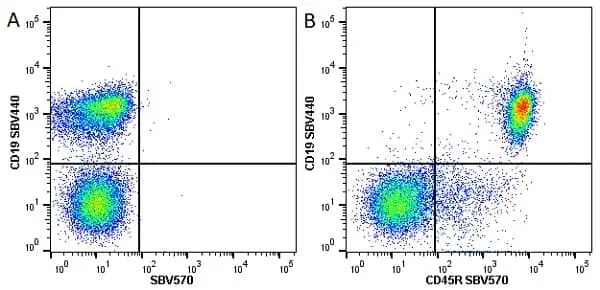
Figure B. StarBright Violet 440 conjugated Rat anti Mouse CD19 antibody, clone 6D5 (MCA1439SBV440) and StarBright Violet 570 conjugated Rat anti Mouse CD45R antibody, clone RA3-6B2 (MCA1258SBV570). All experiments performed on red cell lysed mouse blood gated on live single cell lymphocytes, in the presence of 10% mouse serum.
Data acquired on the ZE5 Cell analyser.

Figure B. StarBright Violet 440 conjugated Rat anti Mouse CD19 antibody, clone 6D5 (MCA1439SBV440) and StarBright Violet 710 conjugated Rat anti Mouse CD45R antibody, clone RA3-6B2 (MCA1258SBV710). All experiments performed on red cell lysed mouse blood gated on live single cell lymphocytes, in the presence of 10% mouse serum.
Data acquired on the ZE5 Cell analyser.

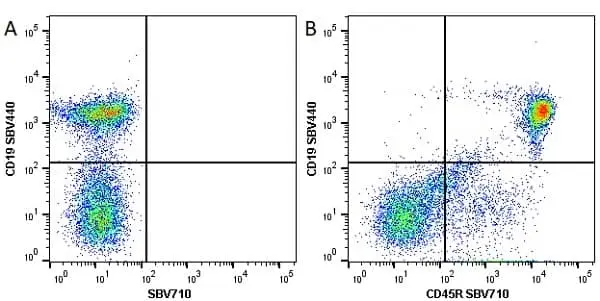
Figure B. StarBright Violet 440 conjugated Rat anti Mouse CD45R antibody, clone RA3-6B2 (MCA1258SBV440) and StarBright Violet 710 conjugated Rat anti Mouse CD19 antibody, clone 6D5 (MCA1439SBV710). All experiments performed on red cell lysed mouse blood gated on live single cell lymphocytes, in the presence of 10% mouse serum.
Data acquired on the ZE5 Cell analyser.
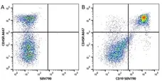
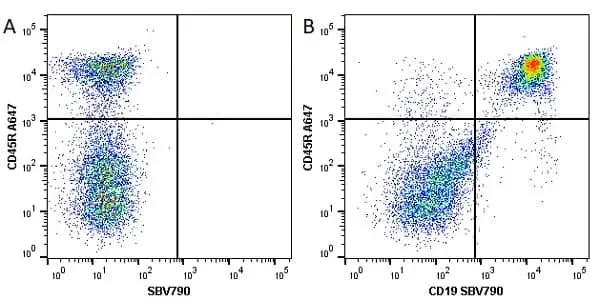
Figure B. Alexa Fluor® 647 conjugated Rat anti Mouse CD45R antibody, clone RA3-6B2 (MCA1258A647) and StarBright Violet 790 conjugated Rat anti Mouse CD19 antibody, clone 6D5 (MCA1439SBV790). All experiments performed on red cell lysed mouse blood gated on live single cell lymphocytes, in the presence of 10% mouse serum.
Data acquired on the ZE5 Cell analyser.
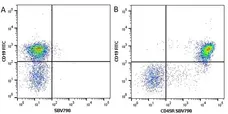
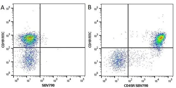
Figure B. FITC conjugated Rat anti Mouse CD19 antibody, clone 6D5 (MCA1439F) and StarBright Violet 790 conjugated Rat anti Mouse CD45R antibody, clone RA3-6B2 (MCA1258SBV790). All experiments performed on red cell lysed mouse blood gated on live single cell lymphocytes, in the presence of 10% mouse serum.
Data acquired on the ZE5 Cell analyser.

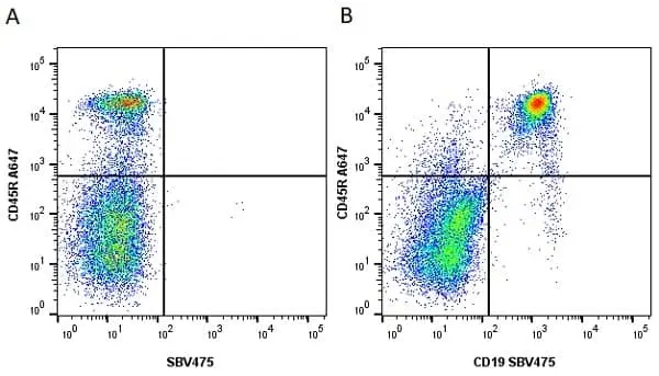
Figure B. Alexa Fluor® 647 conjugated Rat anti Mouse CD45R antibody, clone RA3-6B2 (MCA1258A647) and StarBright Violet 475 conjugated Rat anti Mouse CD19 antibody, clone 6D5 (MCA1439SBV475). All experiments performed on red cell lysed mouse blood gated on live single cell lymphocytes, in the presence of 10% mouse serum.
Data acquired on the ZE5 Cell analyser.
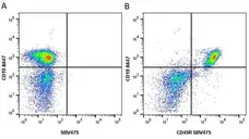
Figure B. Alexa Fluor® 647 conjugated Rat anti Mouse CD19 antibody, clone 6D5 (MCA1439A647) and StarBright Violet 475 conjugated Rat anti Mouse CD45R antibody, clone RA3-6B2 (MCA1258SBV475). All experiments performed on red cell lysed mouse splenocytes gated on live single cells, in the presence of 10% mouse serum.
Data acquired on the ZE5 Cell analyser.
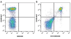
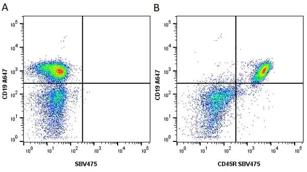
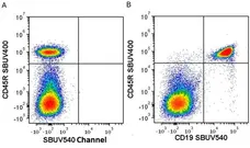
Figure B. Alexa Fluor® 647 conjugated Rat anti Mouse CD45R antibody, clone RA3-6B2 (MCA1258A647) and StarBright UltraViolet 400 conjugated Rat anti Mouse CD19 antibody, clone 6D5 (MCA1439SBUV400). All experiments performed on red cell lysed mouse splenocytes gated on live cells, in the presence of 10% mouse serum.
Data acquired on the ZE5 Cell analyser.
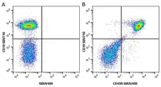
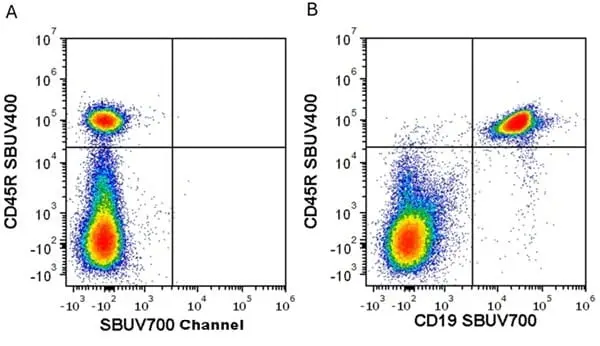
Figure B. StarBright Violet 710 conjugated Rat anti Mouse CD19 antibody, clone 6D5 (MCA1439SBV710) and StarBright UltraViolet 400 conjugated Rat anti Mouse CD45R antibody, clone RA3-6B2 (MCA1258SBUV400). All experiments performed on red cell lysed mouse splenocytes gated on live cells, in the presence of 10% mouse serum.
Data acquired on the ZE5 Cell analyser.
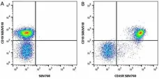
Figure B. StarBright UltraViolet 510 conjugated Rat anti Mouse CD19 antibody, clone 6D5 (MCA1439SBUV510) and StarBright Violet 760 conjugated Rat anti Mouse CD45R antibody, clone RA3-6B2 (MCA1258SBV760). All experiments performed on red cell lysed mouse blood gated on live single cell lymphocytes, in the presence of 10% mouse serum.
Data acquired on the ZE5 Cell analyser.
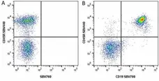
Figure B. StarBright Violet 440 conjugated Rat anti Mouse CD45R antibody, clone RA3-6B2 (MCA1258SBV440) and StarBright Violet 760 conjugated Rat anti Mouse CD19 antibody, clone 6D5 (MCA1439SBV760). All experiments performed on red cell lysed mouse blood gated on live single cell lymphocytes, in the presence of 10% mouse serum.
Data acquired on the ZE5 Cell analyser.
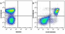
Figure B. StarBright Violet 440 conjugated Rat anti Mouse CD19 antibody, clone 6D5 (MCA1439SBV440) and StarBright UltraViolet 665 conjugated Rat anti Mouse CD45R antibody, clone RA3-6B2 (MCA1258SBUV665). All experiments performed on red cell lysed mouse blood gated on live single cell lymphocytes, in the presence of 10% mouse serum.
Data acquired on the ZE5 Cell analyser.
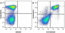
Figure B. StarBright Violet 515 conjugated Rat anti Mouse CD45R antibody, clone RA3-6B2 (MCA1258SBV515) and StarBright UltraViolet 665 conjugated Rat anti Mouse CD19 antibody, clone 6D5 (MCA1439SBUV665). All experiments performed on red cell lysed mouse blood gated on live single cell lymphocytes, in the presence of 10% mouse serum.
Data acquired on the ZE5 Cell analyser.

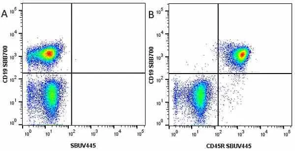
Figure B. StarBright Blue 700 conjugated Rat anti Mouse CD19 antibody, clone 6D5 (MCA1439SBB700) and StarBright UltraViolet 445 conjugated Rat anti Mouse CD45R antibody, clone RA3-6B2 (MCA1258SBUV445). All experiments performed on red cell lysed mouse blood gated on live single cell lymphocytes, in the presence of 10% mouse serum.
Data acquired on the ZE5 Cell analyser.
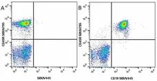
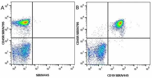
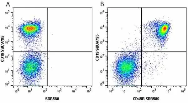
Figure B. StarBright UltraViolet 795 conjugated Rat anti Mouse CD45R antibody, clone RA3-6B2 (MCA1258SBUV795) and StarBright UltraViolet 445 conjugated Rat anti Mouse CD19 antibody, clone 6D5 (MCA1439SBUV445). All experiments performed on red cell lysed mouse blood gated on live single cell lymphocytes, in the presence of 10% mouse serum.
Data acquired on the ZE5 Cell analyser.
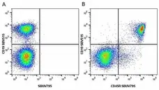
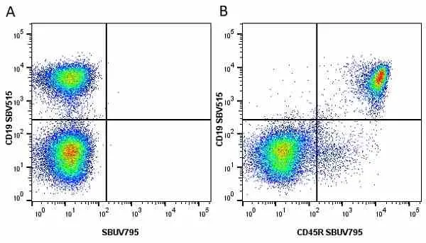
Figure B. StarBright Violet 515 conjugated Rat anti Mouse CD19 antibody, clone 6D5 (MCA1439SBV515) and StarBright UltraViolet 795 conjugated Rat anti Mouse CD45R antibody, clone RA3-6B2 (MCA1258SBUV795). All experiments performed on red cell lysed mouse blood gated on live single cell lymphocytes, in the presence of 10% mouse serum.
Data acquired on the ZE5 Cell analyser.
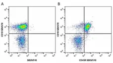
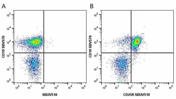
Figure B. StarBright Violet 570 conjugated Rat anti Mouse CD19 antibody, clone 6D5 (MCA1439SBV570) and StarBright UltraViolet 510 conjugated Rat anti Mouse CD45R antibody, clone RA3-6B2 (MCA1258SBUV510). All experiments performed on red cell lysed mouse blood gated on live single cell lymphocytes, in the presence of 10% mouse serum.
Data acquired on the ZE5 Cell analyser.
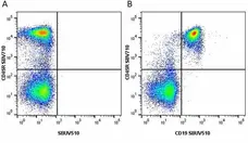
Figure B. StarBright Violet 710 conjugated Rat anti Mouse CD45R antibody, clone RA3-6B2 (MCA1258SBV710) and StarBright UltraViolet 510 conjugated Rat anti Mouse CD19 antibody, clone 6D5 (MCA1439SBUV510). All experiments performed on red cell lysed mouse blood gated on live single lymphocytes, in the presence of 10% mouse serum.
Data acquired on the ZE5 Cell analyser.
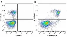
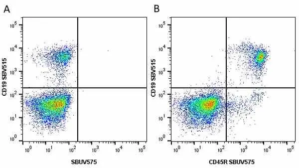
Figure B. StarBright Violet 515 conjugated Rat anti Mouse CD19 antibody, clone 6D5 (MCA1439SBV515) and StarBright UltraViolet 575 conjugated Rat anti Mouse CD45R antibody, clone RA3-6B2 (MCA1258SBUV575). All experiments performed on red cell lysed mouse blood gated on live single cell lymphocytes, in the presence of 10% mouse serum
Data acquired on the ZE5 Cell analyser.
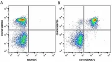
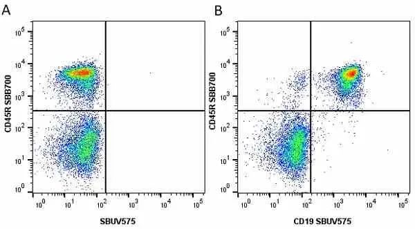
Figure B. StarBright Blue 700 conjugated Rat anti Mouse CD45R antibody, clone RA3-6B2 (MCA1258SBB700) and StarBright UltraViolet 575 conjugated Rat anti Mouse CD19 antibody, clone 6D5 (MCA1439SBUV575). All experiments performed on red cell lysed mouse blood gated on live single cell lymphocytes, in the presence of 10% mouse serum.
Data acquired on the ZE5 Cell analyser.
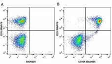
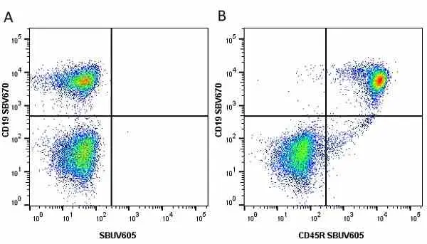
Figure B. StarBright Violet 670 conjugated Rat anti Mouse CD19 antibody, clone 6D5 (MCA1439SBV670) and StarBright UltraViolet 605 conjugated Rat anti Mouse CD45R antibody, clone RA3-6B2 (MCA1258SBUV605). All experiments performed on red cell lysed mouse blood gated on live single cell lymphocytes, in the presence of 10% mouse serum.
Data acquired on the ZE5 Cell analyser.

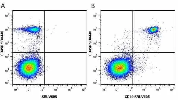
Figure B. StarBright Violet 440 conjugated Rat anti Mouse CD45R antibody, clone RA3-6B2 (MCA1258SBV440) and StarBright UltraViolet 605 conjugated Rat anti Mouse CD19 antibody, clone 6D5 (MCA1439SBUV605). All experiments performed on red cell lysed mouse blood gated on live single cell lymphocytes, in the presence of 10% mouse serum.
Data acquired on the ZE5 Cell analyser.

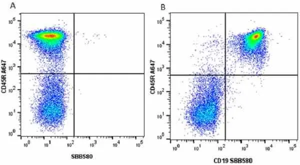
Figure B. Alexa Fluor® 647 conjugated Rat anti Mouse CD45R antibody, clone RA3-6B2 (MCA1258A647) and StarBright Blue 580 conjugated Rat anti Mouse CD19 antibody, clone 6D5 (MCA1439SBB580). All experiments performed on red cell lysed mouse blood gated on live single cell lymphocytes, in the presence of 10% mouse serum.
Data acquired on the ZE5 Cell analyser.
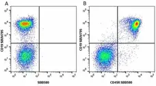
Figure B. StarBright UltraViolet 795 conjugated Rat anti Mouse CD19 antibody, clone 6D5 (MCA1439SBUV795) and StarBright Blue 580 conjugated Rat anti Mouse CD45R antibody, clone RA3-6B2 (MCA1258SBB580). All experiments performed on red cell lysed mouse blood gated on live single cell lymphocytes, in the presence of 10% mouse serum.
Data acquired on the ZE5 Cell analyser.
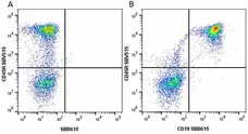
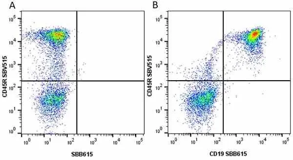
Figure B. StarBright Violet 515 conjugated Rat anti Mouse CD45R antibody, clone RA3-6B2 (MCA1258SBV515) and StarBright Blue 615 conjugated Rat anti Mouse CD19 antibody, clone 6D5 (MCA1439SBB615). All experiments performed on mouse blood gated on live single cells, in the presence of 10% mouse serum.
Data acquired on the ZE5 Cell analyser.
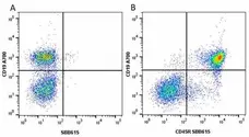
Figure B. Alexa Fluor® 700 conjugated Rat anti Mouse CD19 antibody, clone 6D5 (MCA1439A700) and StarBright Blue 615 conjugated Rat anti Mouse CD45R antibody, clone RA3-6B2 (MCA1258SBB615). All experiments performed on mouse blood gated on live single cells, in the presence of 10% mouse serum.
Data acquired on the ZE5 Cell analyser.
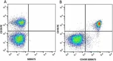
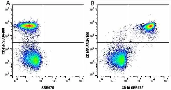
Figure B. RPE conjugated Rat anti Mouse CD19 antibody, clone 6D5 (MCA1439PE) and StarBright Blue 675 conjugated Rat anti Mouse CD45R antibody, clone RA3-6B2 (MCA1258SBB675). All experiments performed on red cell lysed mouse blood gated on live single cell lymphocytes, in the presence of 10% mouse serum.
Data acquired on the ZE5 Cell analyser.
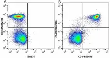
Figure B. StarBright UltraViolet 400 conjugated Rat anti Mouse CD45R antibody, clone RA3-6B2 (MCA1258SBUV400) and StarBright Blue 675 conjugated Rat anti Mouse CD19 antibody, clone 6D5 (MCA1439SBB675). All experiments performed on red cell lysed mouse blood gated on live single cell lymphocytes, in the presence of 10% mouse serum.
Data acquired on the ZE5 Cell analyser.
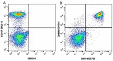
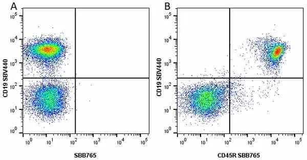
Figure B. StarBright Violet 515 conjugated Rat anti Mouse CD45R antibody, clone 6D5 (MCA1258SBV515) and StarBright Blue 765 conjugated Rat anti Mouse CD19 antibody, clone 6D5 (MCA1439SBB765). All experiments performed on red cell lysed mouse blood gated on live single cell lymphocytes, in the presence of 10% mouse serum.
Data acquired on the ZE5 Cell analyser.

Figure B. StarBright Violet 440 conjugated Rat anti Mouse CD19 antibody, clone 6D5 (MCA1439SBV440) and StarBright Blue 675 conjugated Rat anti Mouse CD45R antibody, clone RA3-6B2 (MCA1258SBB675). All experiments performed on mouse blood gated on live single cells, in the presence of 10% mouse serum.
Data acquired on the ZE5 Cell analyser.

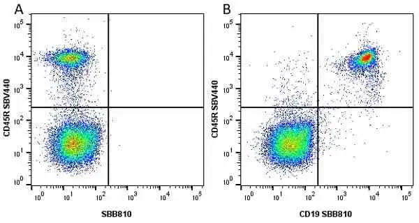
Figure B. StarBright Violet 440 conjugated Rat anti Mouse CD45R antibody, clone RA3-6B2 (MCA1258SBV440) and StarBright Blue 810 conjugated Rat anti Mouse CD19 antibody, clone 6D5 (MCA1439SBB810). All experiments performed on red cell lysed mouse blood gated on live single cell lymphocytes, in the presence of 10% mouse serum.
Data acquired on the ZE5 Cell analyser.
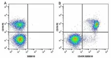
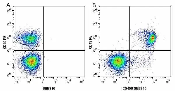
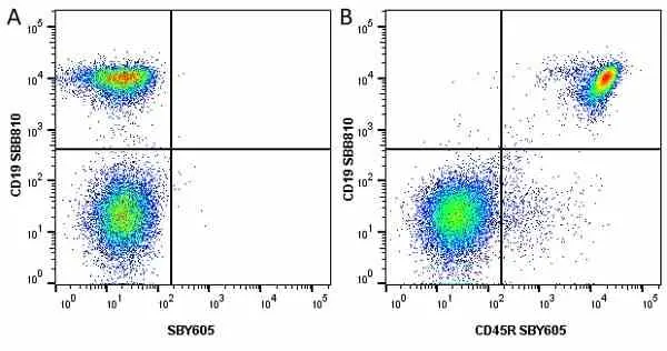
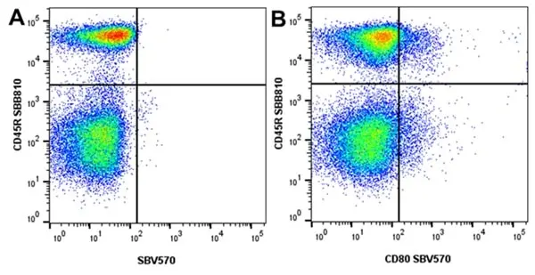
Figure B. RPE conjugated Rat anti Mouse CD19 antibody, clone 6D5 (MCA1439PE) and StarBright Blue 810 conjugated Rat anti Mouse CD45R antibody, clone RA3-6B2 (MCA1258SBB810). All experiments performed on red cell lysed mouse blood gated on live lymphocytes, in the presence of 10% mouse serum.
Data acquired on the ZE5 Cell analyser.
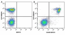
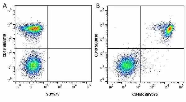
Figure B. StarBright Blue 810 conjugated Rat anti Mouse CD19 antibody, clone 6D5 (MCA1439SBB810). and StarBright Yellow 575 conjugated Rat anti Mouse CD45R (MCA1258SBY575). All experiments performed on red cell lysed Mouse blood gated on live single cell lymphocytes, in the presence of 10% Mouse serum.
Data acquired on the ZE5 Cell analyser.
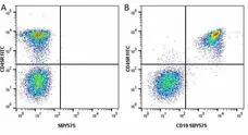
Figure B. FITC conjugated Rat anti Mouse CD45R antibody, clone RA3-6B2 (MCA1258F) and StarBright Yellow 575 conjugated Rat anti Mouse CD19 antibody, clone 6D5 (MCA1439SBY575). All experiments performed on red cell lysed Mouse blood gated on live single cell lymphocytes, in the presence of 10% Mouse serum.
Data acquired on the ZE5 Cell analyser.
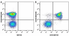
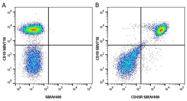
Figure B. StarBright UltraViolet 400 conjugated Rat anti Mouse CD45R antibody, clone RA3-6B2 (MCA1258SBUV400) and StarBright Yellow 720 conjugated Rat anti Mouse CD19 antibody, clone 6D5 (MCA1439SBY720). All experiments performed on mouse blood gated on live single cell lymphocytes, in the presence of 10% mouse serum.
Data acquired on the ZE5 Cell analyser.
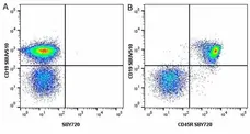
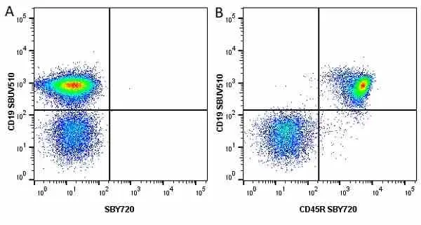
Figure B. StarBright UltraViolet 510 conjugated Rat anti Mouse CD19 antibody, clone 6D5 (MCA1439SBUV510) and StarBright Yellow 720 conjugated Rat anti Mouse CD45R antibody, clone RA3-6B2 (MCA1258SBY720). All experiments performed on mouse blood gated on live single cell lymphocytes, in the presence of 10% mouse serum.
Data acquired on the ZE5 Cell analyser.
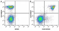
Figure B. StarBright Blue 810 conjugated Rat anti Mouse CD19 antibody, clone 6D5 (MCA1439SBB810) and StarBright Yellow 605 conjugated Rat anti Mouse CD45R antibody, clone RA3-6B2 (MCA1258SBY605). All experiments performed on mouse blood gated on live single cell lymphocytes, in the presence of 10% mouse serum.
Data acquired on the ZE5 Cell analyser.
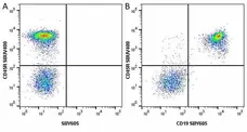
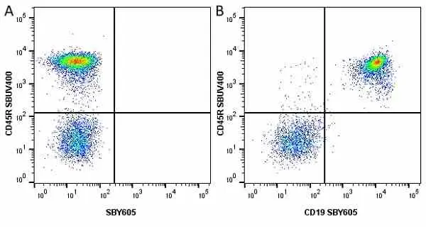
Figure B. StarBright UltraViolet 400 conjugated Rat anti Mouse CD45R antibody, clone RA3-6B2 (MCA1258SBUV400) and StarBright Yellow 605 conjugated Rat anti Mouse CD19 antibody, clone 6D5 (MCA1439SBY605). All experiments performed on mouse blood gated on live single cell lymphocytes, in the presence of 10% mouse serum.
Data acquired on the ZE5 Cell analyser.

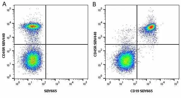
Figure B. StarBright Violet 440 conjugated Rat anti Mouse CD45R antibody, clone RA3-6B2 (MCA1258SBV440) and StarBright Yellow 665 conjugated Rat anti Mouse CD19 antibody, clone 6D5 (MCA1439SBY665). All experiments performed on red cell lysed mouse blood gated on live single cell lymphocytes, in the presence of 10% mouse serum.
Data acquired on the ZE5 Cell analyser.

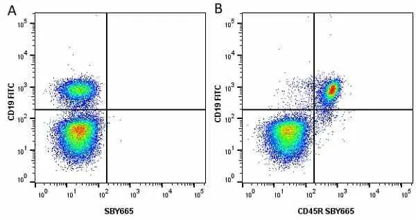
Figure B. FITC conjugated Rat anti Mouse CD19 antibody, clone 6D5 (MCA1439F) and StarBright Yellow 665 conjugated Rat anti Mouse CD45R antibody, clone RA3-6B2 (MCA1258SBY665). All experiments performed on red cell lysed mouse blood gated on live single cell lymphocytes, in the presence of 10% mouse serum.
Data acquired on the ZE5 Cell analyser.
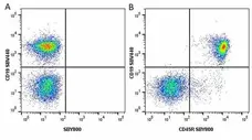
Figure B. StarBright Violet 440 conjugated Rat anti Mouse CD19 antibody, clone 6D5 (MCA1439SBV440) and StarBright Yellow 800 conjugated Rat anti Mouse CD45R antibody, clone RA3-6B2 (MCA1258SBY800). All experiments performed on red cell lysed mouse blood gated on live single cell lymphocytes, in the presence of 10% mouse serum.
Data acquired on the ZE5 Cell analyser.
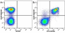
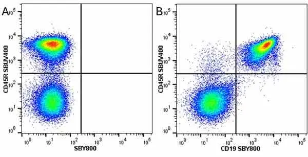
Figure B. StarBright UltaViolet 400 conjugated Rat anti Mouse CD45R antibody, clone RA3-6B2 (MCA1258SBUV400) and StarBright Yellow 800 conjugated Rat anti Mouse CD19 antibody, clone 6D5 (MCA1439SBY800). All experiments performed on red cell lysed mouse blood gated on live single cell lymphocytes, in the presence of 10% mouse serum.
Data acquired on the ZE5 Cell analyser.
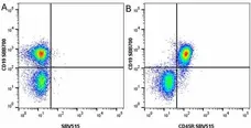
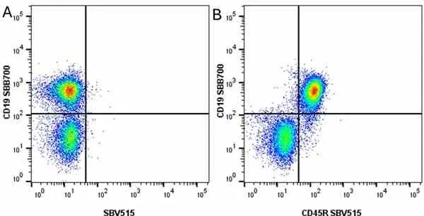
Figure B. StarBright Blue 700 conjugated Rat anti Mouse CD19 antibody, clone 6D5 (MCA1439SBB700) and StarBright Violet 515 conjugated Rat anti Mouse CD45R antibody, clone RA3-6B2 (MCA1258SBV515). All experiments performed on red cell lysed mouse blood gated on live single cell lymphocytes, in the presence of 10% mouse serum.
Data acquired on the ZE5 Cell analyser.
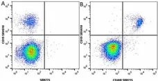
Figure B. StarBright Violet 610 conjugated Rat anti Mouse CD19 antibody, clone 6D5 (MCA1439SBV610) and StarBright Red 775 conjugated Rat anti Mouse CD45R antibody, clone RA3-6B2 (MCA1258SBR775). All experiments performed on red cell lysed mouse blood gated on live single cell lymphocytes, in the presence of 10% mouse serum.
Data acquired on the ZE5 Cell analyser.

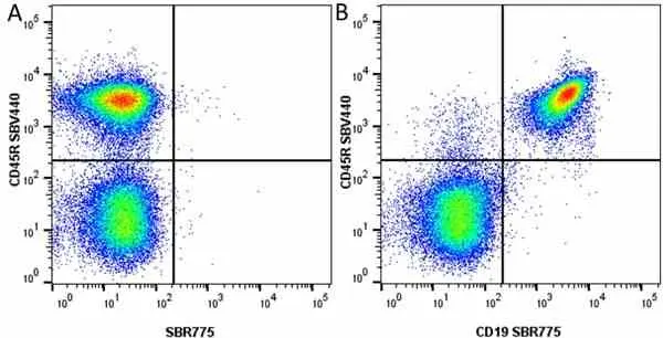
Figure B. StarBright Violet 440 conjugated Rat anti Mouse CD45R antibody, clone RA3-6B2 (MCA1258SBV440) and StarBright Red 775 conjugated Rat anti Mouse CD19 antibody, clone 6D5 (MCA1439SBR775). All experiments performed on red cell lysed mouse blood gated on live single cell lymphocytes, in the presence of 10% mouse serum.
Data acquired on the ZE5 Cell analyser.
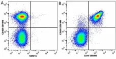
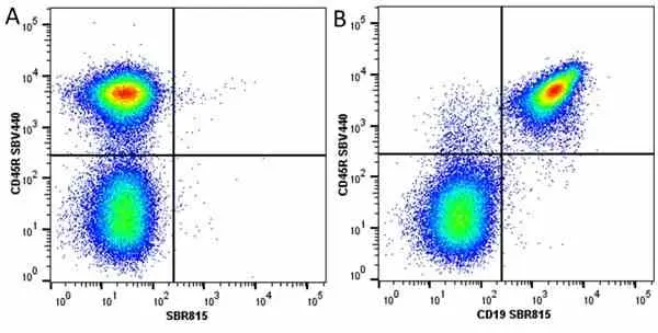
Figure B. StarBright UltraViolet 440 conjugated Rat anti Mouse CD45R antibody, clone RA3-6B2 (MCA1258SBV440) and StarBright Red 815 conjugated Rat anti Mouse CD19 antibody, clone 6D5 (MCA1439SBR815). All experiments performed on mouse blood gated on live single cells, in the presence of 10% mouse serum.
Data acquired on the ZE5 Cell analyser.
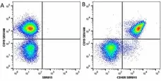
Figure B. StarBright UltraViolet 440 conjugated Rat anti Mouse CD19 antibody, clone 6D5 (MCA1439SBV440) and StarBright Red 815 conjugated Mouse anti Human CD45R antibody, clone RA3-6B2 (MCA1258SBR815). All experiments performed on red cell lysed human blood gated on live single cell lymphocytes, in the presence of 10% human serum.
Data acquired on the ZE5 Cell analyser.
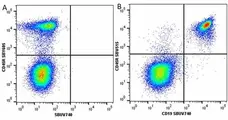
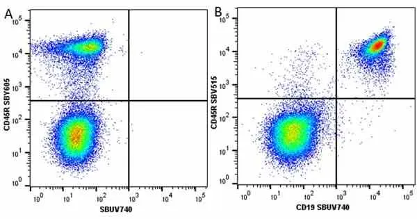
Figure B. StarBright Yellow 605 conjugated Rat anti Mouse CD45R antibody, clone RA3-6B2 (MCA1258SBY605) and StarBright UltraViolet 740 conjugated Rat anti Mouse CD19 antibody, clone 6D5 (MCA1439SBUV740). All experiments performed on red cell lysed mouse blood gated on live single cell lymphocytes, in the presence of 10% mouse serum.
Data acquired on the ZE5 Cell analyser.
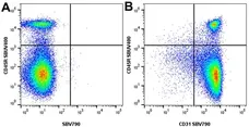
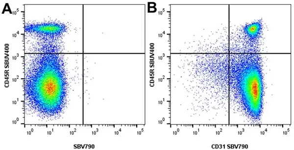
Figure B. StarBright UltraViolet 400 conjugated Rat anti Mouse CD45R antibody, clone RA3-6B2 (MCA1258SBUV400) and StarBright Violet 790 conjugated Rat anti Mouse CD31 antibody, clone ER-MP12 (MCA2388SBV790). All experiments performed on mouse splenocytes, in the presence of 10% mouse serum.
Data acquired on the ZE5 Cell analyser.
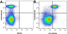
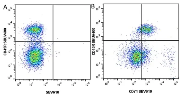
Figure B. StarBright UltraViolet 400 conjugated Rat anti Mouse CD45R antibody, clone RA3-6B2 (MCA1258SBUV400) and StarBright Violet 790 conjugated Rat anti Mouse CD71 antibody, clone YTA74.4 (MCA1033SBV790). All experiments performed on mouse splenocytes, in the presence of 10% mouse serum.
Data acquired on the ZE5 Cell analyser.

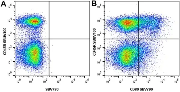
Figure B. StarBright UltraViolet 400 conjugated Rat anti Mouse CD45R antibody, clone RA3-6B2 (MCA1258SBUV400) and StarBright Violet 790 conjugated Rat anti Mouse CD80 antibody, clone RM80 (MCA2462SBV790). All experiments performed on LPS-stimulated (24 Hours) mouse splenocytes, in the presence of 10% mouse serum.
Data acquired on the ZE5 Cell analyser.

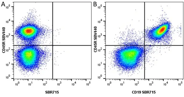
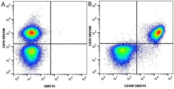
Figure B. StarBright Violet 440 conjugated Rat anti Mouse CD45R antibody, RA3-6B2 (MCA1258SBV440) and StarBright Red 715 conjugated Rat anti Mouse CD19 antibody, clone 6D5 (MCA1439SBR715). All experiments performed on red cell lysed mouse blood gated on live single cell lymphocytes, in the presence of 10% mouse serum.
Data acquired on the ZE5 Cell analyser.
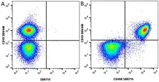
Figure B. StarBright Violet 440 conjugated Rat anti Mouse CD19 antibody, clone 6D5 (MCA1439SBV440) and StarBright Red 715 conjugated Rat anti Mouse CD45R (MCA1258SBR715). ). All experiments performed on red cell lysed mouse blood gated on live single cell lymphocytes, in the presence of 10% mouse serum.
Data acquired on the ZE5 Cell analyser.
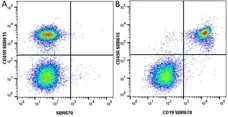
Figure B. StarBright Blue 615 conjugated Rat anti Mouse CD45R antibody, clone RA3-6B2 (MCA1258SBB615) and StarBright Red 670 conjugated Rat anti Mouse CD19 (MCA1439SBR670). All experiments performed on red cell lysed mouse blood gated on live single cell lymphocytes, in the presence of 10% mouse serum.
Data acquired on the ZE5 Cell analyser.
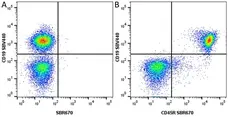
Figure B. StarBright Violet 440 conjugated Rat anti Mouse CD19 antibody, clone 6D5 (MCA1439SBV440) and StarBright Red 670 conjugated Rat anti Mouse CD45R antibody, clone RA3-6B2 (MCA1258SBR670). All experiments performed on red cell lysed mouse blood gated on live single cell lymphocytes, in the presence of 10% mouse serum.
Data acquired on the ZE5 Cell analyser
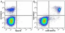
Figure B. StarBright Violet 515 conjugated Rat anti Mouse C19 antibody, clone 6D5 (MCA1439SBV515) and StarBright UltraViolet 740 conjugated Rat anti Mouse CD45R (MCA1258SBUV740). All experiments performed on red cell lysed mouse blood gated on live single cell lymphocytes, in the presence of 10% mouse serum.
Data acquired on the ZE5 Cell analyser

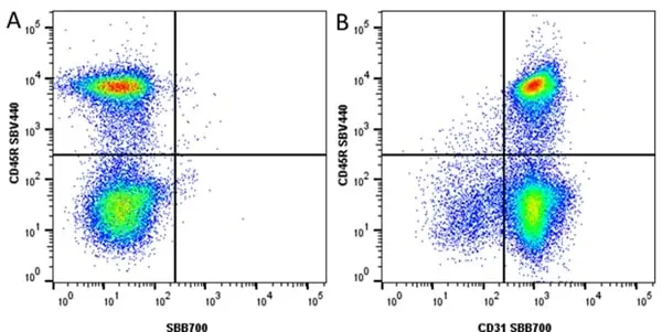
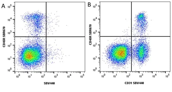
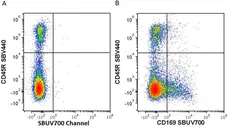
Figure B. StarBright Violet 440 conjugated Rat anti Mouse CD45R antibody, clone RA3-6B2 (MCA1258SBV440) and StarBright Blue 700 conjugated Rat anti Mouse CD31 antibody, clone ER-MP12 (MCA2388SBB700).
All experiments performed on red cell lysed mouse blood gated on live single Lymphocytes, in the presence of 10% mouse serum. Data acquired on the ZE5 Cell Analyzer.
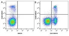
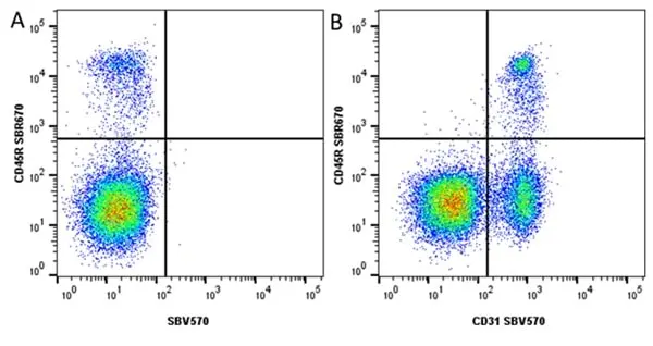
Figure B. StarBright Red 670 conjugated Rat anti Mouse CD45R antibody, clone RA3-6B2 (MCA1258SBR670) and StarBright Violet 570 conjugated Rat anti Mouse CD31 antibody, clone ER-MP12 (MCA2388SBV570).
All experiments performed on red cell lysed mouse blood gated on live single lymphocytes, in the presence of 10% mouse serum. Data acquired on the ZE5 Cell analyser.

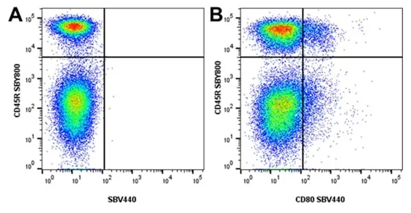
Figure B. StarBright Yellow 800 conjugated Rat anti Mouse CD45R antibody, clone RA3-6B2 (MCA1258SBY800) and StarBright Violet 440 conjugated Rat anti Mouse CD80 antibody, clone RM80 (MCA2462SBV440).
All experiments performed on LPS-stimulated (1 day) mouse splenocytes, in the presence of 10% mouse serum. Data acquired on the ZE5 Cell analyser.
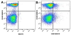
Figure B. StarBright Blue 810 conjugated Rat anti Mouse CD45R antibody, clone RA3-6B2 (MCA1258SBB810) and StarBright Violet 570 conjugated Rat anti Mouse CD80 antibody, clone RM80 (MCA2462SBV570).
All experiments performed on LPS-stimulated (1 day) mouse splenocytes, in the presence of 10% mouse serum. Data acquired on the ZE5 Cell analyser.
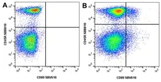
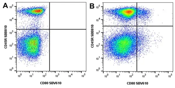
Figure B. StarBright Blue 810 conjugated Rat anti Mouse CD45R antibody, cone RA3-6B2 (MCA1258SBB810) and StarBright Violet 610 conjugated Rat anti Mouse CD80 antibody, clone RM80 (MCA2462SBV610).
All experiments performed on LPS-stimulated (1 day) mouse splenocytes, in the presence of 10% mouse serum. Data acquired on the ZE5 Cell analyser.
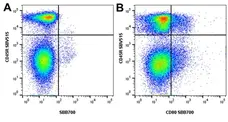
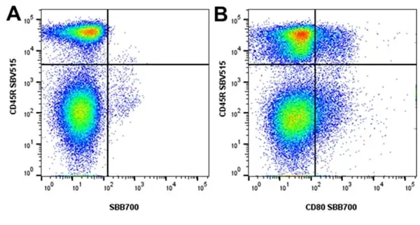
Figure B. StarBright Violet 515 conjugated Rat anti Mouse CD45R antibody, clone RA3-6B2 (MCA1258SBV515) and StarBright Blue 700 conjugated Rat anti Mouse CD80 antibody, clone RM80 (MCA2462SBB700).
All experiments performed on LPS-stimulated (1 day) mouse splenocytes, in the presence of 10% mouse serum. Data acquired on the ZE5 Cell analyser.
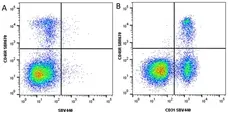
Figure B. StarBright Red 670 conjugated Rat anti Mouse CD45R antibody, clone RA3-6B2 (MCA1258SBR775) and StarBright Violet 440 conjugated Rat anti Mouse CD31 antibody, clone ER-MP12 (MCA2388SBV440). All experiments performed on red blood lysed mouse blood gated on live single cell lymphocytes, in the presence of 10% mouse serum.
Data acquired on the ZE5 Cell analyser.
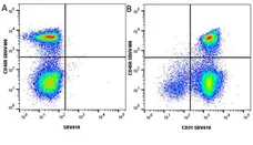
Figure B. StarBright Ulta Violet 400 conjugated Rat anti Mouse CD45R antibody, clone RA3-6B2 (MCA1258SBUV400) and StarBright Violet 610 conjugated Rat anti Mouse CD31 antibody, clone ER-MP12 (MCA2388SBV610). All experiments performed on red blood lysed mouse blood gated on live single cell lymphocytes, in the presence of 10% mouse serum.
Data acquired on the ZE5 Cell analyser.
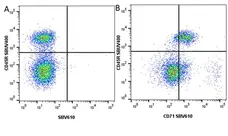
Figure B. StarBright UltraViolet 400 conjugated Rat anti Mouse CD45R antibody, clone RA3-6B2 (MCA1258SBUV400) and StarBright Violet 610 conjugated Rat anti Mouse CD71 antibody, clone YTA74.4 (MCA1033SBV610). mouse splenocytes, in the presence of 10% mouse serum.
Data acquired on the ZE5 Cell analyser.


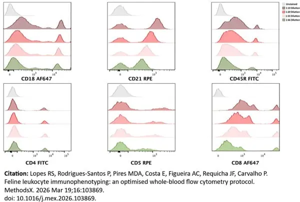
FITC conjugated Mouse anti Canine CD45R antibody, clone RA3-6B2 (MCA1258F) used for feline leukocyte typing by flow cytometry.
Image caption:
Representative leukocyte-gated histograms illustrating antibody titration for feline leukocyte immunophenotyping. All histograms display singlet leukocytes, defined by FSC-A versus SSC-A morphological gating, followed by FSC—H versus FSC-A singlet discrimination. Titration was performed for CD18, CD21, CD45R, CD4, CD5 and CD8 monoclonal antibodies using three antibody volumes: 10 µL (1:10 dilution), 5.0 µL (1:20 dilution), 3.0 µL (1:33 dilution) and 1.5 µL (1:66 dilution). Minimal working volumes were selected based on optimal signal-to-noise ratios.
From: Lopes RS, Rodrigues-Santos P, Pires MDA, Costa E, Figueira AC, Requicha JF, Carvalho P.
Feline leukocyte immunophenotyping: an optimised whole-blood flow cytometry protocol.
MethodsX. 2026 Mar 19;16:103869.
doi: 10.1016/j.mex.2026.103869.
This image is from an open access article distributed under terms of a Creative Commons Attribution License.
Filter by Application:
F IF P Reset| Rat anti Mouse CD45R antibody, clone RA3-6B2 recognizes murine CD45R, a form of the CD45 antigen expressed by B cells and lytically active subsets of NK cells and non-MHC restricted CTL's. Rat anti Mouse CD45R antibody, clone RA3-6B2 immunoprecipitates the high molecular weight form of CD45 (220 kDa). Rat anti Mouse CD45R antibody, clone RA3-6B2 is suitable for plp fixed paraffin embedded tissues (Whiteland et al.1995). |
- Target Species
- Mouse
- Species Cross-Reactivity
-
Target Species Cross Reactivity Human Cat - N.B. Antibody reactivity and working conditions may vary between species.
- Product Form
- Purified IgG - liquid
- Preparation
- Purified IgG prepared by affinity chromatography on Protein G from tissue culture supernatant
- Buffer Solution
- Phosphate buffered saline
- Preservative Stabilisers
- 0.09% sodium azide (NaN3)
- Carrier Free
- Yes
- Immunogen
- Murine leukemia-induced pre-B tumor cells (RAW112)
- Approx. Protein Concentrations
- IgG concentration 1.0mg/ml
- Fusion Partners
- Spleen cells from immunized Lewis rats were fused with cells of the rat S194/5 XX0.BU-1 myeloma cell line
- Regulatory
- For research purposes only
- Guarantee
- 12 months from date of despatch
This product is shipped at ambient temperature. It is recommended to aliquot and store at -20°C on receipt. When thawed, aliquot the sample as needed. Keep aliquots at 2-8°C for short term use (up to 4 weeks) and store the remaining aliquots at -20°C.
Avoid repeated freezing and thawing as this may denature the antibody. Storage in frost-free freezers is not recommended.
Avoid repeated freezing and thawing as this may denature the antibody. Storage in frost-free freezers is not recommended.
This product has been reported to work in the following applications. This information is derived from testing within our laboratories, peer-reviewed publications or personal communications from the originators. Please refer to references indicated for further information. For general protocol recommendations, please visit the antibody protocols page.
| Application Name | Verified | Min Dilution | Max Dilution |
|---|---|---|---|
| Flow Cytometry |  |
1/100 | 1/200 |
| Immunofluorescence | |
||
| Immunohistology - Frozen | |
||
| Immunohistology - Paraffin 1 | |
||
| Immunoprecipitation | |
- 1PLP fixation is recommended for optimal results, see Whiteland et al. for details
Where this product has not been tested for use in a particular technique this does not necessarily exclude its use in such procedures. Suggested working dilutions are given as a guide only. It is recommended that the user titrates the product for use in their own system using appropriate negative/positive controls.
- Flow Cytometry
- Use 10μl of the suggested working dilution to label 106 cells in 100μl.
| Description | Product Code | Applications | Pack Size | List Price | Your Price | Quantity | |
|---|---|---|---|---|---|---|---|
| Rat IgG2a Negative Control | MCA1212 | E F | 1 ml |

|
Log in | ||
| List Price | Your Price | ||||||
|
|
Log in | ||||||
| Description | Rat IgG2a Negative Control | ||||||
| Description | Product Code | Applications | Pack Size | List Price | Your Price | Quantity | |
|---|---|---|---|---|---|---|---|
| Antigen Retrieval Buffer, pH8.0 | BUF025A | P | 500 ml | Log in | |||
| List Price | Your Price | ||||||
| Log in | |||||||
| Description | Antigen Retrieval Buffer, pH8.0 | ||||||
Source Reference
-
Coffman, R.L. (1982) Surface antigen expression and immunoglobulin gene rearrangement during mouse pre-B cell development.
Immunol Rev. 69: 5-23.
References for CD45R antibody
-
Holmes, K.L. et al. (1986) Analysis of neoplasms induced by Cas-Br-M MuLV tumor extracts.
J Immunol. 137 (2): 679-88. -
Spangrude, G.J. et al. (1988) Purification and characterization of mouse hematopoietic stem cells.
Science. 241: 58-62. -
Spangrude, G.J. et al. (1988) Two rare populations of mouse Thy-1lo bone marrow cells repopulate the thymus.
J Exp Med. 167 (5): 1671-83. -
Whiteland, J.L. et al. (1995) Immunohistochemical detection of T-cell subsets and other leukocytes in paraffin-embedded rat and mouse tissues with monoclonal antibodies.
J Histochem Cytochem. 43 (3): 313-20. -
Hawke, S. et al. (1998) Long-term persistence of activated cytotoxic T lymphocytes after viral infection of the central nervous system.
J Exp Med. 187: 1575-82. -
Rosmalen, J.G. et al. (2000) Subsets of macrophages and dendritic cells in nonobese diabetic mouse pancreatic inflammatory infiltrates: correlation with the development of diabetes.
Lab Invest. 80 (1): 23-30. -
Perry, M.J. et al. (2000) Effects of high-dose estrogen on murine hematopoietic bone marrow precede those on osteogenesis.
Am J Physiol Endocrinol Metab. 279: E1159-65. -
Stevenson, P.G. et al. (2002) Uncoupling of virus-induced inflammation and anti-viral immunity in the brain parenchyma.
J Gen Virol. 83: 1735-43.
View The Latest Product References
-
Straubinger, R.K. et al. (2003) Quantitative evaluation of inflammatory and immune responses in the early stages of chronic Helicobacter pylori infection.
Infect Immun. 71: 2693-703. -
Shulga-Morskaya, S. et al. (2004) B cell-activating factor belonging to the TNF family acts through separate receptors to support B cell survival and T cell-independent antibody formation.
J Immunol. 173 (4): 2331-41. -
Gengozian, N. et al. (2005) Characterization of a monoclonal antibody identifying a CD45RA antigen on feline leukocytes.
Vet Immunol Immunopathol. 108: 253-64. -
Herrmann, I. et al. (2006) Streptococcus pneumoniae Infection aggravates experimental autoimmune encephalomyelitis via Toll-like receptor 2.
Infect Immun. 74: 4841-8. -
Itoh, T. et al. (2007) Ddb2 is a haploinsufficient tumor suppressor and controls spontaneous germ cell apoptosis.
Hum Mol Genet. 16: 1578-86. -
McGill, J. et al. (2009) Fetal exposure to ethanol has long-term effects on the severity of influenza virus infections.
J Immunol. 182: 7803-8 -
Ankeny, D.P. et al. (2009) B cells produce pathogenic antibodies and impair recovery after spinal cord injury in mice.
J Clin Invest. 119: 2990-9. -
Lacroix-Lamande, S. et al. (2009) Neonate intestinal immune response to CpG oligodeoxynucleotide stimulation.
PLoS One. 4: e8291. -
Lundqvist, J. et al. (2010) Concomitant infection decreases the malaria burden but escalates relapsing fever borreliosis.
Infect Immun. 78 (5): 1924-30. -
Giuriato, S. et al. (2010) Conditional TPM3-ALK and NPM-ALK transgenic mice develop reversible ALK-positive early B-cell lymphoma/leukemia.
Blood. 115: 4061-70. -
Kleiter, I. et al. (2010) Smad7 in T cells drives T helper 1 responses in multiple sclerosis and experimental autoimmune encephalomyelitis.
Brain. 133: 1067-81. -
Nakaya, T. et al. (2010) Critical role of Pcid2 in B cell survival through the regulation of MAD2 expression.
J Immunol. 185: 5180-7. -
Soejima, M. et al. (2011) Role of innate immunity in a murine model of histidyl-transfer RNA synthetase (Jo-1)-mediated myositis.
Arthritis Rheum. 63: 479-87. -
Bertilaccio, M.T. et al. (2011) Lack of TIR8/SIGIRR triggers progression of chronic lymphocytic leukemia in mouse models.
Blood. 118: 660-9. -
Zhou, Z. et al. (2011) Autoreactive marginal zone B cells enter the follicles and interact with CD4+ T cells in lupus-prone mice.
BMC Immunol. 2011; 12:7. -
Fanning, S. et al. (2012) Bifidobacterial surface-exopolysaccharide facilitates commensal-host interaction through immune modulation and pathogen protection.
Proc Natl Acad Sci U S A. 109 (6): 2108-13. -
Ruf, M.T. et al. (2012) Chemotherapy-Associated Changes of Histopathological Features of Mycobacterium ulcerans Lesions in a Buruli Ulcer Mouse Model.
Antimicrob Agents Chemother. 56: 687-96. -
Carpenter, R.S. et al. (2015) Traumatic spinal cord injury in mice with human immune systems.
Exp Neurol. 271: 432-44. -
Lastrucci, C. et al. (2015) Molecular and cellular profiles of the resolution phase in a damage-associated molecular pattern (DAMP)-mediated peritonitis model and revelation of leukocyte persistence in peritoneal tissues.
FASEB J. 29 (5): 1914-29. -
Gibson-Corley, K.N. et al. (2016) A method for histopathological study of the multifocal nature of spinal cord lesions in murine experimental autoimmune encephalomyelitis.
PeerJ. 4: e1600. -
Thiele Née Schrewe, L. et al. (2020) Functional relevance of the multi-drug transporter abcg2 on teriflunomide therapy in an animal model of multiple sclerosis.
J Neuroinflammation. 17 (1): 9. -
Allen, A.C. et al. (2021) Parallel in vivo. experimental evolution reveals that increased stress resistance was key for the emergence of persistent tuberculosis bacilli.
Nat Microbiol. 6 (8): 1082-93. -
Chanut, F.J.A. et al. (2021) Conditioning Regimens in Long-Term Pre-Clinical Studies to Support Development of Ex Vivo Gene Therapy: Review of Nonproliferative and Proliferative Changes.
Hum Gene Ther. 32 (1-2): 66-76. -
Jaensch, S.M. et al. (2022) Clinicopathologic and immunophenotypic features in dogs with presumptive large granular lymphocyte leukaemia.
Aust Vet J. 100 (11): 527-32. -
Roca, C.P. et al. (2023) A cross entropy test allows quantitative statistical comparison of t-SNE and UMAP representations
Cell Reports Methods. 3 (1): 100390. -
Kohlmeyer, J.L. et al. (2023) CDK4/6-MEK Inhibition in MPNSTs Causes Plasma Cell Infiltration, Sensitization to PD-L1 Blockade, and Tumor Regression.
Clin Cancer Res. 29 (17): 3484-97. -
Lopes, R.S. et al. (2026) Feline leukocyte immunophenotyping: an optimised whole-blood flow cytometry protocol.
MethodsX. 16: 103869.
- Synonyms
- B220
- LY-5
- RRID
- AB_323211
- UniProt
- P06800
- Entrez Gene
- Ptprc
- GO Terms
- GO:0000187 activation of MAPK activity
- GO:0001915 negative regulation of T cell mediated cytotoxicity
- GO:0001916 positive regulation of T cell mediated cytotoxicity
- GO:0001960 negative regulation of cytokine-mediated signaling pathway
- GO:0002925 positive regulation of humoral immune response mediated by circulating immunoglobulin
- GO:0051726 regulation of cell cycle
- GO:0004725 protein tyrosine phosphatase activity
- GO:0005887 integral to plasma membrane
- GO:0005925 focal adhesion
- View More GO Terms
- GO:0006469 negative regulation of protein kinase activity
- GO:0006470 protein dephosphorylation
- GO:0007159 leukocyte cell-cell adhesion
- GO:0008201 heparin binding
- GO:0009897 external side of plasma membrane
- GO:0019901 protein kinase binding
- GO:0030183 B cell differentiation
- GO:0030890 positive regulation of B cell proliferation
- GO:0031953 negative regulation of protein autophosphorylation
- GO:0034113 heterotypic cell-cell adhesion
- GO:0042098 T cell proliferation
- GO:0042100 B cell proliferation
- GO:0043065 positive regulation of apoptosis
- GO:0043395 heparan sulfate proteoglycan binding
- GO:0043410 positive regulation of MAPKKK cascade
- GO:0045059 positive thymic T cell selection
- GO:0045060 negative thymic T cell selection
- GO:0045121 membrane raft
- GO:0045577 regulation of B cell differentiation
- GO:0045588 positive regulation of gamma-delta T cell differentiation
- GO:0046641 positive regulation of alpha-beta T cell proliferation
- GO:0048304 positive regulation of isotype switching to IgG isotypes
- GO:0050732 negative regulation of peptidyl-tyrosine phosphorylation
- GO:0050852 T cell receptor signaling pathway
- GO:0050853 B cell receptor signaling pathway
- GO:0050855 regulation of B cell receptor signaling pathway
- GO:0050857 positive regulation of antigen receptor-mediated signaling pathway
- GO:0051209 release of sequestered calcium ion into cytosol
- GO:0051607 defense response to virus
View more products with CD45R specificity
Please Note: All Products are "FOR RESEARCH PURPOSES ONLY"
View all Anti-Mouse ProductsAlways be the first to know.
When we launch new products and resources to help you achieve more in the lab.
Yes, sign me up