CD4 antibody | YKIX302.9
Rat anti Dog CD4
- Product Type
- Monoclonal Antibody
- Clone
- YKIX302.9
- Isotype
- IgG2a
- Specificity
- CD4


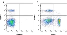
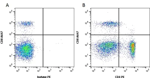
Figure B. Alexa Fluor® 647 conjugated Rat anti Dog CD8 antibody, clone YCATE55.9 (MCA1039A647) and RPE conjugated Rat anti Dog CD4 antibody, clone YKIX302.9 (MCA1038PE). All experiments performed on red cell lysed canine blood gated on lymphoid cells in the presence of 10% dog serum.
Data acquired on the ZE5 Cell Analyzer.
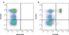
Figure B. Alexa Fluor® 647 conjugated Rat anti Canine CD5 antibody, clone YKIX322.3 (MCA1037A647) and Alexa Fluor® 488 conjugated Rat anti Canine CD4 antibody, clone YKIX302.9 (MCA1038A488). All experiments performed on red cell lysed canine blood gated on lymphoid cells in the presence of 10% dog serum.
Data acquired on the ZE5 Cell Analyzer.
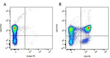
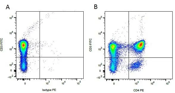
Figure B. FITC conjugated Mouse anti Canine CD3 antibody, clone CA17.2A12 (MCA1774F) and RPE conjugated Rat anti Canine CD4 antibody, clone YKIX302.9 (MCA1038PE). All experiments performed on red cell lysed canine blood gated on mononuclear cells.
Data acquired on the ZE5 Cell Analyzer.
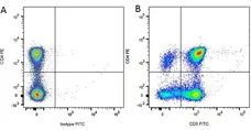
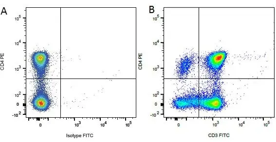
Figure B. RPE conjugated Rat anti Canine CD4 antibody, clone YKIX302.9 (MCA1038PE) and FITC conjugated Mouse anti Canine CD3 antibody, clone CA17.2A12 (MCA1774F). All experiments performed on red cell lysed canine blood gated on mononuclear cells.
Data acquired on the ZE5 Cell Analyzer.
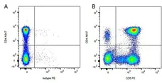
Figure B. Alexa Fluor® 647 conjugated Rat anti Canine CD4 antibody, clone YKIX302.9 (MCA1038A647) and RPE conjugated Rat anti Canine CD5 (MCA1037PE). All experiments performed on red cell lysed canine blood gated on mononuclear cells.
Data acquired on the ZE5 Cell Analyzer.
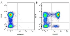
Figure B. RPE conjugated Mouse anti Canine CD5 antibody, clone YKIX322.3 (MCA1037PE) and Alexa Fluor® 647 conjugated Rat anti Canine CD4 antibody, clone YKIX302.9 (MCA1038A647). All experiments performed on red cell lysed canine blood gated on mononuclear cells.
Data acquired on the ZE5 Cell Analyzer.
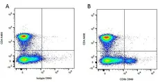
Figure B. Alexa Fluor® 488 conjugated Rat anti Canine CD4 antibody, YKIX302.9 (MCA1038A488) and purified Mouse anti Canine CD8β antibody, clone CA15.4G2 (MCA1775S) detected with DyLight® 649 conjugated Goat anti Mouse IgG1 antibody. All experiments performed on red cell lysed canine blood gated on mononuclear cells.
Data acquired on the ZE5 Cell Analyzer.
Pacific Blue® conjugated Mouse anti Canine CD4 antibody, clone YKIX302.9 (MCA1038PB) used for evaluation of CD4 expression on canine cells by flow cytometry.
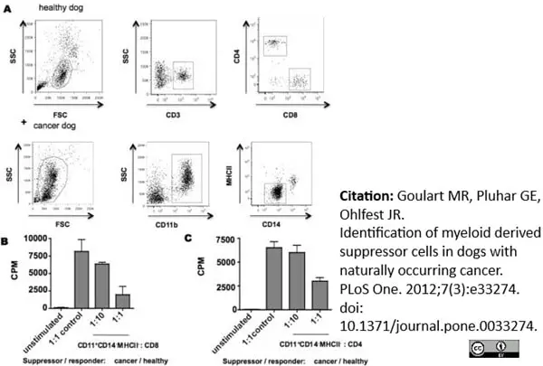
Image caption:
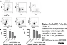
CD11b+CD14−MHCII− cells suppress T cell proliferation. Facs sorted CD11b+CD14−MHCII− cells isolated from a dog with osteosarcoma or healthy PBMCs were co-incubated with mitogen-stimulated CD4+ and CD8+ T cells isolated from a healthy dog for 72 hrs. No stimulated cells were used as negative control. Proliferative responses were measured by 3H-thymidine incorporation from experiments performed in triplicate. CPM, counts per minute. Mean ± SEM are shown.
From: Goulart MR, Pluhar GE, Ohlfest JR (2012)
Identification of Myeloid Derived Suppressor Cells in Dogs with Naturally Occurring Cancer.
PLoS ONE 7(3): e33274.
This image is from an open access article distributed under terms of a Creative Commons Attribution License.
RPE conjugated Mouse anti Canine CD4 antibody, clone YKIX302.9 (MCA1038PE) used for the assessment of CD4 levels on canine cells by flow cytometry.
Image caption:
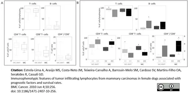
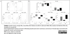
Immunophenotypic profile of tumor infiltrating lymphocyte in canine mammary carcinomas. Analysis of tumor infiltrating T-cells, B-lymphocytes and T-cell subsets from MC-BMT or MC (A), further subcategorized according to the absence (-) or presence (+) of lymph node metastasis (-) (B). Lymphocyte populations and subsets were identified by flow cytometric immunostaining as described in Material and Methods. Data were expressed as percentage of positive cells within gated lymphocytes and CD4+/CD8+ T-cell ratio. Significant differences at p < 0.05 are highlighted by asterisk.
From: From: Estrela-Lima A, Araújo MS, Costa-Neto JM, Teixeira-Carvalho A, Barrouin-Melo SM, Cardoso SV, Martins-Filho OA, Serakides R, Cassali GD.
Immunophenotypic features of tumor infiltrating lymphocytes from mammary carcinomas in female dogs associated with prognostic factors and survival rates.
BMC Cancer. 2010 Jun 4;10:256.
doi: 10.1186/1471-2407-10-256..
This image is from an open access article distributed under terms of a Creative Commons Attribution License.
FITC conjugated Mouse anti Canine CD4 antibody, clone YKIX302.9 (MCA1038F) used to evaluate CD4 expression on canine T-cell subsets by flow cytometry.
Image caption:
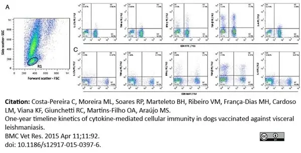
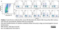
Representative dot plots illustrating the analysis of intracellular cytokine profile in T-cell subsets. (A) Pseudocolor plot distribution of short-term in vitro cultured (control or SLA-Ag stimulated) canine whole blood sample according to cell size (Forward scatter - FSC) and granularity (Side scatter- SSC) used for lymphocyte gate selection (R1) of FSCLowSSCLow events. (B) Pseudocolor plots representing cytokines + (IL-17, TNF-α, IFN-γ, TGF-β and IL-4) CD4+ cells within gated lymphocytes and (C) Pseudocolor plots representing cytokines + (IL-17, TNF-α, IFN-γ, TGF-β and IL-4) CD8+ cells within gated lymphocytes. The frequency of cytokines+ T-cells subsets were calculated by quadrant statistics approach and first reported as percentage of gated lymphocytes prior to the calculation of the SLAg/Control indexes.
From: Costa-Pereira C, Moreira ML, Soares RP, Marteleto BH, Ribeiro VM, França-Dias MH, Cardoso LM, Viana KF, Giunchetti RC, Martins-Filho OA, Araújo MS.
One-year timeline kinetics of cytokine-mediated cellular immunity in dogs vaccinated against visceral leishmaniasis.
BMC Vet Res. 2015 Apr 11;11:92.
This image is from an open access article distributed under terms of a Creative Commons Attribution License.
FITC conjugated Rat anti Dog CD4 antibody, clone YKIX302.9 (MCA1038F) used for identifying CD4 expressing lymphocytes by flow cytometry.
Image caption:
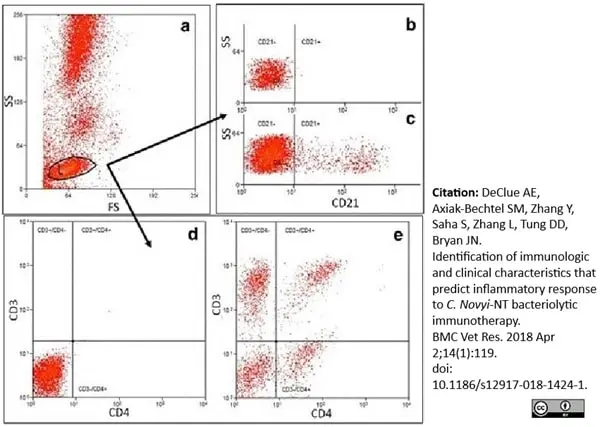
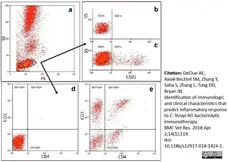
Example gating scheme for identification of CD3+/CD4+ lymphocytes and CD21+ lymphocytes. Lymphocytes were identified and gated using a forward and side scatter plot (a). Gated lymphocytes were applied were then applied to PE (CD3) vs FITC (CD4) plots or an Alexa Fluor 647 (CD21) histogram, respectively. Unstained cells and matched isotype controls from the same manufacturer were used to determine cut offs for negative (b and d) versus positive (c and e) cells. CD3+/CD4+ or CD21 positive cells were then identified. CD3+/CD8+ cells were selected in the same manner as the CD3+/CD4+ cells
From: DeClue AE, Axiak-Bechtel SM, Zhang Y, Saha S, Zhang L, Tung DD, Bryan JN.
Identification of immunologic and clinical characteristics that predict
inflammatory response to C. Novyi-NT bacteriolytic immunotherapy.
BMC Vet Res. 2018 Apr 2;14(1):119.
This image is from an open access article distributed under the terms of the Creative Commons Attribution License.
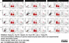
FITC conjugated Rat anti Canine CD4 antibody, clone YKIX302.9 (MCA1038F) used to evaluate CD4 expression on isolated control PBMCs by flow cytometry.
Image caption:
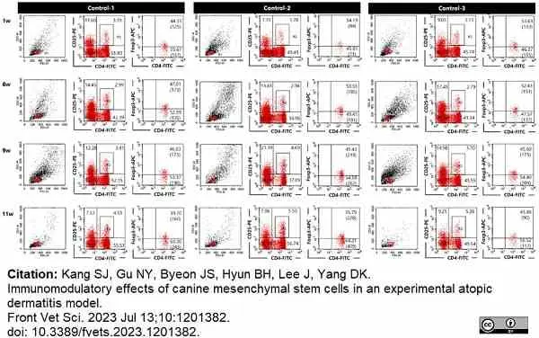
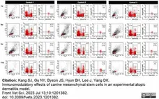
Dot plot of Tregs using flow cytometry at 1, 6, 9 and 11 wk of control group (n=3).
From: Kang SJ, Gu NY, Byeon JS, Hyun BH, Lee J, Yang DK.
Immunomodulatory effects of canine mesenchymal stem cells in an experimental atopic dermatitis model.
Front Vet Sci. 2023 Jul 13;10:1201382.
doi: 10.3389/fvets.2023.1201382.
This image is from an open access article distributed under terms of a Creative Commons Attribution License.
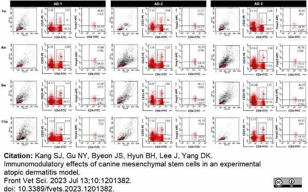
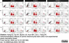
FITC conjugated Rat anti Canine CD4 antibody, clone YKIX302.9 (MCA1038F) used to evaluate CD4 expression on isolated PBMCs from animals suffering atopic dermatitis (AD) by flow cytometry.
Image caption:
Dot plot of Tregs using flow cytometry at 1, 6, 9 and 11wk of AD group (n=3).
From: Kang SJ, Gu NY, Byeon JS, Hyun BH, Lee J, Yang DK.
Immunomodulatory effects of canine mesenchymal stem cells in an experimental atopic dermatitis model.
Front Vet Sci. 2023 Jul 13;10:1201382.
doi: 10.3389/fvets.2023.1201382.
This image is from an open access article distributed under terms of a Creative Commons Attribution License.
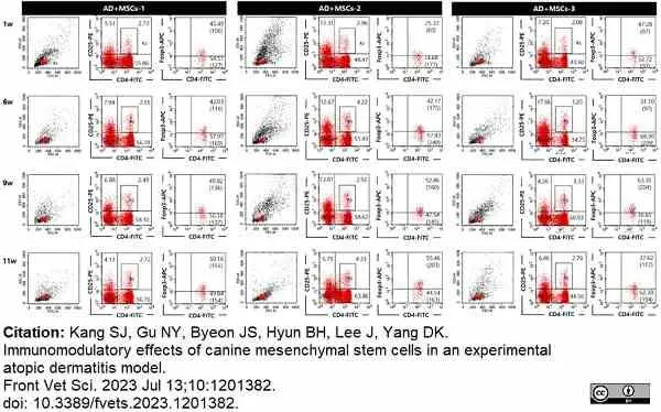
FITC conjugated Rat anti Canine CD4 antibody, clone YKIX302.9 (MCA1038F) used to evaluate CD4 expression on isolated PBMCs from animals suffering atopic dermatitis treated with MSCs (AD + MSC) by flow cytometry.
Image caption:
Dot plot of Tregs using flow cytometry at 1, 6, 9 and 11wk of AD+MSCs group (n=3).
From: Kang SJ, Gu NY, Byeon JS, Hyun BH, Lee J, Yang DK.
Immunomodulatory effects of canine mesenchymal stem cells in an experimental atopic dermatitis model.
Front Vet Sci. 2023 Jul 13;10:1201382.
doi: 10.3389/fvets.2023.1201382.
This image is from an open access article distributed under terms of a Creative Commons Attribution License.
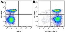
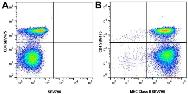
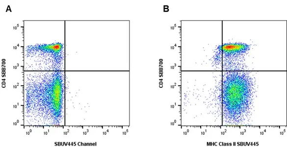
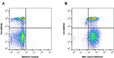
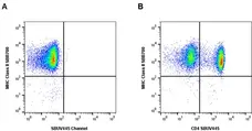
Figure B. StarBright Violet 475 conjugated Rat anti Canine CD4 antibody, clone YKIX302.9 (MCA1038SBV475) and StarBright Violet 790 conjugated Rat anti Canine MHC Class II antibody, clone YKIX334.2 (MCA1044SBV790). All experiments performed on canine PBMCs gated on live single lymphocytes, in the presence of 10% canine serum.
Data acquired on the ZE5 Cell analyser.

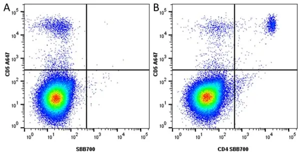
Figure B. Alexa Flour®A647 conjugated Rat anti Dog CD5 antibody, clone YKIX322.3 (MCA1037A647) and StarBright Blue 700 conjugated Rat anti Dog CD4 antibody, clone YKIX302.9 (MCA1038SBB700).
All experiments performed on red cell lysed dog blood gated on live single Lymphocytes, in the presence of 10% dog serum. Data acquired on the Data acquired on the ZE5 Cell Analyzer.
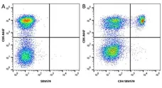
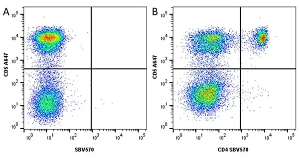
Figure B. Alexa Flour®647 conjugated Rat anti Dog CD5 antibody, clone YKIX322.3 (MCA1037A647) and StarBright Violet 570 conjugated Rat anti Dog CD4 antibody, clone YKIX302.9 (MCA1038SBV570).
All experiments performed on red cell lysed dog blood gated on live single lymphocytes, in the presence of 10% dog serum. Data acquired on the ZE5 Cell analyser.

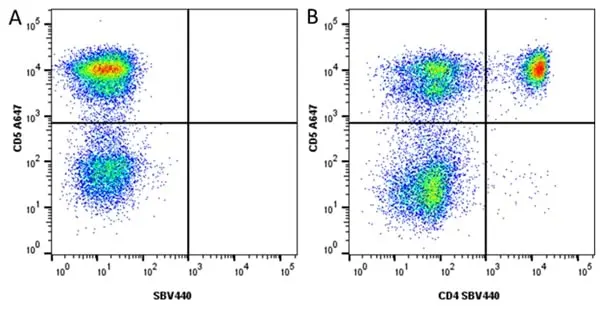
Figure B. Alexa Flour® 647 conjugated Rat anti Dog CD5 antibody, clone YKIX322.3 (MCA1037A647) and StarBright Violet 440 conjugated Rat anti Dog CD4 antibody, clone YKIX302.9 (MCA1038SBV440). All experiments performed on red blood lysed dog blood gated on live single cell lymphocytes, in the presence of 10% dog serum.
Data acquired on the ZE5 Cell analyser.
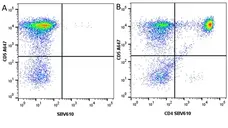
Figure B. Alexa Fluor® 647 conjugated Rat anti Dog CD5 antibody, clone YKIX322.3 (MCA1037A647) and StarBright Violet 610 conjugated Rat anti Dog CD4 antibody, clone YKIX302.9 (MCA1038SBV610). All experiments performed on red blood lysed dog blood gated on live single cell lymphocytes, in the presence of 10% dog serum.
Data acquired on the ZE5 Cell analyser
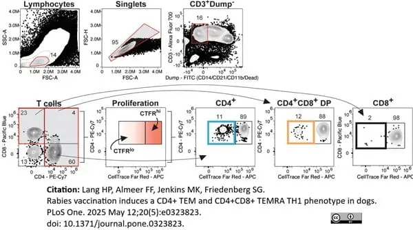
RPE-Cy7 conjugated Mouse anti Canine CD4 antibody, clone YKIX302.9 (MCA1038PECY7) used to identify T cells from canine samples by flow cytometry.
Image caption:
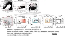
Gating strategy for canine T cells.
Representative contour plots showing the gating strategy to detect CD4+, CD8+, and DP T cell subsets in dogs based on gates on lymphocytes, singlets, CD3+Dump– (CD14, CD21, CD11b, and dead cells). Then, based on CD8 and CD4 expression, T cells are further distinguished by CellTrace Far Red (CTFR) dilution by CTFRHI and CTFRLO gates.
From: Lang HP, Almeer FF, Jenkins MK, Friedenberg SG.
Rabies vaccination induces a CD4+ TEM and CD4+CD8+ TEMRA TH1 phenotype in dogs.
PLoS One. 2025 May 12;20(5):e0323823.
doi: 10.1371/journal.pone.0323823.
This image is from an open access article distributed under terms of a Creative Commons Attribution License.
Data acquired on the ZE5 Cell Analyzer.
Data acquired on the ZE5 Cell Analyzer.
Filter by Application:
F Reset| Rat anti Dog CD4 antibody, clone YKIX302.9, is a monoclonal antibody specific for the canine CD4 cell surface antigen. Clone YKIX302.9 was clustered at the first Canine Leukocyte Antigen Workshop (CLAW) [Cobbold et al. 1992] along with clone CA13.1E4. Rat anti Dog CD4 antibody, clone YKIX302.9 partially depletes circulating T lymphocytes when administered in vivo, but alone is not sufficient to prolong allograft survival in a canine transplant model (Watson et al. 1993). Uniquely amongst mammals, canine CD4 is expressed by neutrophils as well as by lymphocyte subsets (Moore et al. 1992). |
- Target Species
- Dog
- Product Form
- Purified IgG - liquid
- Preparation
- Purified IgG prepared by affinity chromatography on Protein G from tissue culture supernatant
- Buffer Solution
- Phosphate buffered saline
- Preservative Stabilisers
- 0.09% sodium azide (NaN3)
- Carrier Free
- Yes
- Immunogen
- Canine concanavilin A activated T cell blasts.
- Approx. Protein Concentrations
- IgG concentration 1.0 mg/ml
- Fusion Partners
- Spleen cells from immunized DA rats were fused with cells of the Y3/Ag1.2.3 rat myeloma cell line.
- Regulatory
- For research purposes only
- Guarantee
- 12 months from date of despatch
This product is shipped at ambient temperature. It is recommended to aliquot and store at -20°C on receipt. When thawed, aliquot the sample as needed. Keep aliquots at 2-8°C for short term use (up to 4 weeks) and store the remaining aliquots at -20°C.
Avoid repeated freezing and thawing as this may denature the antibody. Storage in frost-free freezers is not recommended.
Avoid repeated freezing and thawing as this may denature the antibody. Storage in frost-free freezers is not recommended.
This product has been reported to work in the following applications. This information is derived from testing within our laboratories, peer-reviewed publications or personal communications from the originators. Please refer to references indicated for further information. For general protocol recommendations, please visit the antibody protocols page.
| Application Name | Verified | Min Dilution | Max Dilution |
|---|---|---|---|
| Flow Cytometry |  |
1/50 | 1/100 |
| Immunohistology - Frozen | |
Where this antibody has not been tested for use in a particular technique this does not necessarily exclude its use in such procedures. Suggested working dilutions are given as a guide only. It is recommended that the user titrates the antibody for use in their own system using appropriate negative/positive controls.
- Flow Cytometry
- Use 10μl of the suggested working dilution to label 106 cells or 100μl whole blood
Source Reference
-
Cobbold, S. & Metcalfe, S. (1994) Monoclonal antibodies that define canine homologues of human CD antigens: summary of the First International Canine Leukocyte Antigen Workshop (CLAW).
Tissue Antigens. 43 (3): 137-54.
References for CD4 antibody
-
Watson, C.J. et al. (1993) CD4 and CD8 monoclonal antibody therapy: strategies to prolong renal allograft survival in the dog.
Br J Surg. 80 (11): 1389-92. -
Gorman, S.D. et al. (1994) Isolation and expression of cDNA encoding the canine CD4 and CD8 alpha antigens.
Tissue Antigens. 43 (3): 184-8. -
Out, T.A. et al. (2002) Local T-cell activation after segmental allergen challenge in the lungs of allergic dogs.
Immunology. 105: 499-508. -
Benyacoub, J. et al. (2003) Supplementation of food with Enterococcus faecium (SF68) stimulates immune functions in young dogs.
J Nutr. 133: 1158-62. -
Bauer. T.R. Jr. et al. (2006) Correction of the disease phenotype in canine leukocyte adhesion deficiency using ex vivo hematopoietic stem cell gene therapy.
Blood. 108: 3313-20. -
Reis, A.B. et al. (2006) Phenotypic features of circulating leucocytes as immunological markers for clinical status and bone marrow parasite density in dogs naturally infected by Leishmania chagasi.
Clin Exp Immunol. 146: 303-11. -
Miranda, S. et al. (2007) Characterization of circulating lymphocyte subpopulations in canine leishmaniasis throughout treatment with antimonials and allopurinol.
Vet Parasitol. 144 (3-4): 251-60. -
Yasuda, N. et al. (2008) CC chemokine receptor 4-positive CD4(+) lymphocytes in peripheral blood increases during maturation in healthy beagles.
J Vet Med Sci. 70 (9): 989-92.
View The Latest Product References
-
Papadogiannakis, E.I. et al. (2009) Determination of intracellular cytokines IFN-gamma and IL-4 in canine T lymphocytes by flow cytometry following whole-blood culture.
Can J Vet Res. 73 (2): 137-43. -
Estrela-Lima, A. et al. (2010) Immunophenotypic features of tumor infiltrating lymphocytes from mammary carcinomas in female dogs associated with prognostic factors and survival rates.
BMC Cancer. 10: 256. -
Boggiatto, P.M. et al. (2010) Immunologic indicators of clinical progression during canine Leishmania infantum infection.
Clin Vaccine Immunol. 17: 267-73. -
Tominaga, M. et al. (2010) Flow cytometric analysis of peripheral blood and tumor-infiltrating regulatory T cells in dogs with oral malignant melanoma.
J Vet Diagn Invest. 22: 438-41. -
Bund, D. et al. (2010) Canine-DCs using different serum-free methods as an approach to provide an animal-model for immunotherapeutic strategies.
Cell Immunol. 263: 88-98. -
Pinheiro, D. (2011) Phenotypic and functional characterization of a CD4(+) CD25(high) FOXP3(high) regulatory T-cell population in the dog.
Immunology. 132: 111-22. -
Araújo, M.S. et al. (2011) Immunological changes in canine peripheral blood leukocytes triggered by immunization with first or second generation vaccines against canine visceral leishmaniasis.
Vet Immunol Immunopathol. 141: 64-75. -
Mitchell, L. et al. (2012) Induction of remission results in spontaneous enhancement of anti-tumor cytotoxic T-lymphocyte activity in dogs with B cell lymphoma.
Vet Immunol Immunopathol. 145 (3-4): 597-603. -
Mitchell, L. et al. (2012) Clinical and immunomodulatory effects of toceranib combined with low-dose cyclophosphamide in dogs with cancer.
J Vet Intern Med. 26: 355-62. -
Aricò, A. et al. (2013) The role of vascular endothelial growth factor and matrix metalloproteinases in canine lymphoma: in vivo and in vitro study.
BMC Vet Res. 9: 94. -
Michael HT et al. (2013) Isolation and characterization of canine natural killer cells.
Vet Immunol Immunopathol. 155 (3): 211-7. -
Figueiredo, M.M. et al. (2014) Expression of Regulatory T Cells in Jejunum, Colon, and Cervical and Mesenteric Lymph Nodes of Dogs Naturally Infected with Leishmania infantum.
Infect Immun. 82: 3704-12. -
Aresu, L. et al. (2014) VEGF and MMP-9: biomarkers for canine lymphoma.
Vet Comp Oncol. 12: 29-36. -
Duz AL et al. (2014) The TcI and TcII Trypanosoma cruzi experimental infections induce distinct immune responses and cardiac fibrosis in dogs.
Mem Inst Oswaldo Cruz. 109 (8): 1005-13. -
Gelain, M.E. et al. (2014) CD44 in canine leukemia: analysis of mRNA and protein expression in peripheral blood.
Vet Immunol Immunopathol. 159 (1-2): 91-6. -
Yamaya, Y. & Watari, T. (2015) Increased proportions of CCR4(+) cells among peripheral blood CD4(+) cells and serum levels of allergen-specific IgE antibody in canine chronic rhinitis and bronchitis.
J Vet Med Sci. 77 (4): 421-5. -
Miller, J. et al. (2015) Humoral and Cellular Immune Response in Canine Hypothyroidism.
J Comp Pathol. 153 (1): 28-37. -
Viana, K.F. et al. (2015) Setting the proportion of CD4+ and CD8+ T-cells co-cultured with canine macrophages infected with Leishmania chagasi.
Vet Parasitol. 211 (3-4): 124-32. -
Costa-Pereira, C. et al. (2015) One-year timeline kinetics of cytokine-mediated cellular immunity in dogs vaccinated against visceral leishmaniasis.
BMC Vet Res. 11 (1): 92. -
Hauck, V. et al. (2016) Increased numbers of FoxP3-expressing CD4(+) CD25(+) regulatory T cells in peripheral blood from dogs with atopic dermatitis and its correlation with disease severity.
Vet Dermatol. 27 (1): 26-e9. -
Riondato, F. et al. (2016) Analytical and diagnostic validation of a flow cytometric strategy to quantify blood and marrow infiltration in dogs with large B-cell lymphoma.
Cytometry B Clin Cytom. 90 (6): 525-30. -
Bonnefont-Rebeix, C. et al. (2016) Characterization of a novel canine T-cell line established from a spontaneously occurring aggressive T-cell lymphoma with large granular cell morphology.
Immunobiology. 221 (1): 12-22. -
Viana, K.F. et al. (2016) Application of rapid in vitro co-culture system of macrophages and T-cell subsets to assess the immunogenicity of dogs vaccinated with live attenuated Leishmania donovani centrin deleted parasites (LdCen-/-).
Parasit Vectors. 9: 250. -
Munhoz.T.D. et al. (2016) Regulatory T cells in dogs with multicentric lymphoma: peripheral blood quantification at diagnosis and after initial stage chemotherapy.
Arq. Bras. Med. Vet. Zootec. 68 (1): 1-9. -
Tagawa, M. et al. (2016) Evaluation of Costimulatory Molecules in Peripheral Blood Lymphocytes of Canine Patients with Histiocytic Sarcoma.
PLoS One. 11 (2): e0150030. -
Schaut, R.G. et al. (2016) Regulatory IgDhi B Cells Suppress T Cell Function via IL-10 and PD-L1 during Progressive Visceral Leishmaniasis.
J Immunol. 196 (10): 4100-9. -
Schaut, R.G. et al. (2016) Recovery of antigen-specific T cell responses from dogs infected with Leishmania (L.) infantum by use of vaccine associated TLR-agonist adjuvant.
Vaccine. 34 (44): 5225-34. -
Deravi, N. et al. (2017) Specific immunotypes of canine T cell lymphoma are associated with different outcomes.
Vet Immunol Immunopathol. 191: 5-13. -
Bahamondes, F. et al. (2017) Omental adipose tissue is a more suitable source of canine Mesenchymal stem cells.
BMC Vet Res. 13 (1): 166. -
Roatt, B.M. et al. (2017) A Vaccine Therapy for Canine Visceral Leishmaniasis Promoted Significant Improvement of Clinical and Immune Status with Reduction in Parasite Burden.
Front Immunol. 8: 217. -
Anai, L.A. et al. (2017) Quantification of Treg cells in peripheral blood and lymph nodes of dogs with multicentric lymphoma
Arq Bras Med Vet Zootec. 69 (6): 1496-502. -
Pellin, M.A. et al. (2017) Safety evaluation of combination doxorubicin and toceranib phosphate (Palladia®) in tumour bearing dogs: a phase I dose-finding study.
Vet Comp Oncol. 15 (3): 919-31. -
Martins, G.C. et al. (2018) Clinical-pathological and immunological biomarkers in dogs with atopic dermatitis.
Vet Immunol Immunopathol. 205: 58-64. -
Withers, S.S. et al. (2018) Multi-color flow cytometry for evaluating age-related changes in memory lymphocyte subsets in dogs.
Dev Comp Immunol. 87: 64-74. -
DeClue, A.E. et al. (2018) Identification of immunologic and clinical characteristics that predict inflammatory response to C. Novyi-NT bacteriolytic immunotherapy.
BMC Vet Res. 14 (1): 119. -
DaSilva, A.V.A. et al. (2018) Morphophysiological changes in the splenic extracellular matrix of Leishmania infantum-naturally infected dogs is associated with alterations in lymphoid niches and the CD4+ T cell frequency in spleens.
PLoS Negl Trop Dis. 12 (4): e0006445. -
Lisiecka. U. et al. (2019) Evaluation of T regulatory lymphocytes and serum concentration of selected cytokines in dogs with perianal tumors.
Vet Immunol Immunopathol. 207: 10-17. -
Akiyama, S. et al. (2019) Th17 cells increase during maturation in peripheral blood of healthy dogs.
Vet Immunol Immunopathol. 209: 17-21. -
Martini, V. et al. (2019) Prognostic role of non-neoplastic lymphocytes in lymph node aspirates from dogs with diffuse large B-cell lymphoma treated with chemo-immunotherapy.
Res Vet Sci. 125: 130-5. -
Aguiar-Soares, R.D.O. et al. (2020) Phase I and II Clinical Trial Comparing the LBSap, Leishmune®, and Leish-Tec® Vaccines against Canine Visceral Leishmaniasis.
Vaccines (Basel). 8 (4): 690. -
Wolf-Ringwall, A. et al. (2020) Prospective evaluation of flow cytometric characteristics, histopathologic diagnosis and clinical outcome in dogs with naïve B-cell lymphoma treated with a 19-week CHOP protocol.
Vet Comp Oncol. 18 (3): 342-52. -
Sayag, D. et al. (2020) Proof-of-concept study: Evaluation of plasma and urinary electrolytes as markers of response to L-asparaginase therapy in dogs with high-grade lymphoma.
Vet Clin Pathol. 49 (3): 476-83. -
Lee, J. et al. (2021) Canine Natural Killer Cell-Derived Exosomes Exhibit Antitumor Activity in a Mouse Model of Canine Mammary Tumor.
Biomed Res Int. 2021: 6690704. -
Grudzien, M. et al. (2021) A newly established canine NK-type cell line and its cytotoxic properties.
Vet Comp Oncol. 19 (3): 567-77. -
Lee, S.H. et al. (2021) Safety and immunological effects of recombinant canine IL-15 in dogs.
Cytokine. 148: 155599. -
Knebel, A. et al. (2021) Measurement of canine Th17 cells by flow cytometry.
Vet Immunol Immunopathol. 243: 110366. -
Konno, H. et al. (2022) An experimental challenge model for Leishmania donovani in beagle dogs, showing a similar pattern of parasite burden in the peripheral blood and liver.
Parasitol Res. 121 (12): 3569-79. -
Kanei, T. et al. (2022) Expression and functional analysis of chemokine receptor 7 in canine lymphoma cell lines.
J Vet Med Sci. 84 (1): 25-30. -
do Prado Duzanski, A. et al. (2022) Cell-mediated immunity and expression of MHC class I and class II molecules in dogs naturally infected by canine transmissible venereal tumor: Is there complete spontaneous regression outside the experimental CTVT?
Res Vet Sci. 145: 193-204. -
Karayannopoulou, M. et al. (2022) Effect of major versus minor mastectomy on host immunity in canine mammary cancer
Vet Immunol Immunopathol. Feb 24: 110403. -
Bragato, J.P. et al. (2022) miRNA-21 regulates CD69 and IL-10 expression in canine leishmaniasis.
PLoS One. 17 (3): e0265192. -
Riccardo, F. et al. (2022) Antigen mimicry as an effective strategy to induce CSPG4-targeted immunity in dogs with oral melanoma: a veterinary trial.
J Immunother Cancer. 10(5):e004007. -
Matralis, D.T. et al. (2023) Intracellular IFN-γ and IL-4 levels of CD4 + and CD8 + T cells in the peripheral blood of naturally infected (Leishmania infantum) symptomatic dogs before and following a 4-week treatment with miltefosine and allopurinol: a double-blinded, controlled and cross-sectional study.
Acta Vet Scand. 65 (1): 2. -
Hamouzová, P. et al. (2023) Lymphocyte immunophenotyping in dogs with lymphopenia of common causes.
Vet Immunol Immunopathol. 261: 110620. -
Tarone, L. et al. (2023) A chimeric human/dog-DNA vaccine against CSPG4 induces immunity with therapeutic potential in comparative preclinical models of osteosarcoma.
Mol Ther. 31 (8): 2342-59. -
Yamauchi, A. et al. (2023) Negative Influence of Aging on Differentiation and Proliferation of CD8(+) T-Cells in Dogs.
Vet Sci. 10 (9): 541. -
Bencze, M. et al. (2023) Receptor interacting protein kinase-3 mediates both myopathy and cardiomyopathy in preclinical animal models of Duchenne muscular dystrophy.
J Cachexia Sarcopenia Muscle. Nov 01 [Epub ahead of print]. -
Martini, V. et al. (2018) A retrospective study of flow cytometric characterization of suspected extranodal lymphomas in dogs.
J Vet Diagn Invest. 30 (6): 830-6. -
DeClue, A.E. et al. (2020) Transportation and Routine Veterinary Interventions Alter Immune Function in the Dog.
Top Companion Anim Med. 39: 100408. -
Lee, G.W. et al. (2021) Case Report: Long-Term Survival of a Dog With Chronic Lymphocytic Leukemia Treated With Chlorambucil, Prednisolone, and Imatinib.
Front Vet Sci. 8: 625527. -
Sainz, Á. et al. (2021) Effect of chemically modified tetracycline-8 (CMT-8) on hematology, blood chemistry, cytokines and peripheral blood lymphocyte subsets of healthy dogs.
Res Vet Sci. 136: 200-8. -
Kang, S.J. et al. (2023) Immunomodulatory effects of canine mesenchymal stem cells in an experimental atopic dermatitis model.
Front Vet Sci. 10: 1201382. -
Sheng, R. et al. (2023) Prognostic significance of CD25 expression in dogs with a noninvasive diagnosis of B-cell lymphoma treated with CHOP chemotherapy.
Vet Comp Oncol. 21 (1): 28-35. -
Miguelena Chamorro, B. et al. (2023) Characterization of Canine Peyer's Patches by Multidimensional Analysis: Insights from Immunofluorescence, Flow Cytometry, and Single-Cell RNA Sequencing.
Immunohorizons. 7 (11): 788-805. -
Stokol, T. et al. (2024) Flow cytometric-based detection of CD80 is a useful diagnostic marker of acute myeloid leukemia in dogs.
Front Vet Sci. 11: 1405297. -
Rogato, F. et al. (2024) Leukemia cutis as a prominent clinical sign in a dog with acute myeloid leukemia.
Vet Clin Pathol. 53 (4): 448-457. -
De Maria, R. et al. (2025) Development and activity of canine B7-H3-CAR.CIK lymphocytes against sarcomas: preclinical evidence and perspectives for human clinical translation.
Cancer Immunol Immunother. 74 (10): 306. -
Lang, H.P. et al. (2025) Rabies vaccination induces a CD4+ TEM and CD4+CD8+ TEMRA TH1 phenotype in dogs.
PLoS One. 20 (5): e0323823. -
Szydłowski, P. et al. (2025) Tregitopes derived from canine proteins can enhance T regulatory lymphocytes frequency in dog peripheral blood mononuclear cells (PBMC) culture in vitro.
J Leukoc Biol. 117 (11) [Epub ahead of print]. -
Foos, K.M. et al. (2026) Generation of functional canine TIL products for solid tumors.
Front Immunol. 17: 1810955. -
Blockeel, A. et al. (2025) CD94 as a novel marker for immunophenotyping of leukemia and lymphoma in dogs.
Front Vet Sci. 12: 1716800.
- UniProt
- P33705
- Entrez Gene
- CD4
- GO Terms
- GO:0007155 cell adhesion
- GO:0016021 integral to membrane
- GO:0006955 immune response
- GO:0045058 T cell selection
MCA1038GA
If you cannot find the batch/lot you are looking for please contact our technical support team for assistance.
View more products with CD4 specificity
Please Note: All Products are "FOR RESEARCH PURPOSES ONLY"
View all Anti-Dog ProductsAlways be the first to know.
When we launch new products and resources to help you achieve more in the lab.
Yes, sign me up