MBP antibody | 12
Rat anti MBP (aa82-87)
- Product Type
- Monoclonal Antibody
- Clone
- 12
- Isotype
- IgG2a
- Specificity
- MBP
- Region
- (aa82-87)


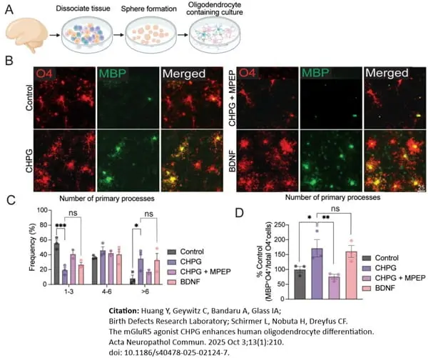
Rat anti Bovine MBP antibody, clone 12 (MCA409S) used to identify rat oligodendrocyte progenitors in vitro by immunofluorescence.
Image caption:
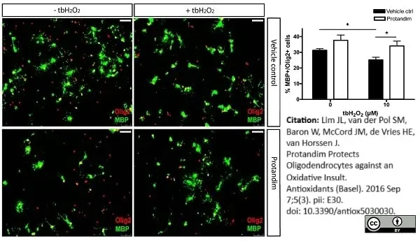
Protandim promotes differentiation of primary rat OPCs under oxidative stress. After 2 day de-differentiation of OLs with growth factors bFGF-2 and PDGF-AA, primary rat OPCs were incubated with 30 μg/mL Protandim or vehicle control (EtOH) for 24 h. After removal of medium, cells were subsequently exposed to either control medium or medium with 10 μM tert-butyl hydrogen peroxide (tbH2O2) for 5 days. MBP and Olig2 expression were assayed by immunocytochemistry. Data are presented as percentage of control and expressed as the mean ± SEM of 3 independent experiments. Statistics reflect student's t-test, one-tailed; * p<0.05.
From: Lim JL, van der Pol SM, Baron W, McCord JM, de Vries HE, van Horssen J.
Protandim Protects Oligodendrocytes against an Oxidative Insult.
Antioxidants (Basel). 2016 Sep 7;5(3). pii: E30.
doi: 10.3390/antiox5030030.
This is an open access article distributed under the terms of a Creative Commons Attribution License.
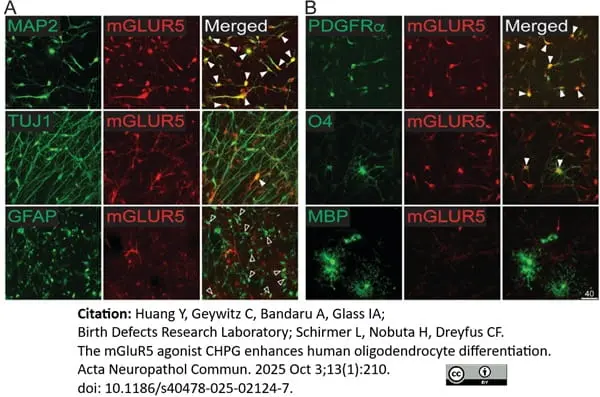
Rat anti MBP antibody, clone 12 (MCA409S) used to identify rat oligodendrocyte progenitors in vitro by immunofluorescence.
Image caption:
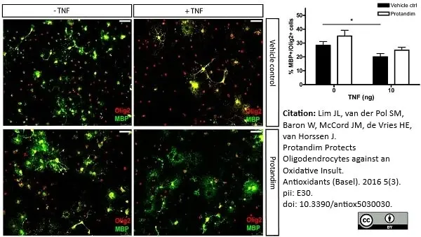
Protandim marginally promotes differentiation of primary rat OPCs in the presence of TNF. After 2 days of de-differentiation, primary rat OPCs were incubated with 30 μg/mL Protandim or vehicle control (EtOH) for 24 h. After removal of medium, cells were subsequently exposed to either control medium or medium with 10 ng/mL TNF for 5 days. MBP and Olig2 expression were assayed by immunocytochemistry. Data are presented as percentage of control and expressed as the mean ± SEM of 3 independent experiments. Statistics reflect student's t-test, one-tailed;*p<0.05.
From: Lim JL, van der Pol SM, Baron W, McCord JM, de Vries HE, van Horssen J.
Protandim Protects Oligodendrocytes against an Oxidative Insult.
Antioxidants (Basel). 2016 Sep 7;5(3). pii: E30.
doi: 10.3390/antiox5030030.
This is an open access article distributed under the terms of a Creative Commons Attribution License.

Rat anti Bovine MBP antibody, clone 12 (MCA409S) used for the detection of myelin basic protein in murine myelinating cell cultures using immunofluorescence.
Image caption:
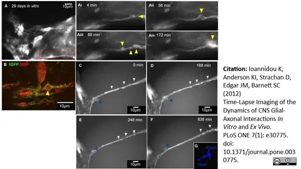
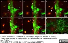
Time-lapse imaging of fluorescently labelled cells in association with neurites in vitro. A–E) Myelinating cultures generated from a mix of wild type/beta-actin mice were visualised using time-lapse microscopy (Nikon TE2000 (60×, 0.75NA) over 16 hr in 5 min intervals on DIV 27 after the addition of wild type neurospheres previously infected with lentivirus carrying dsRed/GFP gene and addition of cyto-GFP cells. Two cell types were followed over time, one that expressed DS red/cyto-GFP and the other cyto-GFP. A–E) Strongly positive green cells typical of cyto-GFP morphologically resembled oligodendrocytes in contact with neurite bundles. The membrane appears to ruffle and form flaps/bubbles (arrow). In addition, the soma changes its location with respect to the neurite processes, over time, by moving closer to the neurite bundle. C–E) Dynamic imaging over 7.5 hours of a dsred/GFP labelled cell (asterisk) which was engulfed by a cell resembling a microglial cell (yellow arrow). This fluorescence was very much weaker than the cells generated from the beta-actin cyto-GFP mouse. Time frames obtained with 40× magnification (long distance working lens) and without perfect focus.
From: Ioannidou K, Anderson KI, Strachan D, Edgar JM, Barnett SC (2012)
Time-Lapse Imaging of the Dynamics of CNS Glial-Axonal Interactions In Vitro and Ex Vivo.
PLoS ONE 7(1): e30775.
doi: 10.1371/journal.pone.0030775.
This is an open access article distributed under the terms of a Creative Commons Attribution License.

Rat anti Bovine MBP antibody, clone 12 (MCA409S) used for the detection of myelin basic protein in murine myelinating cell cultures using immunofluorescence.
Image caption:
Time-lapse imaging of the putative assembly of myelin membrane. A) Neurospheres expressing farns-GFP were added to shiverer myelinating cultures on DIV 19 and time-lapse imaging (Nikon TE2000) was performed on 29 DIV, over 24 hr with 4 min time intervals. Ai–iv) Magnified view of the inset in A illustrates a farns-GFP process looping around a presumptive neurite and forming a membranous protrusions or ‘bubble’ (yellow arrow head). This membrane bubble appears to moves along the neurite over time. See Video S3. B) The cells from the Petri dish imaged with confocal microscopy were immunostained with anti-GFP and anti-MBP to confirm differentiation of cyto-GFP labelled oligodendrocytes.C–F) Time-lapse sequence of the same culture for a period of 30 hours, with 3 min time interval, on 24 DIV revealed membrane cuffs (arrowheads) extending and joining up over a neurite. After about 13 hours, the farns-GFP-positive cuffs were observed to form a single, united thick membrane sheath over a neurite. G) Manual tracking of the pathway of a weakly GFP-positive cell which was possibly associated with the membranous fragments.
From: Ioannidou K, Anderson KI, Strachan D, Edgar JM, Barnett SC (2012)
Time-Lapse Imaging of the Dynamics of CNS Glial-Axonal Interactions In Vitro and Ex Vivo.
PLoS ONE 7(1): e30775.
doi: 10.1371/journal.pone.0030775.
This is an open access article distributed under the terms of a Creative Commons Attribution License.
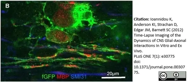
Rat anti Bovine MBP antibody, clone 12 (MCA409S) used for the detection of Myelin basic protein in murine myelinating cell cultures by immunofluorescence.
Image caption:
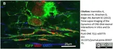
B) MBP staining of cells in the Petri dish using a Zeiss 710 (×63, 1.4NA) after imaging confirms that farns-GFP expressing cells belong to the oligodendroglial lineage.
From: Ioannidou K, Anderson KI, Strachan D, Edgar JM, Barnett SC (2012)
Time-Lapse Imaging of the Dynamics of CNS Glial-Axonal Interactions In Vitro and Ex Vivo.
PLoS ONE 7(1): e30775
doi: 10.1371/journal.pone.0030775.
This is an open access article distributed under the terms of a Creative Commons Attribution License.

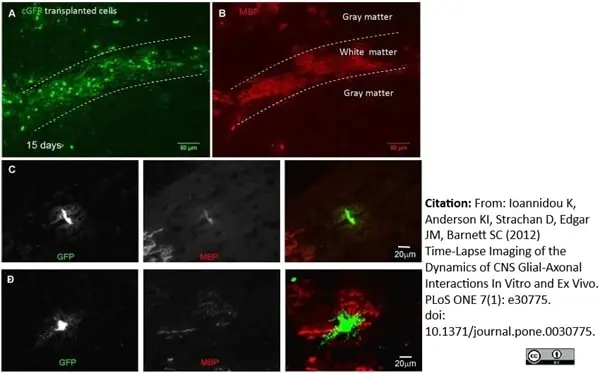
Rat anti Bovine MBP antibody, clone 12 (MCA409S) used for the detection of myelin basic protein in murine myelinating cell cultures using immunofluorescence.
Image caption:
Immunohistochemistry of transplanted neurospheres demonstrate that cyto-GFP labelled cells form early and mature myelinating oligodendrocytes. Cyto-GFP-expressing neurospheres were transplanted into a shiverer mouse 3, 7 or 15 days post-transplantation, and 10 µm thick frozen sections were cut and immunolabelled with antibodies to GFP and MBP. Low magnification image of a dorso-vental section of spinal cord, 15 days post-transplantation showing GFP (A) and MBP (B) immunostaining. Transplanted cells were located in both grey and white matter (dorsal columns are delineated by the dotted lines) and expressed MBP-positive myelin sheaths. C) A pre-myelinating cell in which multiple fine GFP positive processes emanate from a central cell body. The soma is also lightly stained with MBP, confirming the identity of the cell as that of the oligodendroglial lineage. MBP-positive myelin sheaths, belonging to a second cell are seen in the bottom left hand corner of the images. D) An early myelinating cell in which short MBP-positive profiles are present at the periphery of the GFP-positive soma. All images were obtained using epifluorescence Olympus microscope (FV10 ASW). Representative images from at least 30 separate experiments.
From: Ioannidou K, Anderson KI, Strachan D, Edgar JM, Barnett SC (2012)
Time-Lapse Imaging of the Dynamics of CNS Glial-Axonal Interactions In Vitro and Ex Vivo.
PLoS ONE 7(1): e30775.
doi: 10.1371/journal.pone.0030775.
This is an open access article distributed under the terms of a Creative Commons Attribution License.
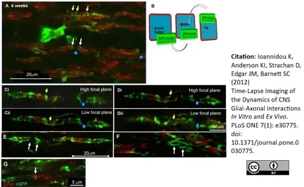
Rat anti MBP antibody, clone 12 (MCA409S) used for the detection of Myelin basic protein in murine spinal cord using immunofluorescence.
Image caption:
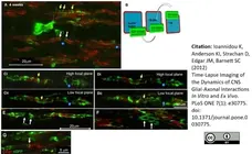
Confocal images of transplanted cyto-GFP expressing cells in the shiverer spinal cord. Four weeks after transplantation of cyto-GFP expressing neurospheres, fixed sections of the shiverer spinal cord were immunolabelled for MBP (red) and GFP (green). A) A cyto-GFP labelled cell appears to extend spirals of cytoplasm around an MBP-positive myelin-like sheath (yellow arrows). Below the cell body, cyto-GFP is seen at the lateral edges (in relation to the long axis of the sheaths, white arrow) of adjacent sheaths and probably represents the cytoplasm filled paranodal loops on either side of the node of Ranvier (asterisks). B) Schematic of visualisation of the sections in C and D. Ci–ii and Di–ii) Spiral of GFP cytoplasm was followed by focussing up and down through the plane of view where they crossed up, traversed the axonal surface, then crossed down again representing the looping as shown in the schematic in B. E–G) 3D reconstruction of cyto-GFP structures (E), illustrates cyto-GFP either side of a space typical of a node of Ranvier (white arrows). F) is a tilted perspective of E) and shows the cyto-GFP form complete rings (white arrows representing the same position in E), consistent with the morphology of paranodal loops. I) Asymmetric caspr positive structures in association with cyto-GFP, at either side of a heminode. On the left, caspr forms a single vertical line and co-localises with cyto-GFP from the myelinating cell. On the right, caspr appears like a loose coil, consistent with its pattern of expression in non-myelinated axons. All images were acquired using an Olympus FV1000 confocal microscope (×60, 1.35NA). Representative images from at least 10 separate experiments.
From: Ioannidou K, Anderson KI, Strachan D, Edgar JM, Barnett SC (2012)
Time-Lapse Imaging of the Dynamics of CNS Glial-Axonal Interactions In Vitro and Ex Vivo.
PLoS ONE 7(1): e30775.
doi: 10.1371/journal.pone.0030775.
This is from an open access article distributed under the terms of a Creative Commons Attribution License.
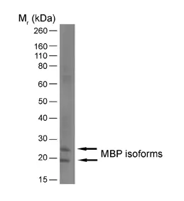
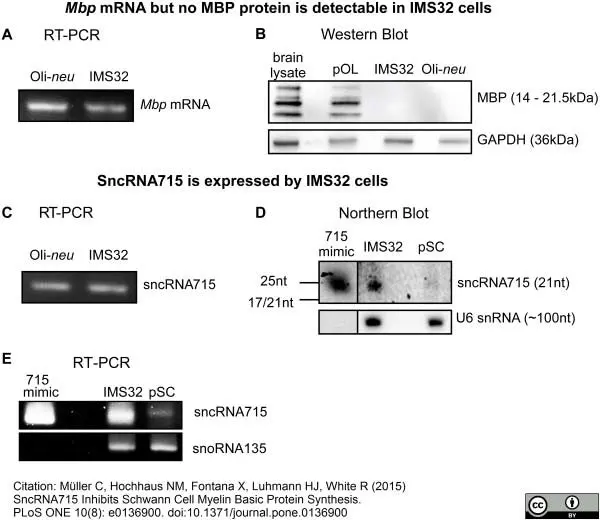
Rat anti MBP antibody, clone 12 (MCA409S) used for the evaluation of myelin basic protein expression in mouse brain lysates by western blotting
Image caption:
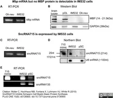
MBP and sncRNA715 Expression in Schwann cells. A, Reverse transcription PCR (RT-PCR) on RNA extracted from Oli-neu or IMS32 cells using Mbp-specific primers. The 88nt long amplicon for Mbp was visualized in an ethidium bromide-stained 4% agarose gel. B, Western Blots of lysates from P18 mouse brain (brain lysate), primary oligodendrocytes (pOL, 7DIV), IMS32 and Oli-neu cells using MBP and GAPDH (loading control) specific antibodies. C, Reverse transcription PCR (RT-PCR) on RNA extracted from Oli-neu or IMS32 cells using a sncRNA715-specific primer assays. PCR products (~60-nt long due to the use of hairpin primers in the RT reaction) were visualized in an ethidium bromide stained 4% agarose gel. D, Northern Blots with RNA from IMS32 and undifferentiated primary Schwann cells (pSC) shows expression of sncRNA715 in IMS32 and a lower expression in pSC. Synthetic sncRNA715 (715-mimic) and U6 snRNA were used as positive control and loading control, respectively. E, RT-PCR on RNA from IMS32 and undifferentiated pSC confirms lower expression of sncRNA715 in pSC compared to IMS32 cells shown in D. 715-mimic was used as positive control and snoRNA135 as loading control.
From: Müller C, Hochhaus NM, Fontana X, Luhmann HJ, White R (2015)
SncRNA715 Inhibits Schwann Cell Myelin Basic Protein Synthesis.
PLoS ONE 10(8): e0136900.
This is from an open access article distributed under the terms of a Creative Commons Attribution License.
Rat anti Bovine MBP antibody, clone 12 (MCA409S) used for the evaluation of myelin basic protein expression in mouse Schwann cells using immunofluorescence and western blotting
Image caption:
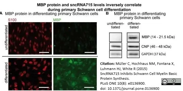
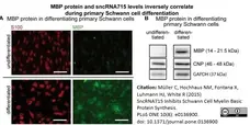
Inverse correlation of MBP and sncRNA715 in primary Schwann cells. A, Primary Schwann cells derived from sciatic nerves of P3 Wistar rats were cultured in non-differentiating (untreated) or differentiating (+NRG1 +dbcAMP) conditions. MBP protein can only be detected by immunocytochemistry in differentiated Schwann cells. Scale bar represents 50μm. B, Western Blots of undifferentiated and differentiated primary Schwann cells show MBP protein only present in differentiated Schwann cells. CNP is expressed in both maturation stages of primary Schwann cells. GAPDH serves as loading control.
From: Müller C, Hochhaus NM, Fontana X, Luhmann HJ, White R (2015)
SncRNA715 Inhibits Schwann Cell Myelin Basic Protein Synthesis.
PLoS ONE 10(8): e0136900.
doi: 10.1371/journal.pone.0136900.
This is from an open access article distributed under the terms of a Creative Commons Attribution License.

Rat anti MBP antibody, clone 12 (MCA409S) used for the evaluation of myelin basic protein expression in mouse sciatic nerve lysates by western blotting
Image caption:
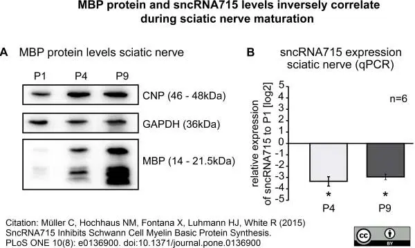
Inverse correlation of MBP and sncRNA715 in the sciatic nerve. A&B, The sciatic nerve was lysed from mice at postnatal day 1, 4 and 9 and myelin proteins as well as sncRNA715 expression was analyzed by Western blotting (A) and qPCR (B), respectively. MBP and CNP Western blots show increasing levels in differentiating sciatic nerves (A) while sncRNA715 levels decrease during differentiation, P-values P4: 0,0313, P9: 0,0313 (B, log2 values are plotted, sncRNA715 levels at P4 and P9 were quantified relative to P1 using snoRNA135 as a reference gene). Number of experiments (n) are indicated and bar graphs represent mean values ± s.e.m. (Wilcoxon signed-rank test, *P< 0.05, GraphPad Prism5 was used for statistical analysis).
From: Müller C, Hochhaus NM, Fontana X, Luhmann HJ, White R (2015)
SncRNA715 Inhibits Schwann Cell Myelin Basic Protein Synthesis.
PLoS ONE 10(8): e0136900.
This is from an open access article distributed under the terms of a Creative Commons Attribution License.
Rat anti MBP antibody, clone 12 (MCA409S) used for the evaluation of myelin basic protein expression in mouse Schwann cell lysates by western blotting
Image caption:
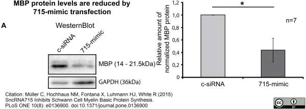
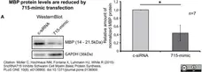
Regulation of MBP levels by sncRNA715. Rat primary Schwann cells were differentiated as described and transfected with synthetic sncRNA715 (715-mimic) or control siRNA (c-siRNA). A, Representative Western Blots for MBP and GAPDH showing reduced MBP levels in 715-mimic transfected cells. B, Densitometric analysis of 7 experiments (n = 7) as shown in (A). Normalized MBP/GAPDH values related to control-siRNA transfected cells are plotted. P-value for 715-mimic: 0.0469. Bar graphs represent mean values ± s.e.m. (Wilcoxon signed-rank test, n = 7, *P< 0.05, GraphPad Prism5 was used for statistical analysis).
From: Müller C, Hochhaus NM, Fontana X, Luhmann HJ, White R (2015)
SncRNA715 Inhibits Schwann Cell Myelin Basic Protein Synthesis.
PLoS ONE 10(8): e0136900.
This is from an open access article distributed under the terms of a Creative Commons Attribution License.
Rat anti Bovine MBP antibody, clone 12 (MCA409S) used for the detection of myelin basic protein by immunofluorescence on formalin fixed, paraffin embedded murine tissue sections.
Image caption:
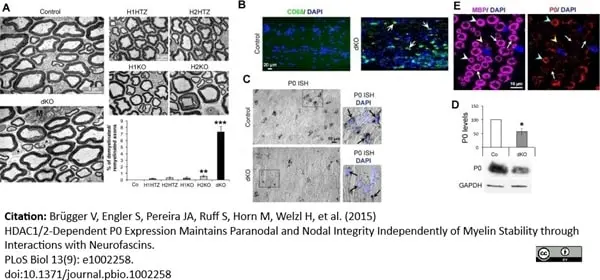
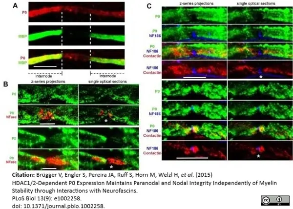
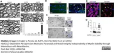
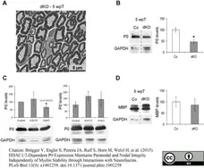
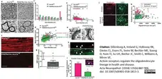
Demyelination/remyelination and decreased P0 expression in dKO mice. (A) Electron micrographs of ultrathin cross sections of control, dKO, H1HTZ, H2HTZ, H1KO, and H2KO sciatic nerves, at 8 wk post-tamoxifen, and percentage of demyelinated/remyelinated axons (3 animals per group, at least 700 axons counted per mouse), identifying a demyelination/remyelination phenotype in dKO sciatic nerves. Asterisks indicate demyelinated axons and “M” macrophages. (B) CD68 (green) immunofluorescence in longitudinal cryosections of control and dKO sciatic nerves labeled with DAPI (blue = nuclei) showing increased presence of macrophages in dKO sciatic nerves, consistent with the demyelination phenotype. Arrows indicate macrophages. (C) In situ hybridization (ISH) of P0 on longitudinal cryosections of control and dKO sciatic nerves identifying a reduction of P0 at the transcript level at 5 wk post-tamoxifen, before the onset of demyelination and when macrophages are not present in the nerve. Pictures on the right are magnifications of black boxes depicted on the left. Black arrows show SC nuclei. (D) Western blot of P0 and quantification normalized to GAPDH (loading control) in sciatic nerve lysates of control and dKO mice at 8 wk post-tamoxifen (3 mice per group), showing reduced P0 protein levels in dKO sciatic nerves. (E) Confocal images of MBP (magenta) and P0 (red) coimmunofluorescence in paraffin cross sections of dKO mice at 8 wk post-tamoxifen, showing reduced P0 levels in most myelin rings, while MBP levels remain high. Nuclei are labeled in blue by DAPI. A single optical section is shown. White arrows indicate MBP positive/P0 negative myelin rings, the yellow arrowhead shows an MBP/P0 double negative SC, and blue arrowheads MBP/P0 double positive myelin rings. Three animals per group were used for each experiment. P-values (two-tailed unpaired (A) or paired (D) Student's t test): * = p <0.05, ** = p <0.01, *** = p <0.001, error bars = SEM.
From: Brügger V, Engler S, Pereira JA, Ruff S, Horn M, Welzl H, et al. (2015)
HDAC1/2-Dependent P0 Expression Maintains Paranodal and Nodal Integrity Independently of Myelin Stability through Interactions with Neurofascins.
PLoS Biol 13(9): e1002258.
doi:10.1371/journal.pbio.1002258.
This is from an open access article distributed under the terms of a Creative Commons Attribution License.
Rat anti Bovine MBP antibody, clone 12 (MCA409S) used for the detection of myelin basic protein in mice by immunofluorescence.
Image caption:
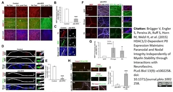
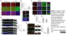
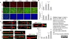
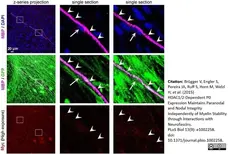
P0 rescues myelination, Caspr, and neurofascins in plp-dKO DRG cultures. Confocal coimmunofluorescence images of (A) MBP (red) and neurofilament (NF, green), or (C) P0 (red) and MBP (green), or (D) Caspr (red), total neurofascins (NFasc, green), and MBP (white), or (F) MBP (red), NF (white), and either GFP, P0, or Myc (green), or (H) Caspr (red) and NFasc186 (false-colored green) in myelinated control and plp-dKO DRG cultures with (A,C,D,F,H) or without (A) tamoxifen. Briefly, A–D demonstrate demyelination, loss of P0 and paranodal/nodal defects in plp-dKO DRG cultures, mimicking the in vivo phenotype of dKO sciatic nerves. In (F,H), plp-dKO DRG cultures were transduced with doxycycline-inducible lentiviruses expressing either GFP, P0, or P0-myc. (B,G) Quantification of MBP fluorescence intensity normalized to NF. (E,I) Percentage of intact nodes/paranodes expressing Caspr and high levels of NFasc. F–I show that exogenously delivered P0 significantly rescues demyelination and paranodal/ nodal defects of plp-dKO DRG cultures. In (C), white arrows indicate MBP positive/P0 negative fibers, and magenta arrowheads MBP/P0 double positive fibers. Merges MBP/P0 (C) or Caspr/NFasc (D) appear yellow. In (D), dashed lines delineate the paranodal region. In (H), control and plp-dKO were transduced with lentiviruses expressing GFP, and plp-dKO + P0 with lentiviruses expressing P0 (white). Z-series projections (A,C,F) and single optical sections (D,H) are shown. In (D,H), arrows indicate heminodes or full nodes. Images on the right are magnifications of white boxes depicted on the left images. At least three control and three plp-dKO embryos were used for quantification (average of three coverslips per embryos), representative images are shown. Nuclei are labeled in blue with DAPI. P-values (unpaired [B,E,I] or paired [G] two-tailed Student's t test, unless stated otherwise in the figure): * = p < 0.05, *** = p < 0.001, error bars = SEM.
From: Brügger V, Engler S, Pereira JA, Ruff S, Horn M, Welzl H, et al. (2015)
HDAC1/2-Dependent P0 Expression Maintains Paranodal and Nodal Integrity Independently of Myelin Stability through Interactions with Neurofascins.
PLoS Biol 13(9): e1002258.
doi: 10.1371/journal.pbio.1002258.
This is from an open access article distributed under the terms of a Creative Commons Attribution License.
Rat anti Bovine MBP antibody, clone 12 (MCA409S) used for the detection of myelin basic protein by immunofluorescence.
Image caption:
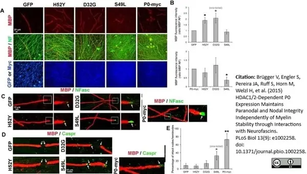
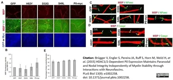
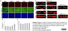
In contrast to the S49L P0 mutant, D32G and H52Y P0 mutants rescue myelination of HDAC1/2 plp-dKO DRG but not paranodal/nodal integrity. Coimmunofluorescence of MBP (red) and (A) neurofilament (NF, green), and Myc or GFP fluorescence (blue), or (C) neurofascins (NFasc, green), or (D) Caspr (green) in myelinated HDAC1/2 plp-dKO DRG cultures transduced with lentiviruses expressing either GFP, H52Y-myc, D32G-myc, S49L-myc or P0-myc, and treated with tamoxifen for 10 d after completion of myelination. A–B show that H52Y and D32G but not S49L P0 mutants are able to rescue myelination of plp-dKO DRG cultures, similarly to P0-myc, and C–D show that S49L, but not H52Y or D32G, P0 mutant is able to partially rescue paranodal/nodal defects of plp-dKO DRG cultures. In (C), pictures on the right are magnifications of the white boxes depicted on left images. Arrows indicate paranodes/nodes. In (B), quantification of MBP fluorescence intensity normalized to NF and compared to GFP or P0-myc (set to 1). DRG of six plp-dKO embryos were quantified (three plp-dKO embryos per graph, four coverslips per plp-dKO). In (C,D), DRG of three plp-dKO embryos were analyzed and representative pictures are shown. In (E), the graph represents the percentage of intact (Caspr-positive or high NFasc levels) nodes and heminodes. DRG of three plp-dKO embryos were quantified, four coverslips per plp-dKO, 80 to 300 nodes/heminodes counted per plp-dKO per virus. P-values (paired (B) and unpaired (E) two-tailed (unless stated otherwise in the figure) Student's t test): * = p < 0.05, ** = p < 0.01, error bars = SEM.
From: Brügger V, Engler S, Pereira JA, Ruff S, Horn M, Welzl H, et al. (2015)
HDAC1/2-Dependent P0 Expression Maintains Paranodal and Nodal Integrity Independently of Myelin Stability through Interactions with Neurofascins.
PLoS Biol 13(9): e1002258.
doi: 10.1371/journal.pbio.1002258.
This is from an open access article distributed under the terms of a Creative Commons Attribution License.
Rat anti MBP antibody, clone 12 (MCA409S) used for the detection of myelin basic protein by western blotting.
Image caption:
No demyelination and unchanged MBP levels, but decreased P0 levels at 5 wk post-tamoxifen in dKO and at 8 wk post-tamoxifen in H2KO sciatic nerves. (A) Electron micrograph of ultrathin cross sections of dKO sciatic nerve at 5 wk post-tamoxifen. Sciatic nerves of 3 dKO mice were analyzed and no demyelinated or remyelinated axon or macrophage were found. (B–D) Western blot of P0 (B,C) and MBP (D) in control and dKO sciatic nerve lysates at 5 wk post-tamoxifen (B,D), and in control, H1HTZ, H1KO, H2HTZ, and H2KO at 8 wk post-tamoxifen (C), and quantification of protein levels normalized to the loading control GAPDH in mutants compared to controls (= 100%) (3 animals per genotype were used). In (B,D), the dashed lines indicate that lysates were run on the same gel but not on consecutive lanes. P-values (unpaired (B,C,D) or paired (C, HDAC2 single mutants) two-tailed (unless stated otherwise in the figure) Student's t test): * = p < 0.05, error bars = SEM.
From: Brügger V, Engler S, Pereira JA, Ruff S, Horn M, Welzl H, et al. (2015)
HDAC1/2-Dependent P0 Expression Maintains Paranodal and Nodal Integrity Independently of Myelin Stability through Interactions with Neurofascins.
PLoS Biol 13(9): e1002258.
This is from an open access article distributed under the terms of a Creative Commons Attribution License.

Rat anti MBP antibody, clone 12 (MCA409S) used for the detection of myelin basic protein by western blotting.
Image caption:
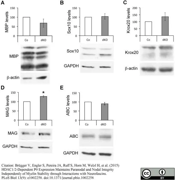
Further characterization of dKO phenotype at the molecular level. Western blots of MBP (A), Sox10 (B), Krox20 (C), MAG (D), and ABC (E) in lysates of control (Co) and dKO sciatic nerves at 8 wk post-tamoxifen, and quantification of protein levels normalized to GAPDH or beta-actin loading control in dKOs compared to controls (= 100%). For each experiment, three control and three dKO animals were used. P-values (paired two-tailed Student's t test): * = p < 0.05, error bars = SEM.
From: Brügger V, Engler S, Pereira JA, Ruff S, Horn M, Welzl H, et al. (2015)
HDAC1/2-Dependent P0 Expression Maintains Paranodal and Nodal Integrity Independently of Myelin Stability through Interactions with Neurofascins.
PLoS Biol 13(9): e1002258.
This is from an open access article distributed under the terms of a Creative Commons Attribution License.
Rat anti Bovine MBP antibody, clone 12 (MCA409S) used for the detection of murine myelin basic protein by immunofluorescence.
Image caption:
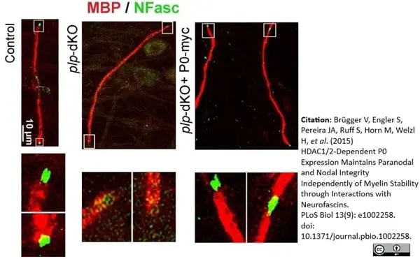
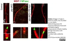
P0-myc rescues neurofascins in plp-dKO DRG cultures. Coimmunofluorescence of total neurofascins (NFasc, green) and MBP (red) in control or plp-dKO myelinated DRG cultures transduced with lentiviruses expressing GFP (Control and plp-dKO) or P0-myc (plp-dKO + P0-myc). Z-series projections of confocal stacks are shown. Images on the right are magnifications of white boxes depicted on the left images highlighting heminodes or nodes. DRG of three control and three plp-dKO embryos were analyzed and representative images are shown.
From: Brügger V, Engler S, Pereira JA, Ruff S, Horn M, Welzl H, et al. (2015) HDAC1/2-Dependent P0 Expression Maintains Paranodal and Nodal Integrity Independently of Myelin Stability through Interactions with Neurofascins.
PLoS Biol 13(9): e1002258.
doi: 10.1371/journal.pbio.1002258.
This is from an open access article distributed under the terms of a Creative Commons Attribution License.

Rat anti Bovine MBP antibody, clone 12 (MCA409S) used for the detection of myelin basic protein by immunofluorescence.
Image caption:
In contrast to the S49L P0 mutant, D32G and H52Y P0 mutants rescue myelination of HDAC1/2 plp-dKO DRG but not paranodal/nodal integrity. Coimmunofluorescence of MBP (red) and (A) neurofilament (NF, green), and Myc or GFP fluorescence (blue), or (C) neurofascins (NFasc, green), or (D) Caspr (green) in myelinated HDAC1/2 plp-dKO DRG cultures transduced with lentiviruses expressing either GFP, H52Y-myc, D32G-myc, S49L-myc or P0-myc, and treated with tamoxifen for 10 d after completion of myelination. A–B show that H52Y and D32G but not S49L P0 mutants are able to rescue myelination of plp-dKO DRG cultures, similarly to P0-myc, and C–D show that S49L, but not H52Y or D32G, P0 mutant is able to partially rescue paranodal/nodal defects of plp-dKO DRG cultures. In (C), pictures on the right are magnifications of the white boxes depicted on left images. Arrows indicate paranodes/nodes. In (B), quantification of MBP fluorescence intensity normalized to NF and compared to GFP or P0-myc (set to 1). DRG of six plp-dKO embryos were quantified (three plp-dKO embryos per graph, four coverslips per plp-dKO). In (C,D), DRG of three plp-dKO embryos were analyzed and representative pictures are shown. In (E), the graph represents the percentage of intact (Caspr-positive or high NFasc levels) nodes and heminodes. DRG of three plp-dKO embryos were quantified, four coverslips per plp-dKO, 80 to 300 nodes/heminodes counted per plp-dKO per virus. P-values (paired (B) and unpaired (E) two-tailed (unless stated otherwise in the figure) Student's t test): * = p < 0.05, ** = p < 0.01, error bars = SEM.
From: Brügger V, Engler S, Pereira JA, Ruff S, Horn M, Welzl H, et al. (2015)
HDAC1/2-Dependent P0 Expression Maintains Paranodal and Nodal Integrity Independently of Myelin Stability through Interactions with Neurofascins.
PLoS Biol 13(9): e1002258.
doi: 10.1371/journal.pbio.1002258.
This is from an open access article distributed under the terms of a Creative Commons Attribution License.
Rat anti Bovine MBP antibody, clone 12 (MCA409S) used for the detection of murine myelin basic protein by immunofluorescence.
Image caption:
No difference of MBP levels or of percentage of intact nodes/heminodes between control DRG cultures transduced with lentiviruses carrying GFP, H52Y, D32G, S49L P0 mutants, or P0-myc. Coimmunofluorescence of MBP (red) and (A) neurofilament (NF, green) and Myc or GFP fluorescence (blue), or (C) neurofascins (NFasc, green), or (D) Caspr (green) in myelinated HDAC1/2 control DRG cultures transduced with lentiviruses expressing either GFP, H52Y-myc, D32G-myc, S49L-myc, or P0-myc, and treated with tamoxifen for 10 d after completion of myelination. Arrows indicate paranodes/nodes. In (B), quantification of MBP fluorescence intensity normalized to NF and compared to GFP (set to 1). DRG of three control embryos were quantified, four coverslips per embryo were analyzed, and representative pictures are shown. In (E), the graph represents the percentage of intact (high NFasc levels) nodes and heminodes. DRG of three control embryos were quantified, four coverslips per control, 40 to 80 nodes/heminodes counted per control per virus. Error bars = SEM.
From: Brügger V, Engler S, Pereira JA, Ruff S, Horn M, Welzl H, et al. (2015) HDAC1/2-Dependent P0 Expression Maintains Paranodal and Nodal Integrity Independently of Myelin Stability through Interactions with Neurofascins.
PLoS Biol 13(9): e1002258.
doi: 10.1371/journal.pbio.1002258.
This is from an open access article distributed under the terms of a Creative Commons Attribution License.

Rat anti Bovine MBP antibody, clone 12 (MCA409S) used for the evaluation of myelin basic protein expression in mouse brain by immunofluorescence.
Image caption:
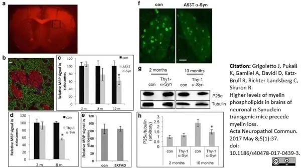
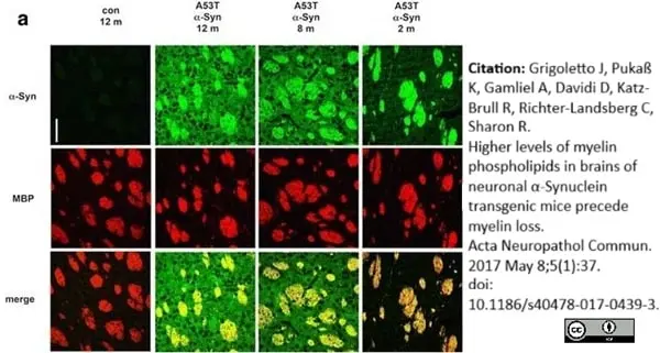
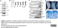
Age-dependent loss of MBP and P25α signals in striosomes (patches). a A coronal brain section (6 μm, paraffin embedded) from a 12 month-old A53T α-Syn mouse, stained with anti-MBP antibody. Black square shows area of interest. b Higher magnification of the area of interest from (a) in a consecutive section, double stained with anti-MBP (red) and anti-tyrosine hydroxylase (TH, green) antibodies. Scale bar, 100 μm. c Quantification of MBP signal inside striosomes. Bars represent mean ± SD of n = 4 A53T and control mice at 2, 8 and 12 months of age. *, <0.05, one-way ANOVA. d Quantification of MBP signal in striosomes of Thy-1 α-Syn tg and age-matched control mouse brains determined at 2 and 8 months of age. Bars represent mean ± SD of n = 4 mice. *, &ly;0.05, one-way ANOVA. e Quantification of MBP signal in striosomes of 12 month-old 5XFAD and control mice. f Coronal brain sections containing rostral-dorsal striatum (6 μm, paraffin embedded) of 12 month-old A53T α-Syn mouse and age-matched control immunoreacted with anti-P25α antibody and showing a striosome. Scale bar, 10 μm. g Samples of high-speed supernatant (50 μg protein) obtained from whole Thy-1 α-Syn and control mouse brains at 2 and 10 months of age analyzed by Western blotting and immunoreacted with anti-P25α antibody. h Graph showing quantitation of blots obtained in (G) mean ± SD of n = 4 mice. *, <0.05, one-way ANOVA.
From: Grigoletto J, Pukaß K, Gamliel A, Davidi D, Katz-Brull R, Richter-Landsberg C, Sharon R.
Higher levels of myelin phospholipids in brains of neuronal α-Synuclein transgenic mice precede myelin loss.
Acta Neuropathol Commun. 2017 May 8;5(1):37.
doi: 10.1186/s40478-017-0439-3.
This is from an open access article distributed under the terms of a Creative Commons Attribution License.
Rat anti Bovine MBP antibody, clone 12 (MCA409S) used for the evaluation of myelin basic protein expression in mouse brain by western blotting.
Image caption:
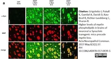
Myelin proteins, membrane flotation and histology. a Western blot showing myelin proteins in preparations of purified myelin from A53T α-Syn and control mouse brains at 4–6 months of age (n = 5–6 mice). b Bars showing mean ± SD of the indicated myelin protein levels, in whole brain protein extracts, of A53T α-Syn at 12–14 months (n = 8–9 mice). Presented as a percent of control age-matched mouse brain, set at 100% (vertical line) *,<0.05, one-way ANOVA. c Flotation assay showing the distribution of detergent-soluble myelin membrane particles into a 8–25% nycodenz gradient. Purified myelin preparations of A53T α-Syn and control mouse brains at 12 months of age, analyzed in parallel. Aliquots of gradient fractions analyzed by Western blotting using the specified antibodies. Representative blot of n = 3. 2', 3'-cyclic nucleotide 3'-phosphodiesterase (CNPase); myelin associated glycoprotein (MAG); myelin basic protein (MBP); myelin oligodendrocyte glycoprotein (MOG); proteolipid protein (PLP). d The caudate nucleus in coronally sectioned human brain hemisphere (100 μm), including striosomes (dark strips) and matrix (light staining) of a 90-year old female without PD (non-PD) and a 68-year-old female PD patient with advanced disease (neuropathological stage 5). Sections were stained for myelin using a modified Pal-Weigert method. Scale bar, 500 μm. Stained brain sections were provided by the Braak laboratory (University of Ulm, Ulm, Germany). e Coronal brain sections (6 μm, paraffin embedded) of A53T α-Syn and control mice at 12 months of age stained with Luxol Fast Blue/Periodic Acid Schiff, showing the corpus callosum (cc) and striatum (st). Scale bar, 500 μm.
From: Grigoletto J, Pukaß K, Gamliel A, Davidi D, Katz-Brull R, Richter-Landsberg C, Sharon R.
Higher levels of myelin phospholipids in brains of neuronal α-Synuclein transgenic mice precede myelin loss.
Acta Neuropathol Commun. 2017 May 8;5(1):37.
doi: 10.1186/s40478-017-0439-3.
This is from an open access article distributed under the terms of a Creative Commons Attribution License.
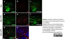
Rat anti Bovine MBP antibody, clone 12 (MCA409S) used for the evaluation of myelin basic protein expression in mouse oligodendrocytes by immunofluorescence.
Image caption:
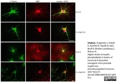
Effects of α-Syn on oligodendrocyte differentiation. Oligodendrocyte progenitor cells were either untreated (Co) or incubated with recombinant human (rh)α-Syn (10 μg/ml) 2 h after plating for the indicated time. Cells were subjected to indirect immunofluorescence staining using antibodies against α-tubulin (green) and MBP (red). Nuclei were stained with DAPI (blue). Scale bar: 20 μm.
From: Grigoletto J, Pukaß K, Gamliel A, Davidi D, Katz-Brull R, Richter-Landsberg C, Sharon R.
Higher levels of myelin phospholipids in brains of neuronal α-Synuclein transgenic mice precede myelin loss.
Acta Neuropathol Commun. 2017 May 8;5(1):37.
doi:10.1186/s40478-017-0439-3.
This is from an open access article distributed under the terms of a Creative Commons Attribution License.
Rat anti Bovine MBP antibody, clone 12 (MCA409S) used for the evaluation of myelin basic protein expression in mouse oligodendrocytes by immunofluorescence and western blotting.
Image caption:
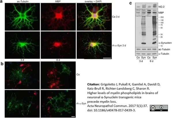
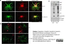
α-Syn impairs oligodendrocyte maturation. Oligodendrocyte progenitor cells were either untreated (Co) or incubated with rh α-Syn (10 μg/ml) 2 h after plating for 3 or 6 days. Cells were subjected to immunocytochemistry using antibodies: a anti-acetylated α-tubulin (green) and anti-MBP (red); b anti-proteoglycan NG-2 (green) and anti-MBP (red). Nuclei were stained with DAPI (blue). Scale bar: 20 μm. c Exogenously applied α-Syn led to an increase in NG-2 and a decrease in MBP levels. Western blot analysis of cell extracts was carried out with antibodies indicated on the right. Numbers on the right indicate molecular weights in kDa.
From: Grigoletto J, Pukaß K, Gamliel A, Davidi D, Katz-Brull R, Richter-Landsberg C, Sharon R.
Higher levels of myelin phospholipids in brains of neuronal α-Synuclein transgenic mice precede myelin loss.
Acta Neuropathol Commun. 2017 May 8;5(1):37.
doi: 10.1186/s40478-017-0439-3.
This is from an open access article distributed under the terms of a Creative Commons Attribution License.
Rat anti Bovine MBPantibody, clone 12 (MCA409S) used for the evaluation of myelin basic protein expression in mouse striatum by immunofluorescence.
Image caption:
Age-dependent accumulation of α-Syn toxicity in the striatum. a Coronal brain sections containing rostral-dorsal striatum (6 μm, paraffin embedded) of A53T α-Syn mouse at 2, 8 and 12 months of age, or control brain section at 12 months old, immunoreacted with anti-α-Syn antibody (syn - 303). Scale bar, 50 μm.
From: Grigoletto J, Pukaß K, Gamliel A, Davidi D, Katz-Brull R, Richter-Landsberg C, Sharon R.
Higher levels of myelin phospholipids in brains of neuronal α-Synuclein transgenic mice precede myelin loss.
Acta Neuropathol Commun. 2017 May 8;5(1):37.
doi: 10.1186/s40478-017-0439-3.
This is from an open access article distributed under the terms of a Creative Commons Attribution License.
Rat anti Bovine MBP antibody, clone 12 (MCA409S) used to evaluate MBP expression in mouse brain by immunofluorescence and western blotting.
Image caption:
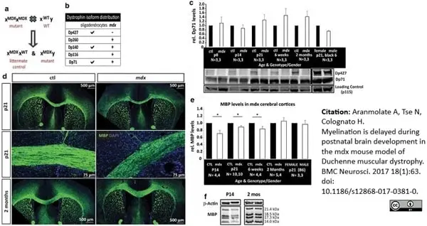
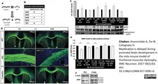
Myelination is delayed during postnatal brain development in the mdx mouse model of DMD. a Breeding strategy to generate litters with mdx males and heterozygous mdx/+ females (controls), b dystrophin isoform expression in oligodendrocytes and mdx mice, c western blot analysis of dystrophin protein content in lysates obtained from mdx and control cerebral cortices at postnatal day 8 (p8), p14, p21, 6 weeks, and 2 months. Comparable levels of Dp71 are found in both mdx and control lysates, while Dp427 is absent from mdx lysates. Cortical lysates from wildtype male and female mice (B6, Black 6) reveals that Dp427 and Dp71 levels are similar in males and females. Representative western blots are shown, including those for p115 as a loading control, d MBP immunohistochemistry of mdx and control coronal floating brain sections. Tiled confocal microscopy images reveal less MBP immunoreactivity in mdx cerebral cortices at postnatal day 21 (p21) but not at 2 months, e quantification of mean MBP densitometries from western blots comparing protein lysates from cerebral cortices of control and mdx mice at p14, p21, 6wks and 2 months. In addition, cortical lysates from wildtype mice (B6, Black 6) were analyzed to ensure that MBP levels did not differ by gender. Immunoblots to detect β-actin were used as loading controls. MBP levels in mdx cerebral cortices were decreased at p14, p21, and 6 weeks but were normal by 2 months (*p<0.05), f representative western blots of cortical lysates from postnatal day (p14) and 2 month old (2 mos) mdx and control mice
From: Aranmolate A, Tse N, Colognato H.
Myelination is delayed during postnatal brain development in the mdx mouse model of Duchenne muscular dystrophy.
BMC Neurosci. 2017 Aug 14;18(1):63.
doi: 10.1186/s12868-017-0381-0.
This is from an open access article distributed under the terms of a Creative Commons Attribution License
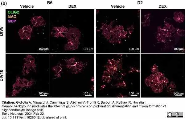
Rat anti Bovine MBP antibody, clone 12 (MCA409S) used for the demonstration of MBP expression by cells in the murine corpus callosum by immunofluorescence.
Image caption:
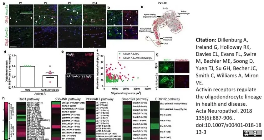
Activin receptor signaling regulates myelin membrane compaction/maturation. a Electron micrographs of myelinated axons in P16 Acvr1bfl/fl and PDGFRa-Cre; Acvr1bfl/fl mice. Scale bars 1 μm. b Average inner tongue thickness per axon diameter per animal in Acvr1bfl/fl (magenta) and PDGFRa-Cre; Acvr1bfl/fl mice (green). n = 3 mice per genotype. c Dot plot of inner tongue thickness per axon diameter for all myelinated axons for all animals, in Acvr1bfl/fl (magenta) and PDGFRa-Cre; Acvr1bfl/fl mice (green). d Myelin thickness versus axon diameter in Acvr1bfl/fl (magenta) and PDGFRa-Cre; Acvr1bfl/fl mice (green). Extra sum of squares F test between slopes, ***P = 0.0014. e Electron micrographs of compact myelin layers in P16 Acvr1bfl/fl and PDGFRa-Cre; Acvr1bfl/fl mice. f Images of myelin basic protein (MBP) in corpus callosum of Acvr1bfl/fl and PDGFRa-Cre; Acvr1bfl/fl mice at P16. Scale bar 25 μm. g MBP intensity in PDGFRa-Cre; Acvr1bfl/fl mice normalized to background and to levels in Acvr1bfl/fl mice. s.e.m. for variance in Acvr1bfl/fl samples indicated. One sample t test against theoretical mean of 1, **P = 0.0097. h Image of PDGFRa-Cre; Acvr1bfl/fl corpus callosum showing MAG+ (green), MBP negative (red) myelin sheaths (arrows). i Percentage of total axonal area (neurofilament (NF)+) co-localizing with compaction marker Caspr in caudal corpus callosum at P16 in Acvr1bfl/fl and PDGFRa-Cre; Acvr1bfl/fl mice. Two-tailed Student's t test, **P = 0.0038, n = 2–4 mice per group. Inset example of Caspr clusters (green) at paranodes along axon (NF+; purple) (arrow). j Images of cultured mature oligodendrocytes treated with vehicle control or activin-A (10 ng ml−1) stained with Phalloidin-Alexa-568 (red) and MBP (green). Scale bar 20 μm. k Phalloidin intensity (arbitrary units; A.U.) in MBP+ sheets of mature oligodendrocytes plotted against oligodendrocyte size (pixels squared; px2) in control (magenta) or activin-A (10 ng ml−1) treated (green) conditions
From: Dillenburg A, Ireland G, Holloway RK, Davies CL, Evans FL, Swire M, Bechler ME, Soong D, Yuen TJ, Su GH, Becher JC, Smith C, Williams A, Miron VE.
Activin receptors regulate the oligodendrocyte lineage in health and disease.
Acta Neuropathol. 2018 135(6):887-906..
doi:10.1007/s00401-018-1813-3
This is an open access article distributed under the terms of a Creative Commons Attribution License.
Rat anti MBP antibody, clone 12 (MCA409S) used for the demonstration of MBP expression by cells in organotypic cerebellar slice cultures by immunofluorescence.
Image caption:
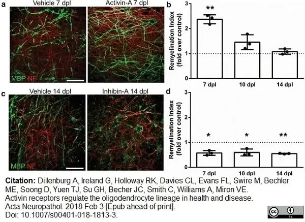
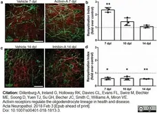
Activin receptor signaling regulates remyelination. a Representative images of organotypic cerebellar slice cultures at 7 days post lysolecithin-induced demyelination, treated with vehicle control or activin receptor agonist activin-A during remyelination, immunostained against myelin basic protein (MBP; green), and axonal neurofilament-H (NF; red). Scale bar 50 μm. b Mean remyelination index ± s.e.m. in activin-A-treated explants at 7, 10, and 14 days post lysolecithin (dpl) normalized to vehicle control from the respective time point. n = 3 animals, one-sample t test compared to theoretical mean of 1 (control), **P = 0.0057. c Representative images of slice cultures at 14 dpl treated with vehicle control or an inhibitor of activin receptor signaling inhibin-A during remyelination, immunostained against myelin basic protein (MBP; green) and axonal neurofilament-H (NF; red). Scale bar 50 μm.
From: Dillenburg A, Ireland G, Holloway RK, Davies CL, Evans FL, Swire M, Bechler ME, Soong D, Yuen TJ, Su GH, Becher JC, Smith C, Williams A, Miron VE.
Activin receptors regulate the oligodendrocyte lineage in health and disease.
Acta Neuropathol. 2018 135(6):887-906..
doi: 10.1007/s00401-018-1813-3.
This is an open access article distributed under the terms of a Creative Commons Attribution License.
Rat anti MBP antibody, clone 12 (MCA409S) used to evaluate MBP expression in murine cell culture lysates by western blotting.
Image caption:
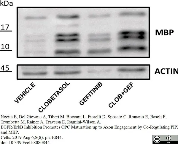
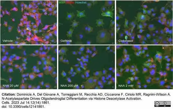
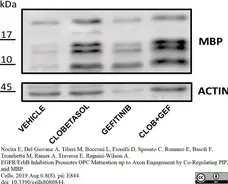
Epidermal growth factor receptor (EGFR) gene silencing upregulates MBP expression and increases Clobetasol-mediated Oli-neuM differentiation.
Immunoblot analyses of MBP expression with the indicated treatment. Left panel: Representative immunoblot analysis using anti-MBP or anti-Actin antibody as indicated in Material and Methods.
From: Nocita E, Del Giovane A, Tiberi M, Boccuni L, Fiorelli D, Sposato C, Romano E, Basoli F, Trombetta M, Rainer A, Traversa E, Ragnini-Wilson A.
EGFR/ErbB Inhibition Promotes OPC Maturation up to Axon Engagement by Co-Regulating PIP2 and MBP.
Cells. 2019 Aug 6;8(8). pii: E844.
doi: 10.3390/cells8080844.
This image is from an open access article distributed under the terms of a Creative Commons Attribution License.
Rat anti MBP antibody, clone 12 (MCA409S) used for the demonstration of MBP expression by cells in the murine corpus callosum by immunofluorescence.
Image caption:
Activin receptor signaling regulates myelin membrane compaction/maturation. a Electron micrographs of myelinated axons in P16 Acvr1bfl/fl and PDGFRa-Cre; Acvr1bfl/fl mice. Scale bars 1 μm. b Average inner tongue thickness per axon diameter per animal in Acvr1bfl/fl (magenta) and PDGFRa-Cre; Acvr1bfl/fl mice (green). n = 3 mice per genotype. c Dot plot of inner tongue thickness per axon diameter for all myelinated axons for all animals, in Acvr1bfl/fl (magenta) and PDGFRa-Cre; Acvr1bfl/fl mice (green). d Myelin thickness versus axon diameter in Acvr1bfl/fl (magenta) and PDGFRa-Cre; Acvr1bfl/fl mice (green). Extra sum of squares F test between slopes, ***P = 0.0014. e Electron micrographs of compact myelin layers in P16 Acvr1bfl/fl and PDGFRa-Cre; Acvr1bfl/fl mice. f Images of myelin basic protein (MBP) in corpus callosum of Acvr1bfl/fl and PDGFRa-Cre; Acvr1bfl/fl mice at P16. Scale bar 25 μm. g MBP intensity in PDGFRa-Cre; Acvr1bfl/fl mice normalized to background and to levels in Acvr1bfl/fl mice. s.e.m. for variance in Acvr1bfl/fl samples indicated. One sample t test against theoretical mean of 1, **P = 0.0097. h Image of PDGFRa-Cre; Acvr1bfl/fl corpus callosum showing MAG+ (green), MBP negative (red) myelin sheaths (arrows). i Percentage of total axonal area (neurofilament (NF)+) co-localizing with compaction marker Caspr in caudal corpus callosum at P16 in Acvr1bfl/fl and PDGFRa-Cre; Acvr1bfl/fl mice. Two-tailed Student's t test, **P = 0.0038, n = 2–4 mice per group. Inset example of Caspr clusters (green) at paranodes along axon (NF+; purple) (arrow). j Images of cultured mature oligodendrocytes treated with vehicle control or activin-A (10 ng ml−1) stained with Phalloidin-Alexa-568 (red) and MBP (green). Scale bar 20 μm. k Phalloidin intensity (arbitrary units; A.U.) in MBP+ sheets of mature oligodendrocytes plotted against oligodendrocyte size (pixels squared; px2) in control (magenta) or activin-A (10 ng ml−1) treated (green) conditions.
From: Dillenburg A, Ireland G, Holloway RK, Davies CL, Evans FL, Swire M, Bechler ME, Soong D, Yuen TJ, Su GH, Becher JC, Smith C, Williams A, Miron VE.
Activin receptors regulate the oligodendrocyte lineage in health and disease.
Acta Neuropathol. (2018) 135(6):887-906.
doi: 10.1007/s00401-018-1813-3.
This is an open access article distributed under the terms of a Creative Commons Attribution License.
Rat anti MBP antibody, clone 12 (MCA409S) used for the detection of murine myelin basic protein by immunofluorescence.
Image caption:
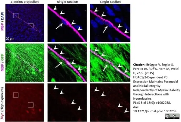
Validation of Myc staining specificity in myelinated plp-dKO DRG cultures efficiently transduced with lentiviruses expressing GFP. Coimmunofluorescence of MBP (Magenta, rat antibody) and Myc (red, mouse antibody), and GFP fluorescence (green) in myelinated plp-dKO DRG cultures transduced with lentiviruses expressing GFP and treated with tamoxifen for 10 d. Even at high exposure, Myc staining did not cross-react with MBP staining. To avoid cross-reactivity, we used multiple labeling (adsorbed against many animal species, including rat for antimouse and mouse for antirat) secondary antimouse and antirat antibodies. Antibody concentrations and staining protocol (buffers, incubation times and temperature, washes) were the same as for stainings presented in Fig 9A. Nuclei are labeled in blue with DAPI. Pictures on the right (single optical sections) are magnifications of the white boxes depicted on left images (z-series projections). Arrows indicate Schwann cell nuclei of myelinated fibers, arrowheads indicate MBP staining. DRG of three plp-dKO embryos were analyzed. None of the MBP-positive fibers were labeled by Myc staining.
From: Brügger V, Engler S, Pereira JA, Ruff S, Horn M, Welzl H, et al. (2015)
HDAC1/2-Dependent P0 Expression Maintains Paranodal and Nodal Integrity Independently of Myelin Stability through Interactions with Neurofascins.
PLoS Biol 13(9): e1002258.
doi: 10.1371/journal.pbio.1002258.
This is from an open access article distributed under the terms of a Creative Commons Attribution License.
Mouse anti Human Myelin Basic Protein antibody, clone 12 (MCA409S) used to demonstrate MBP expressing cells in human duodenal biopsy specimens by immunohistochemistry on cryostat sections.
Image caption:
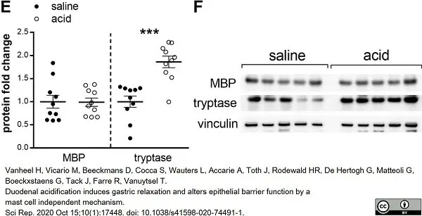
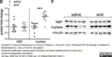
Duodenal acid induces tryptase expression, without changing mast cell counts or ultrastructure. Duodenal acid induces tryptase expression, without changing mast cell counts or ultrastructure. (E) Protein expression of MBP and tryptase was measured by western blot after intraduodenal saline (black dots) and acid (white dots) perfusion (n = 10 for both groups). (F) Representative western blot of five saline perfused and five acid perfused subjects. Bands were cropped from different parts of the same gel, or from different gels.
MBP, eosinophilic major basic protein.
From: Tack J, Farre R, Vanuytsel T.
Duodenal acidification induces gastric relaxation and alters epithelial barrier function by a mast cell independent mechanism.
Sci Rep. 2020 Oct 15;10(1):17448.
This image is from an open access article distributed under terms of a Creative Commons Attribution License.
Rat anti Myelin Basic Protein antibody, clone 12 (MCA409S) used to demonstrate rat myelin basic protein expressing oligodendrocytes by immunofluorescence.
Image caption:
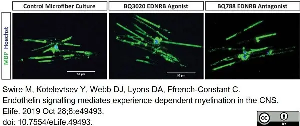
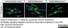
EDNRB enhances myelin sheath number in vitro.
Representative images of MBP positive oligodendrocytes in microfiber culture. Scale bar = 50 μm.
From: Swire M, Kotelevtsev Y, Webb DJ, Lyons DA, Ffrench-Constant C.
Endothelin signalling mediates experience-dependent myelination in the CNS.
Elife. 2019 Oct 28;8:e49493.
This image is from an open access article distributed under terms of a Creative Commons Attribution License.

Rat anti MBP antibody, clone 12 (MCA409S) used to label MBP expressing cells in mouse cerebellar cultures by immunofluorescence.
Image caption:
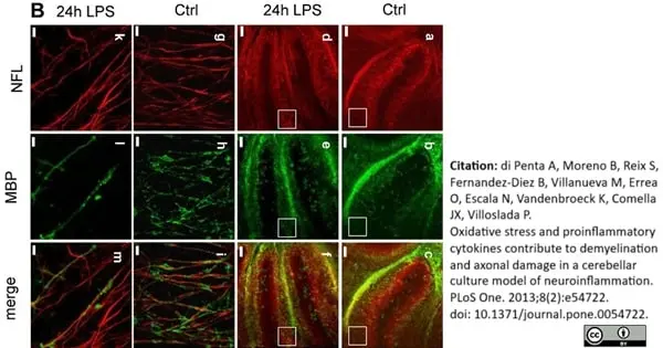
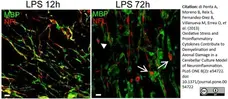
Microglial activation induces demyelination in mouse cerebellar cultures.
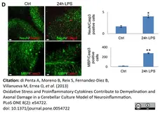
B) Immunofluorescence for NfH (red) and MBP (green) in cerebellar cultures treated with LPS (15 μg/ml: panels d-f and k-m) or control slices (Ctrl, panels a-c and g-i). Panels g-m show a higher magnification (×60) of images in a-f (white boxes in panels a-f). Scale bars = 100 μm (panels a-f) and 5 μm (panels g-m). The graph represent the percentage of myelinated axons (double staining for MBP and NfH) compared to unmyelinated axons (NfH).
From: di Penta A, Moreno B, Reix S, Fernandez-Diez B, Villanueva M, Errea O, et al. (2013)
Oxidative Stress and Proinflammatory Cytokines Contribute to Demyelination and Axonal Damage in a Cerebellar Culture Model of Neuroinflammation.
PLoS ONE 8(2): e54722.
doi: 10.1371/journal.pone.0054722
This image is from an open access article distributed under terms of a Creative Commons Attribution License.
Rat anti MBP antibody, clone 12 (MCA409S) used to label MBP expressing cells in mouse cerebellar cultures by immunofluorescence.
Image caption:
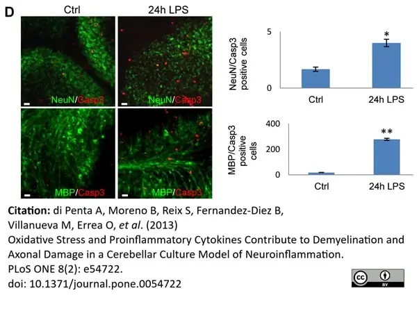
Microglial activation induces demyelination in mouse cerebellar cultures.
Cerebellar cultures were treated with LPS (15 µg/ml) for 24 h and then immunostained for MBP/Casp3 or NeuN/Casp3 colabeling. Scale bar = 10μm.
From: di Penta A, Moreno B, Reix S, Fernandez-Diez B, Villanueva M, Errea O, et al. (2013)
Oxidative Stress and Proinflammatory Cytokines Contribute to Demyelination and Axonal Damage in a Cerebellar Culture Model of Neuroinflammation.
PLoS ONE 8(2): e54722.
doi: 10.1371/journal.pone.0054722
This image is from an open access article distributed under terms of a Creative Commons Attribution License.
Rat anti Bovine MBP antibody, clone 12 (MCA409S) used to label MBP expressing cells in mouse cerebellar cultures by immunofluorescence.
Image caption:
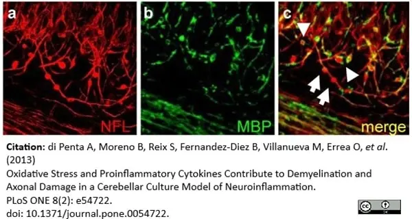
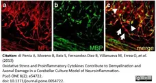
Microglial activation induces axonal damage in mouse cerebellar cultures.
Immunostaining for NfL (red) and MBP (green) in the same conditions as in A. Arrows indicate axonal beads and arrowheads indicate axonal transection (end-bulbs). Scale bar = 5 μm.
From: di Penta A, Moreno B, Reix S, Fernandez-Diez B, Villanueva M, Errea O, et al. (2013)
Oxidative Stress and Proinflammatory Cytokines Contribute to Demyelination and Axonal Damage in a Cerebellar Culture Model of Neuroinflammation.
PLoS ONE 8(2): e54722.
doi: 10.1371/journal.pone.0054722.
This image is from an open access article distributed under terms of a Creative Commons Attribution License.
Rat anti MBP antibody, clone 12 (MCA409S) used to label MBP expressing cells in mouse cerebellar cultures by immunofluorescence.
Image caption:
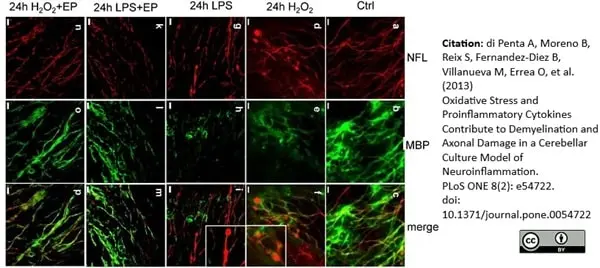
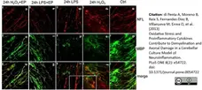
Role of Ethyl pyruvate in preventing microglia activation in cerebellar cultures.
Comparative effects of LPS, H2O2 and EP in demyelination and axonal damage in cerebellar cultures: Immunostaining for NfL (red, panels a, d, g, k and n) and MBP (green, panels b, e, h, l and o) in untreated organotypic cultures (control; panels a-c) or those treated for 24 h with H2O2 (panels d-f), LPS (panels g-i), LPS plus EP (panels k-m) or H2O2 plus EP (panels n-p). Co-localization is shown in the merged panels c, f, i, m and p. Insets show a higher magnification of the areas in panels f and i. Scale bar = 10 μm.
From: di Penta A, Moreno B, Reix S, Fernandez-Diez B, Villanueva M, Errea O, et al. (2013)
Oxidative Stress and Proinflammatory Cytokines Contribute to Demyelination and Axonal Damage in a Cerebellar Culture Model of Neuroinflammation.
PLoS ONE 8(2): e54722.
doi: 10.1371/journal.pone.0054722.
This image is from an open access article distributed under terms of a Creative Commons Attribution License
Rat anti MBP antibody, clone 12 (MCA409S) used to label MBP expressing cells in mouse cerebellar cultures by immunofluorescence.
Image caption:
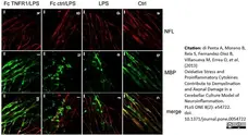
TNF-α blockade modulates microglia activation and demyelination.
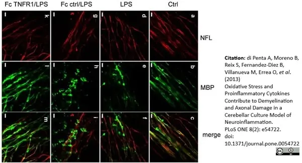
Role of TNFα blockade after LPS stimulation in demyelination of cerebellar cultures: Immunofluorescence for NfL (red) and MBP (green) in cultures untreated (ctrl, panels a-c), cultures treated with LPS (panels d-f), LPS plus control Fc (panels g-i) or LPS plus Fc-TNFR1 (15 μg/ml, panels k-m) for 24 h,. Scale bar = 5 μm.
From: di Penta A, Moreno B, Reix S, Fernandez-Diez B, Villanueva M, Errea O, et al. (2013)
Oxidative Stress and Proinflammatory Cytokines Contribute to Demyelination and Axonal Damage in a Cerebellar Culture Model of Neuroinflammation.
PLoS ONE 8(2): e54722..
doi: 10.1371/journal.pone.0054722
This image is from an open access article distributed under terms of a Creative Commons Attribution License.
Rat anti Bovine MBP antibody, clone 12 (MCA409S) used to label MBP expressing cells in mouse cerebellar cultures by immunofluorescence.
Image caption:
Demyelination and impaired axonal transport were maintained after 24 h of LPS treatment. Cerebellar cultures were stimulated with LPS (15 μg/ml) for 0, 1, 3, 6, 12, 24, 48, 72 and 96 h, and stained for NfH (red) and MBP (green). The time points 12 h and 96 h after LPS challenge are shown. Arrows indicate axonal beads and arrowheads indicate axonal transection (end-bulbs). Scale bar = 10 μm.
From: di Penta A, Moreno B, Reix S, Fernandez-Diez B, Villanueva M, Errea O, et al. (2013)
Oxidative Stress and Proinflammatory Cytokines Contribute to Demyelination and Axonal Damage in a Cerebellar Culture Model of Neuroinflammation.
PLoS ONE 8(2): e54722.
doi: 10.1371/journal.pone.0054722
This image is from an open access article distributed under terms of a Creative Commons Attribution License.
Rat anti Bovine MBP antibody, clone 12 (MCA409S) used to label MBP expressing cells in mouse cerebellar cultures by immunofluorescence.
Image caption:
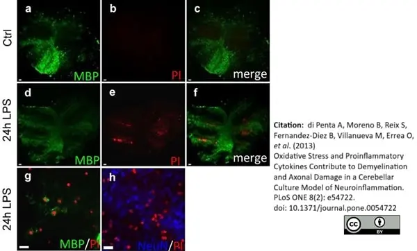
Microglial activation induces oligodendrocyte death in mouse cerebellar cultures. A) Cerebellar cultures were treated with LPS (15 μg/ml) for 24 h and then immunostained for MBP (green) or NeuN (blue) and counterstained with propidium iodide (red). The graph shows the number of PI-MBP-positive cells. Higher magnification images of white (g) and grey (h) matter in cultures treated with LPS. Scale bar = 100 µm (panels a-f) and 10 μm (panels g and h). B) Cerebellar cultures were incubated for 24 h in the presence (LPS) or absence (Ctrl) of LPS (15 μg/ml)..
From: di Penta A, Moreno B, Reix S, Fernandez-Diez B, Villanueva M, Errea O, et al. (2013)
Oxidative Stress and Proinflammatory Cytokines Contribute to Demyelination and Axonal Damage in a Cerebellar Culture Model of Neuroinflammation.
PLoS ONE 8(2): e54722.
doi: 10.1371/journal.pone.0054722
This image is from an open access article distributed under terms of a Creative Commons Attribution License.
Mouse anti Human Myelin Basic Protein antibody, clone 12 (MCA409S) used for the identification of MBP in murine brain by immunohistochemistry on formalin fixed, paraffin embedded tissue sections.
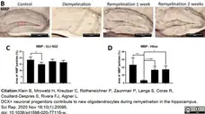
Image caption:
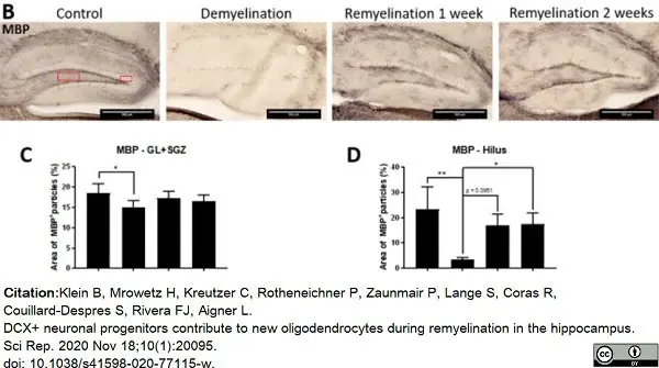
Experimental design and analysis of de- and remyelination in the dorsal hippocampal DG in wildtype mice. (B) Myelination in the hippocampal DG was analyzed by MBP staining. The rectangles indicate the regions of interest: Hilus and granular layer (GL) plus subgranular zone (SGZ). (C) In the GL plus SGZ, a significant demyelination after cuprizone treatment was observed. (D) In the hilus of the dorsal hippocampal DG, the myelin changes were more pronounced. Values are shown as means + S.D. (n = 6 per group). Statistical significance was evaluated using (C) a one-way ANOVA followed by a Tukey post-hoc test or (D) a Kruskal–Wallis test followed by a Dunn’s post-hoc test. The p-values are indicated in the graphs: *p <0.05, and **p <0.01. Bars: (B) 500μm.
From: Klein B, Mrowetz H, Kreutzer C, Rotheneichner P, Zaunmair P, Lange S, Coras R, Couillard-Despres S, Rivera FJ, Aigner L.
DCX+ neuronal progenitors contribute to new oligodendrocytes during remyelination in the hippocampus.
Sci Rep. 2020 Nov 18;10(1):20095.
doi: 10.1038/s41598-020-77115-w.
This image is from an open access article distributed under terms of a Creative Commons Attribution License.

Rat anti Bovine MBP antibody, clone 12 (MCA409S) used to demonstrate myelin expression in cultured brain neuronal cells by immunofluorescence.
Image caption:
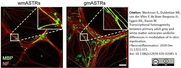
WmASTRs are less supportive to in vitro myelination. In vitro myelination cultures that depend on a feeding layer of astrocytes (ASTRs) are stained for myelin basic protein (MBP, green) and neurofilament-H (NF, red), a myelin and axonal marker, respectively. Representative images of myelinating cultures on either adult white matter (wm) ASTRs or adult grey matter (gm) ASTRs are shown.
From: Werkman IL, Dubbelaar ML, van der Vlies P, de Boer-Bergsma JJ, Eggen BJL, Baron W.
Transcriptional heterogeneity between primary adult grey and white matter astrocytes underlie differences in modulation of in vitro myelination.
J Neuroinflammation. 2020 Dec 11;17(1):373.
doi: 10.1186/s12974-020-02045-3.
This image is from an open access article distributed under terms of a Creative Commons Attribution License.

Rat anti Bovine MBP antibody, clone 12 (MCA409S) used to demonstrate myelin expression in cultured brain neuronal cells by immunofluorescence.
Image caption:
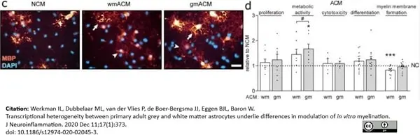
Secreted factors from wmASTRs inhibit myelin membrane formation.
Oligodendrocyte progenitors cells (OPCs) were cultured with PDGF-AA and FGF2 for 24 h to assess proliferation or differentiated into mature oligodendrocytes (OLGs) for 6 days after growth factor withdrawal to assess differentiation (% MBP-positive cells), and myelin membrane formation (% myelin membranes formed by MBP-positive cells). Assays were performed in the presence or absence of astrocyte (ASTR)-conditioned medium (ACM) or ASTR-derived extracellular matrix (ECM) coatings obtained from primary adult grey matter (gm) or white matter (wm) ASTRs. Representative images of MBP-positive OLGs (red, arrow indicates a MBP-positive cell with myelin membrane, arrowhead a MBP-positive cell without myelin membrane) in the presence of non-conditioned medium (NCM) or ACM are shown in c; Quantification of assays of 4–12 independent cell culture preparations (black dots) with 4–12 different batches of ACM (d) are shown. Bars represent the relative means to NCM are shown in d.
From: Werkman IL, Dubbelaar ML, van der Vlies P, de Boer-Bergsma JJ, Eggen BJL, Baron W.
Transcriptional heterogeneity between primary adult grey and white matter astrocytes underlie differences in modulation of in vitro myelination.
J Neuroinflammation. 2020 Dec 11;17(1):373.
doi: 10.1186/s12974-020-02045-3.
This image is from an open access article distributed under terms of a Creative Commons Attribution License.
Rat anti Bovine MBP antibody, clone 12 (MCA409S) used to demonstrate myelin in human brain using immunofluorescence.
Image caption:
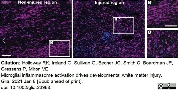
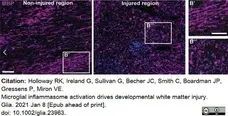
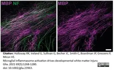
Microglial inflammasome activation in human perinatal brain injury.
Non‐injured and injured regions of human infant brain stained for MBP (purple) and counterstained with Hoechst (blue). Scale bars, 100 μm.
From: Holloway RK, Ireland G, Sullivan G, Becher JC, Smith C, Boardman JP, Gressens P, Miron VE.
Microglial inflammasome activation drives developmental white matter injury.
Glia. 2021 69(5):1268-1280. .
doi: 10.1002/glia.23963.
This image is from an open access article distributed under terms of a Creative Commons Attribution License.
Rat anti Bovine MBP antibody, clone 12 (MCA409S) used to demonstrate myelin in mouse brain by immunofluorescence.
Image caption:
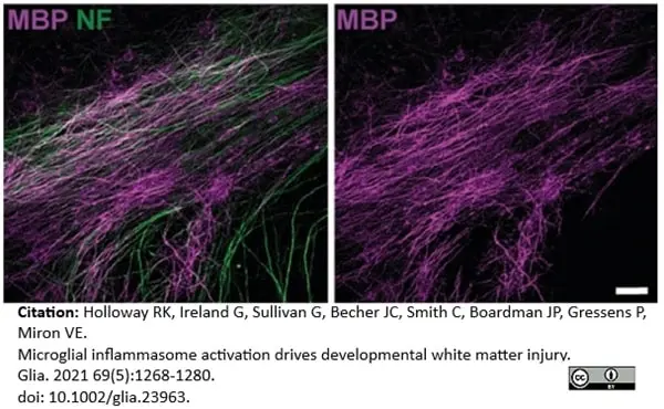
MBP (purple) and NF (green) in explants with no injury at 10 DIV following treatment with Vehicle or Follistatin. Scale bar, 20 μm.
From: Holloway RK, Ireland G, Sullivan G, Becher JC, Smith C, Boardman JP, Gressens P, Miron VE.
Microglial inflammasome activation drives developmental white matter injury.
Glia. 2021 69(5):1268-1280.
doi: 10.1002/glia.23963.
This image is from an open access article distributed under terms of a Creative Commons Attribution License.
Rat anti Bovine MBP antibody, clone 12 (MCA409S) used to demonstrate myelin in mouse brain using immunofluorescence.
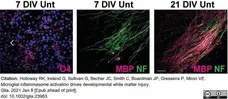
Image caption:
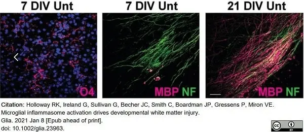
Untreated explants (Unt) mirror in vivo. oligodendrocyte maturation, at 7 DIV showing abundance of oligodendrocyte precursors (O4+; magenta) and paucity of myelin (MBP+; magenta) on axons (NF+; green), and myelination then being robust by 21 DIV. Scale bar, 25 μm.
From: Holloway RK, Ireland G, Sullivan G, Becher JC, Smith C, Boardman JP, Gressens P, Miron VE.
Microglial inflammasome activation drives developmental white matter injury.
Glia. 2021 69(5):1268-1280.
doi: 10.1002/glia.23963.
This image is from an open access article distributed under terms of a Creative Commons Attribution License.

Rat anti Bovine MBP antibody, clone 12 (MCA409S) used to demonstrate myelin in murine CNS cultures by immunofluorescence.
Image caption:
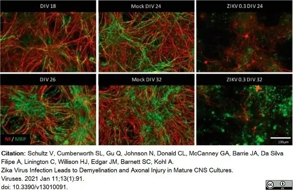
ZIKV infection causes damage to mature, myelinated CNS cultures. (A) Mouse CNS myelinating cultures were either mock- or ZIKV-infected (MOI 0.3) at DIV 18 (upper panel) or DIV 26 (lower panel) and analyzed by IF at 6 dpi. Mature myelin basic protein (MBP, green signal) and axon (NF, red signal) staining are shown, representative of four biological replicates (n = 4).
From: Schultz V, Cumberworth SL, Gu Q, Johnson N, Donald CL, McCanney GA, Barrie JA, Da Silva Filipe A, Linington C, Willison HJ, Edgar JM, Barnett SC, Kohl A.
Zika Virus Infection Leads to Demyelination and Axonal Injury in Mature CNS Cultures.
Viruses. 2021 Jan 11;13(1):91.
doi: 10.3390/v13010091.
This image is from an open access article distributed under terms of a Creative Commons Attribution License.
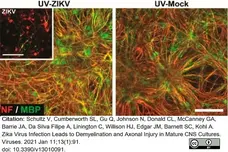
Rat anti Bovine MBP antibody, clone 12 (MCA409S) used to demonstrate Myelin in murine CNS cultures by immunofluorescence.
Image caption:
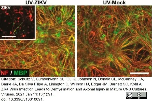
ZIKV inSupernatant of mock- or ZIKV-infected immature mouse CNS cultures was collected at 6 dpi and UV-treated to inactivate viral particles. UV-treated supernatant was mixed 3:1 with fresh medium. Analysis by IF at 6 dpt onto immature cultures. Representative images of myelin staining (MBP, green signal; PLP, red signal) and axon (NF, red signal) staining at 6 dpt (DIV 24) are shown (n = 2). Inset images (upper panels) depict ZIKV (ZIKV E, green signal) infected CNS cells and pathology in cultures used for supernatant collection (6 dpi, DIV 24), with myelin (MBP, green signal; PLP, red signal) and neurofilament (NF, red) shown. Scale bar = 100 μm.
From: Schultz V, Cumberworth SL, Gu Q, Johnson N, Donald CL, McCanney GA, Barrie JA, Da Silva Filipe A, Linington C, Willison HJ, Edgar JM, Barnett SC, Kohl A.
Zika Virus Infection Leads to Demyelination and Axonal Injury in Mature CNS Cultures.
Viruses. 2021 Jan 11;13(1):91.
doi: 10.3390/v13010091.
This image is from an open access article distributed under terms of a Creative Commons Attribution License.
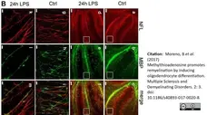
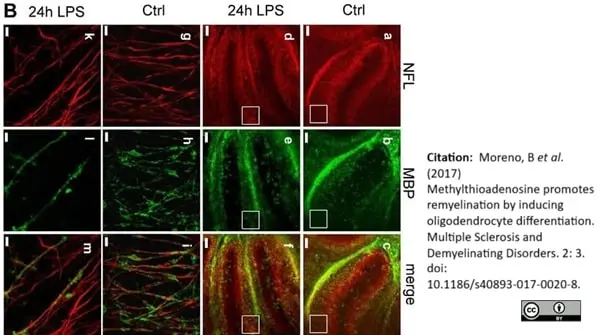
Rat anti Bovine MBP antibody, clone 12 (MCA409S) used for the demonstration of MBP expression in mouse organotypic brain cultures by immunofluorescence.
Image caption:
Methylthioadenosine (MTA) dampens neuroinflammation, and prevents demyelination and axonal loss in mouse cerebellar cultures. A Organotypic cultures were treated with MTA (192 μM) or the placebo for 24 h at seven DIV and thereafter, they were stimulated with LPS (15 μg/ml) for 24 h. Immunofluorescent staining for NFL (red) and MBP (green) was analyzed in the slices, and in time-matched untreated control slices (arrow, axonal swelling; arrowhead, end bulb). Panels a, b and c scale bars: 50 μm. Panels d-i scale bar: μ μm.
From: Moreno, B et al. (2017)
Methylthioadenosine promotes remyelination by inducing oligodendrocyte differentiation.
Multiple Sclerosis and Demyelinating Disorders. 2: 3.
10.1186/s40893-017-0020-8.
This is from an open access article distributed under the terms of a Creative Commons Attribution License.
Rat anti Bovine MBP antibody, clone 12 (MCA409S) used to indicate MBP expression in murine sciatic nerve by immunofluoresence.
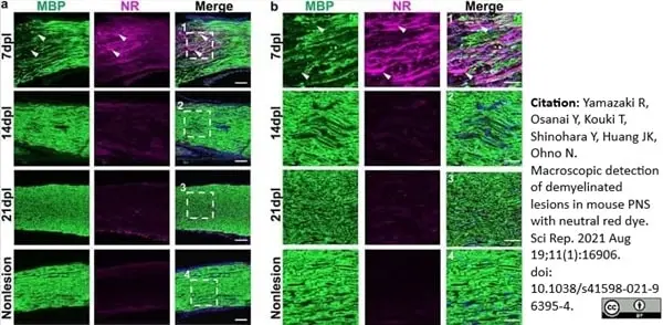
Image caption:
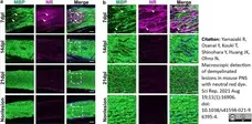
Neutral red (NR) signals were gradually decreased during remyelination. (a) Representative confocal images of NR (magenta) and immunofluorescence staining for myelin with anti-MBP (green) in areas with lysophosphatidylcholine injection at 7, 14, 21 dpl and a nonlesion area at 7 dpl (n = 3 mice per timepoint examined). (b1–4) Enlarged images of (a1–4) (white dotted square). The demyelinated lesion with weak MBP immunoreactivity is shown in 7 dpl image (a, b; arrowhead).
From: Yamazaki R, Osanai Y, Kouki T, Shinohara Y, Huang JK, Ohno N.
Macroscopic detection of demyelinated lesions in mouse PNS with neutral red dye.
Sci Rep. 2021 Aug 19;11(1):16906.
doi: 10.1038/s41598-021-96395-4.
This image is from an open access article distributed under terms of a Creative Commons Attribution License.
Rat anti MBP antibody, clone 12 (MCA409S) used to lable mouse myelin basic protein on cultured oligodendrocytes by immunofluorescence.
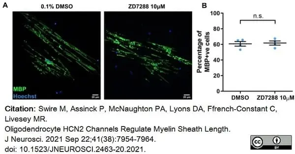
Image caption:
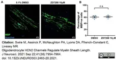
HCN channels regulate the length of myelin sheaths formed in oligodendrocyte-purified cultures. A, Representative MBP-positive oligodendrocytes after 14 d cultured on electrospun microfibers in the presence of either 0.1% DMSO or 10 μm ZD7288. Scale bars, 10 μm. B, Mean ± SE percentage of MBP-positive cells on microfibers: DMSO, 60.77 ± 2.972%; ZD7288, 61.76 ± 2.659%; n = 4 independent cultures; p = 0.8857, Mann–Whitney test.
From: Swire M, Assinck P, McNaughton PA, Lyons DA, Ffrench-Constant C, Livesey MR.
Oligodendrocyte HCN2 Channels Regulate Myelin Sheath Length.
J Neurosci. 2021 Sep 22;41(38):7954-64.
doi: 10.1523/JNEUROSCI.2463-20.2021
This image is from an open access article distributed under terms of a Creative Commons Attribution License.
Rat anti MBP antibody, clone 12 (MCA409S used to label MBP by immunofluorescence.
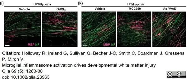
Image caption:
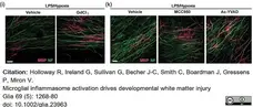
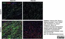
Inflammasome inhibition enhances developmental myelination after injury.
(i) MBP (magenta) and NF (green) in LPS/Hypoxia-exposed explants at 10 DIV following treatment with Vehicle or GdCl3. Scale bar, 25 μm. (k) MBP (magenta) and NF (green) in LPS/Hypoxia-exposed explants at 10 DIV following treatment with Vehicle, NLRP3 inhibitor MCC950, or caspase-1 inhibitor Ac-YVAD. Scale bar, 20 μm
From: Holloway RK, Ireland G, Sullivan G, Becher JC, Smith C, Boardman JP, Gressens P, Miron VE.
Microglial inflammasome activation drives developmental white matter injury.
Glia. 2021 May;69(5):1268-1280.
doi: 10.1002/glia.23963..
This image is from an open access article distributed under terms of a Creative Commons Attribution License.
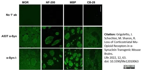
Rat anti Bovine MBP antibody, clone 12 (MCA409S) used to label myelin in murine corticostriatial brain tissue by immunofluorescence on formalin fixed, paraffin embedded sections.
Image caption:
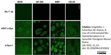
Lower MOR levels in corticostriatal WMT of A53T α-Syn tg mouse brains.
(C). Paraffin-embedded coronal sections (6μm) of α-Syn-/- (C57BL/6JOlaHsd) and A53T α-Syn mouse brains at 2 months of age, containing the dorsal striatum (representative images). Sections immunoreacted either with anti-MOR, anti-NF-200, anti-MBP, or anti calbindin 28 (CB-28) antibodies. The consecutive section of the α-Syn-/- brain were stained with the corresponding secondary antibody as a control (no 1°). Bar = 50μm.
From: Grigoletto, J. Schechter, M. Sharon, R.
Loss of Corticostriatal Mu-Opioid Receptors in α-Synuclein Transgenic Mouse Brains.
Life 2022, 12, 63.
doi: 10.3390/life12010063.
This image is from an open access article distributed under terms of a Creative Commons Attribution License.
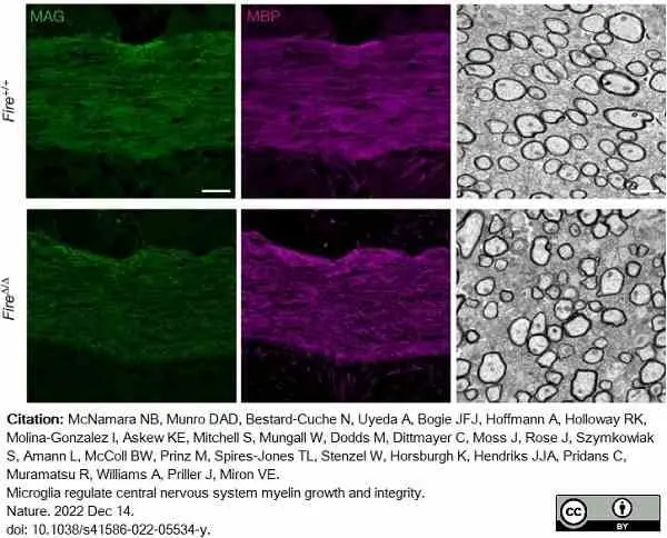
Rat anti MBP antibody, clone 12 (MCA409S) used to label myelin in mouse corpus callosum by immunofluorescence.
Image caption:
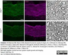
Images of corpus callosum from Fire+/+ and FireΔ/Δ mice stained for the myelin proteins MAG (green) and MBP (magenta) (left and middle) and imaged by electron microscopy (right).
From: McNamara NB, Munro DAD, Bestard-Cuche N, Uyeda A, Bogie JFJ, Hoffmann A, Holloway RK, Molina-Gonzalez I, Askew KE, Mitchell S, Mungall W, Dodds M, Dittmayer C, Moss J, Rose J, Szymkowiak S, Amann L, McColl BW, Prinz M, Spires-Jones TL, Stenzel W, Horsburgh K, Hendriks JJA, Pridans C, Muramatsu R, Williams A, Priller J, Miron VE.
Microglia regulate central nervous system myelin growth and integrity.
Nature. 2022 Dec 14.
doi: 10.1038/s41586-022-05534-y.
This image is from an open access article distributed under terms of a Creative Commons Attribution License.
Rat anti MBP antibody, clone 12 (MCA409S) used to label human iOPCs in vitro using immunofluorescence.
Image caption:
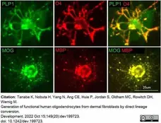
Successful generation of iOPCs from human fibroblasts.
O4+ iOPCs express the additional oligodendrocyte marker PLP1 (top row) and, following a 30-day differentiation period in media lacking growth factors, isolated iOPCs express the terminal differentiation markers MOG and MBP.
From: Tanabe K, Nobuta H, Yang N, Ang CE, Huie P, Jordan S, Oldham MC, Rowitch DH, Wernig M.
Generation of functional human oligodendrocytes from dermal fibroblasts by direct lineage conversion. Development. 2022 Oct 15;149(20):dev199723.
doi: 10.1242/dev.199723.
This image is from an open access article distributed under terms of a Creative Commons Attribution License.
Rat anti MBP antibody, clone 12 (MCA409S) used to label iOPCs transplanted to mouse brain by immunofluorescence. in vitro using immunofluorescence.
Image caption:
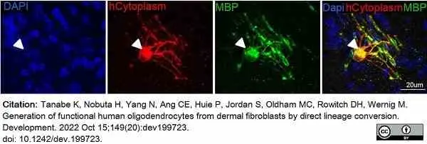
Engraftment and myelination in vivo and disease modeling capability of iOPCs.
MBP (green) and the human-specific antibody SC121 (hCytoplasm, red) detected iOPC-derived cells with a typical morphology of mature oligodendrocytes 12 weeks after transplantation.
From: Tanabe K, Nobuta H, Yang N, Ang CE, Huie P, Jordan S, Oldham MC, Rowitch DH, Wernig M.
Generation of functional human oligodendrocytes from dermal fibroblasts by direct lineage conversion. Development. 2022 Oct 15;149(20):dev199723.
doi: 10.1242/dev.199723.
This image is from an open access article distributed under terms of a Creative Commons Attribution License.
Rat anti MBP antibody, clone 12 (MCA409S) used to identify MBP expressing cells by immunofluorescence.
Image caption:
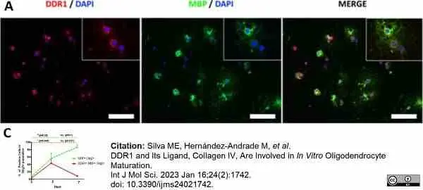
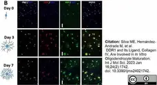
DDR1 is expressed during oligodendrogenic progression. Neural stem cells (NSCs) were obtained from the adult rat subventricular zone, expanded as neurospheres, and thereafter enzymatically dissociated and plated as individual cells. After exposure to pericyte-conditioned culture medium (PC-CM), NSCs differentiated into mature OLs (day 7). (A) Fluorescence images displaying DDR1 (red) and MBP (green) and DDR1-MBP colocalization (merge panel) in NSCs after x days of exposure to PC-CM. DAPI was used for nuclear counterstaining (blue). Scale bar = 50 μm. (C) Quantitative analysis showing MBP+ (green line) and DDR1+ and MBP+ (red line) cells among the total population of Olig2+ cells after 0, 3, and 7 days of exposure to PC-CM. The data show that the DDR1 levels peaked at day 3 and declined at day 7, whereas MBP expression continued to increase during oligodendrogenic progression. The data were obtained from three independent experiments and were analyzed by analysis of variance (ANOVA) followed by Tukey’s post hoc test. # and * p < 0.05, n.s.: not significant. * and # indicate significant differences between DDR1+ MBP+ cells and MBP+ cells, respectively, and the control group.
From: Silva ME, Hernández-Andrade M, Abasolo N, et al.
DDR1 and Its Ligand, Collagen IV, Are Involved in In Vitro Oligodendrocyte Maturation.
Int J Mol Sci. 2023 Jan 16;24(2):1742.
doi: 10.3390/ijms24021742.
This image is from an open access article distributed under terms of a Creative Commons Attribution License.
Rat anti MBP antibody, clone 12 (MCA409S) used to identify MBP expressing cells by immunofluorescence.
Image caption:
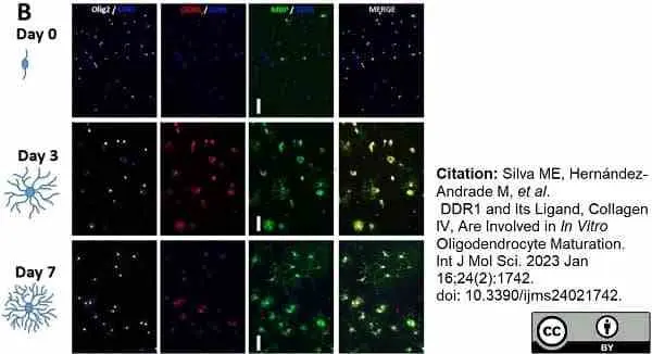
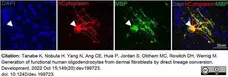
DDR1 is expressed during oligodendrogenic progression. Neural stem cells (NSCs) were obtained from the adult rat subventricular zone, expanded as neurospheres, and thereafter enzymatically dissociated and plated as individual cells. After exposure to pericyte-conditioned culture medium (PC-CM), NSCs differentiated into mature OLs (day 7). (B) Fluorescence images displaying DDR1+, MBP+, and Olig2+ cells after 0, 3, and 7 days of incubation with PC-CM. DAPI was used for nuclear counterstaining (blue). Scale bar = 20 μm.
From: Silva ME, Hernández-Andrade M, Abasolo N, et al.
DDR1 and Its Ligand, Collagen IV, Are Involved in In Vitro Oligodendrocyte Maturation.
Int J Mol Sci. 2023 Jan 16;24(2):1742.
doi: 10.3390/ijms24021742.
This image is from an open access article distributed under terms of a Creative Commons Attribution License.

Rat anti MBP antibody, clone 12 (MCA409S) used to label myelin in rat brain by immunohistochemistry on formalin fixed cryosections.
Image caption:
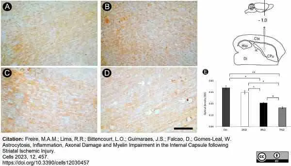
Myelin impairment in the internal capsule following endothelin-1 (ET-1) injection. Control group shows a homogeneous MBP+ labeling (A). A progressive impairment is observed at 1PLD (B), 3PLD (C) and 7PLD (D), with a noticeable rarefaction of reactivity in the latter survival time. Vacuolization, a hallmark of tissue degeneration, is also evident at 7PLD (arrows). Progressive loss of MBP labeling across time points was confirmed by densitometric analysis (E) (* p < 0.05; ** p < 0.01, ANOVA, Tukey post hoc test). Drawings at the left side of the figure show the anatomical localization of the ET-1 injection in the striatum. Ctx: cortex; CPu: caudate putamen (striatum); Di: diencephalon; Hip: hippocampus; ic: internal capsule. Scale bar: 100 μm.
From: Freire, M.A.M.; Lima, R.R.; Bittencourt, L.O.; Guimaraes, J.S.; Falcao, D.; Gomes-Leal, W.
Astrocytosis, Inflammation, Axonal Damage and Myelin Impairment in the Internal Capsule following Striatal Ischemic Injury.
Cells 2023, 12, 457.
doi:10.3390/cells12030457
This image is from an open access article distributed under terms of a Creative Commons Attribution License.

Rat anti Bovine myelin basic protein antibody, clone 12 (MCA409S) used to evaliate MBP expression in human brain tissue by immunofluorescence.
Image caption:
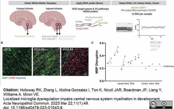
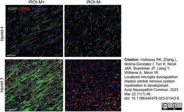
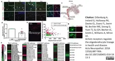
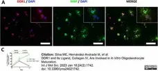
Digital spatial transcriptomic profiling of human developmental white matter. A Diagram of experimental approach. Regions of interest (ROI) were assessed in infant brain white matter samples (cerebellum), with control ROI (cROI) in control infants compared to ROI in injured infants (iROI) which were normally myelinating (iROI-M+) or showed reduced myelination (iROI-M−). A probe library targeting 1825 genes in 55 pathways was applied to the tissue, and 12 ROIs were selected per sample based on levels of myelin basic protein (MBP; green) and microglia/macrophages (CD68; red), counterstained with Hoechst (cyan). Probes were cleaved with UV light and RNA counts performed per ROI. B Example ROIs from cROI, iROI-M+, and iROI-M− stained for MBP (green) and CD68 (red) and counterstained for Hoechst (blue). Scale bar, 65 μm. C MBP Disruption as measured by average MBP Sphericity per infant in injured iROI-M+ (grey), iROI-M− (pink) and cROI (green).
From: Holloway RK, Zhang L, Molina-Gonzalez I, Ton K, Nicoll JAR, Boardman JP, Liang Y, Williams A, Miron VE.
Localized microglia dysregulation impairs central nervous system myelination in development.
Acta Neuropathol Commun. 2023 Mar 22;11(1):49.
doi: 10.1186/s40478-023-01543-8.
This image is from an open access article distributed under terms of a Creative Commons Attribution License.
Rat anti Bovine myelin basic protein antibody, clone 12 (MCA409S) used to evaliate MBP expression in human brain tissue by immunofluorescence.
Image caption:
Representative images of myelin (MBP; green) and microglia/macrophages (CD68; red) in infant with good myelination in iROI-M− (infant 4) versus infant with poor myelination, in iROI-M− (infant 3), counterstained for Hoechst (blue) . Scale bar, 100μm.
From: Holloway RK, Zhang L, Molina-Gonzalez I, Ton K, Nicoll JAR, Boardman JP, Liang Y, Williams A, Miron VE.
Localized microglia dysregulation impairs central nervous system myelination in development.
Acta Neuropathol Commun. 2023 Mar 22;11(1):49.
doi: 10.1186/s40478-023-01543-8.
This image is from an open access article distributed under terms of a Creative Commons Attribution License.

Rat anti Bovine myelin basic protein antibody, clone 12 (MCA409S) used to evaliate MBP expression in human brain tissue by immunofluorescence.
Image caption:
Interferon-responsive microglia functionally impede myelination by mature oligodendrocytes. A Diagram of treatment of neonatal microglia cultures with IFNγ and LPS, or vehicle control, and application of conditioned media to myelinating cultures of oligodendrocytes and neurons. B Representative images of cultures exposed to conditioned media (CM) from vehicle (PBS)-treated microglia or IFNγ and LPS-treated microglia, with myelin indicated by MBP (red), axons indicated by NF (green), and nuclei counterstained with Hoechst (blue). Scale bar, 25 μm. C Percentage of oligodendrocytes (MBP +) which have myelinating processes (grey) versus non-myelinating processes (pink) following treatment with CM from vehicle-treated or IFNγ/LPS-treated microglia, or with IFNγ/LPS directly applied to myelinating cultures. Unpaired 2-tailed Student’s t-test, *P = 0.0428 for non-myelinating processes, and *P = 0.0428 for myelinating processes, for IFNγ/LPS CM versus Veh CM. N = 3 biologically independent repeats. D Percentage of MBP area in myelinating cultures exposed to vehicle (Veh) or IFNγ/LPS-treated microglia CM. Unpaired 2-tailed Student’s t-test, *P = 0.0455. N = 3 biologically independent repeats. E MBP Disruption as measured by sphericity in myelinating cultures exposed to Veh or or IFNγ/LPS treated microglia CM. Unpaired 2-tailed Student’s t-test, ***P = 0.0001. N = 3 biologically independent repeats
From: Holloway RK, Zhang L, Molina-Gonzalez I, Ton K, Nicoll JAR, Boardman JP, Liang Y, Williams A, Miron VE.
Localized microglia dysregulation impairs central nervous system myelination in development.
Acta Neuropathol Commun. 2023 Mar 22;11(1):49.
doi: 10.1186/s40478-023-01543-8.
This image is from an open access article distributed under terms of a Creative Commons Attribution License.
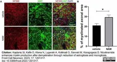
Rat anti Bovine myelin basic protein antibody, clone 12 (MCA409S) used to identify myelin in mouse brain sections by immunofluorescence.
Image caption:
Nicotinamide (NAM) increases myelination of axons in ex vivo organotypic brain slices. (A) Representative confocal images of the cortical area of organotypic brain slices from postnatal day 4 mice, immunolabeled with an antibody against NF200 (red) to mark axons and an antibody against MBP (green) to mark myelin. DAPI was used for nuclear staining (blue). White arrows point at the myelinated axons (yellow). (B) Quantification of the percentage of NF200-positive area that co-localized with MBP, indicates the percentage of myelinated axons. Two groups were used: vehicle-treated (EtOH) for the control and NAM-treated (0.1 mM) for 6 days. For both groups n = 4. Data are shown as mean ± SEM. Student’s t-test was used to determine statistical significance. *p <0.05. Scale bar: 50 μm.
From: Kaplanis SI, Kaffe D, Ktena N, Lygeraki A, Kolliniati O, Savvaki M, Karagogeos D.
Nicotinamide enhances myelin production after demyelination through reduction of astrogliosis and microgliosis.
Front Cell Neurosci. 2023 Aug 17;17:1201317.
doi: 10.3389/fncel.2023.1201317.
This image is from an open access article distributed under terms of a Creative Commons Attribution License.
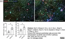
Rat anti Bovine myelin basic protein antibody, clone 12 (MCA409S) used to label rat oligodendrocytes by immunofluorescence.
Image caption:
Representative images of the differentiated rat oligodendrocytes (MBP+) in the hiPSC-EC co-culture system in differentiation condition for 2 days. The quantification shows a significant increased cell density of MBP+ oligodendrocytes and an increased percentage of them in total Olig2+ cells. Scale bar, 200 μm. All data are presented as mean ± SE, n = 4, *p <05. Unpaired t test.
From: Ma D, Zhang H, Yin L, Xu H, Wu L, Shaji R, Rezai F, Mulla A, Kaur S, Tan S, Kysela B, Wang Y, Chen Z, Zhao C, Gu Y.
Human iPSC-derived endothelial cells promote CNS remyelination via BDNF and mTORC1 pathway.
Glia. 2024 Jan;72(1):133-55.
doi: 10.1002/glia.24466.
This image is from an open access article distributed under terms of a Creative Commons Attribution License.
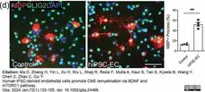
Rat anti Bovine myelin basic protein antibody, clone 12 (MCA409S) used to label rat oligodendrocytes by immunofluorescence.
Image caption:
(d) Representative images of the rat MBP+ cells in hiPSC-EC co-culture system showing differentiated oligodendrocytes with long branches in the presence of hiPSC-ECs. The quantification shows an increased percentage of the MBP+ cells with long branches in the presence of hiPSC-ECs. All data are presented as mean ± SE, n = 4. *p <05. Unpaired t test. Scale bar, 50 μm.
From: Ma D, Zhang H, Yin L, Xu H, Wu L, Shaji R, Rezai F, Mulla A, Kaur S, Tan S, Kysela B, Wang Y, Chen Z, Zhao C, Gu Y.
Human iPSC-derived endothelial cells promote CNS remyelination via BDNF and mTORC1 pathway.
Glia. 2024 Jan;72(1):133-55.
doi: 10.1002/glia.24466.
This image is from an open access article distributed under terms of a Creative Commons Attribution License.Unpaired t test. Scale bar, 50 μm.

Rat anti Bovine myelin basic protein antibody, clone 12 (MCA409S) used to label rat oligodendrocytes by immunofluorescence.
Image caption:
(c) Representative set of bright field images of the OPCs in the presence of control IgG, or anti-BDNF antibody, or hiPSC-ECs plus IgG or hiPSC-EC plus anti-BDNF antibody for 4 days. Scale bar, 50 μm. (d) Immunocytochemical assessment of OPC proliferation with Ki67 and Olig2 co-labelling in cultures under the same conditions as in (c). hiPSC-ECs increased the total Olig2+ cell number per field and the percentage of Ki67+ OPCs in Olig2+ cells, while the neutralizing anti-BDNF antibody did not altered the effects. Scale bar, 50 &mum. (e) Immunocytochemical assessment of OPC differentiation with MBP labelling in Olig2+ cells under the same conditions as in (c), but for 6 days. hiPSC-ECs increased the total MBP+ cell number per field and the percentage of MBP+ cells in Olig2+ cells, neutralizing anti-BDNF antibody partially reduced the enhancing effect. Scale bar, 40 μm. (f) Immunocytochemistry shows the pS6 labelling in Olig2+ cells in the conditions as in (c), the intensity of pS6 labelling and the percentage of pS6+ cells in total oligodendrocyte linage cells (Olig2+) were both increased in the presence of hiPSC-ECs, neutralizing anti-BDNF antibody did not attenuate the effect. Scale bar, 50 μm.
From: Ma D, Zhang H, Yin L, Xu H, Wu L, Shaji R, Rezai F, Mulla A, Kaur S, Tan S, Kysela B, Wang Y, Chen Z, Zhao C, Gu Y.
Human iPSC-derived endothelial cells promote CNS remyelination via BDNF and mTORC1 pathway.
Glia. 2024 Jan;72(1):133-55.
doi: 10.1002/glia.24466.
This image is from an open access article distributed under terms of a Creative Commons Attribution License.
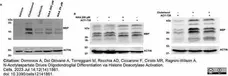
Rat anti MBP antibody, clone 12 (MCA409S) used to evaluate MBP expression in murine oligodendroglial cell lysates by western blotting.
Image caption:
Western blot analyses following treatment with A) NAA 20 or 200 μM or Clobetasol 10 μM or Gefitinib 1 μM like positive controls or vehicle alone (DMSO <0.5%) B) NAA 200 μM, Clobetasol 10 μM or ACY-738 10 μM, alone or in double treatment with ACY-738. Treatments are indicated above the blots. The name of protein detected is indicated to theright of each blot, and the molecular weight indicated by the marker is indicated to the left. All gels and blots were run under the same experimental conditions. For sample preparation typically, 2.75 × 10⁵ Oli-neuM cells were seeded in 6-well plates in GM media and cells were grown to 70% confluence. Treatments were performed for 48h before protein extraction and the western blotting procedure.
From:Dominicis A, Del Giovane A, Torreggiani M, Recchia AD, Ciccarone F, Ciriolo MR, Ragnini-Wilson A.
N-Acetylaspartate Drives Oligodendroglial Differentiation via Histone Deacetylase Activation.
Cells. 2023 Jul 14;12(14):1861.
doi: 10.3390/cells12141861.
This image is from an open access article distributed under terms of a Creative Commons Attribution License.

Rat anti MBP antibody, clone 12 (MCA409S) used to evaluate MBP expression in murine oligodendroglial cell lysates by western blotting.
Image caption:
MBP expression and Oli-neuM cell differentiation are stimulated by 200 μM NAA. Oli-neuM cells were plated 24 h in GM and, after media change, treated in DM with the indicated concentration of NAA, 1 μM Gefitinib, 10 μM Clobetasol, or Vehicle (DMSO 0.5% max) for 48 h prior to being processed. (A) Representative IF images are shown for anti-MBP immunostaining (green, FITC). Nuclei were stained with Hoechst (blue) and actin with phalloidin (red, TRITC). Scale bar, 10 μm.
From:Dominicis A, Del Giovane A, Torreggiani M, Recchia AD, Ciccarone F, Ciriolo MR, Ragnini-Wilson A.
N-Acetylaspartate Drives Oligodendroglial Differentiation via Histone Deacetylase Activation.
Cells. 2023 Jul 14;12(14):1861.
doi: 10.3390/cells12141861.
This image is from an open access article distributed under terms of a Creative Commons Attribution License.
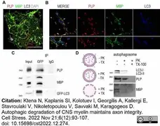
Rat anti MBP antibody, clone 12 (MCA409S) used to evaluate MBP expression in mouse oligodendrocytes by immunofluorescence and western blotting.
Image caption:
Myelin proteins PLP and MBP are detected in the autophagosomes.
(A) Representative confocal image of a DIV2 primary OL, immunolabeled for MBP (green), PLP (red), LC3 (blue) and DAPI (grey). Rectangular boxes indicate areas magnified to the right. Scale bar: 30 μm. (B) Magnification images are of a single z-stack (0,5μm thick) and white arrows indicate the colocalized puncta in myelin membrane (top) and cytoplasmic processes (bottom). Scale bars: 2 μm. (C) Immunoprecipitation with antibodies against GFP in forebrain lysates from adult GFP-LC3 mice identifies MBP and PLP as interactors of LC3. (D) Shematic representation of the proteinase K protection assay and western blot analysis of isolated autophagosomes after proteinase K assay. octylphenol ethoxylate is used as a negative control. Autophagic marker LC3-II was protected from PK digestion, unless octylphenol ethoxylate was present. Both PLP and MBP seem to be protected from PK treatment.
From: Ktena N, Kaplanis SI, Kolotuev I, Georgilis A, Kallergi E, Stavroulaki V, Nikoletopoulou V, Savvaki M, Karagogeos D.
Autophagic degradation of CNS myelin maintains axon integrity.
Cell Stress. 2022 Nov 21;6(12):93-107.
doi: 10.15698/cst2022.12.274.
This image is from an open access article distributed under terms of a Creative Commons Attribution License.
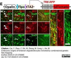
Rat anti MBP antibody, clone 12 (MCA409S) used to identify myelin in murine brain samples by immunofluorescence.
Image caption:
I(E) OL labeling by tTA2. High magnification images of the boxed region showing co-localization of RFP with MBP staining, which further demonstrated the myelination ability of labeled OLs.
From: Cai, Y. Zhao, Z. Shi, M. Zheng, M. Gong, L. He, M.
Embryonic origins of forebrain oligodendrocytes revisited by combinatorial genetic fate mapping.
bioRxiv 2024.01.23.576886.
doi:10.1101/2024.01.23.576886.
This image is from an open access article distributed under terms of a Creative Commons Attribution License.
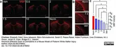
Rat anti MBP antibody, clone 12 (MCA409S) used to label myelin basic protein in mouse coronal sections by immunofluorescence.
Image caption:
Clemastine improves hypoxia-induced myelination deficits.
(a) Representative images of coronal sections stained for myelin basic protein (MBP) at P14. Scale bars, 500 um. (b) Representative images of the corpus callosum and striatum and (c) quantification of staining for MBP in the striatum. N= at least 4 mice/condition from at least 3 independent experiments per condition. *p<0.05, ANOVA followed by Tukey’s post hoc test. For all graphs and images, hypoxia-exposed animals were: Veh (vehicle-treated), 0.5 (clemastine 0.5 mg/kg/day), 2 (clemastine 2 mg/kg/day), 7.5 (clemastine 7.5 mg/kg/day), 10 (clemastine 10 mg/kg/day). Norm, normoxia-exposed animals. Error bars represent standard deviation.
From: Elizabeth Odell, Nora Jabassini, Björn Schniedewind, Sarah E. Pease-Raissi, Adam Frymoyer, Uwe Christians, Ari J. Green, Jonah R. Chan, Bridget E.L. Ostrem
Minimum Effective Dose of Clemastine in a Mouse Model of Preterm White Matter Injury
bioRχiv 2024.02.08.578953
doi: 10.1101/2024.02.08.57895
This image is from an open access article distributed under terms of a Creative Commons Attribution License.

Rat anti MBP antibody, clone 12 (MCA409S) used to stain cultured oligodendrocytes by immunofluorescence.
Image caption:
Prolonged dexamethasone exposure on mature oligodendrocyte (OL) does not affect OL maturation or myelin production. (b) Representative fluorescence images of OLs obtained from B6 and D2 mice and stained for different maturation markers after DEX or vehicle treatment.
From: Gigliotta A, Mingardi J, Cummings S, Alikhani V, Trontti K, Barbon A, Kothary R, Hovatta I.
Genetic background modulates the effect of glucocorticoids on proliferation, differentiation and myelin formation of oligodendrocyte lineage cells.
Eur J Neurosci. 2024 Feb 22.[ Epub ahead of print].
doi: 10.1111/ejn.16285..
This image is from an open access article distributed under terms of a Creative Commons Attribution License.
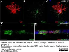
Rat anti MBP antibody, clone 12 (MCA409S) used to stain cultured mouse oligodendrocytes by immunofluorescence.
Image caption:
Prickle1 is expressed by oligodendrocytes and is concentrated at CNS paranodes. (A–C) Prickle1 (green) is localized to the cell body and processes of cultured mouse oligodendrocytes labeled with antibodies against myelin basic protein (MBP, red) at 2 days in vitro (DIV), but is excluded from myelin-like sheets. Nuclei were labeled with Hoechst dye (blue). (D and E) Prickle1 (green) is concentrated at paranodes in P60 spinal cord. In panel E, the axon is immunolabeled for the 200 kDA neurofilament subunit (NFH, blue). The distribution of Prickle1 overlaps with the paranodal marker Caspr (red). Scale bars (A–C): 10 μm, (D and E) 5 μm.
From:Jarjour AA, Velichkova AN, Boyd A, Lord KM, Torsney C, Henderson DJ, Ffrench-Constant C.
The formation of paranodal spirals at the ends of CNS myelin sheaths requires the planar polarity protein Vangl2.
Glia. 2020 Sep;68(9):1840-58.
doi: 10.1002/glia.23809.
This image is from an open access article distributed under terms of a Creative Commons Attribution License.
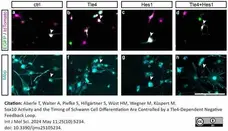
Rat anti MBP antibody, clone 12 (MCA409S) used to label rat Schwann cells by immunofluorescence.
Image caption:
Forced expression of Tle4 and Hes1 in primary rat Schwann cells represses their differentiation. (a–h) Representative images of transduced primary rat Schwann cells differentiated for 4 days and stained for Mbp (cyan). Fluorescence of retrovirally expressed EGFP and tdTomato is shown in green and magenta. Scale bar: 100 μm. Arrowheads mark double-transduced cells.
From: Aberle T, Walter A, Piefke S, Hillgärtner S, Wüst HM, Wegner M, Küspert M.
Sox10 Activity and the Timing of Schwann Cell Differentiation Are Controlled by a Tle4-Dependent Negative Feedback Loop.
Int J Mol Sci. 2024 May 11;25(10):5234.
doi: 10.3390/ijms25105234.
This image is from an open access article distributed under terms of a Creative Commons Attribution License.
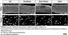
Rat anti MBP antibody, clone 12 (MCA409S) used to identify MBP expressing cells in mouse corpus callosum by immunofluorescence.
Image caption:
Mild reductions in the myelin protein amounts in mutant mice with Sox8 and Sox10 deletions. (c–j) Exemplary immunohistochemical stainings of the corpus callosum from the control (c,g), Sox8cko (d,h), Sox10cko (e,i), and dcko (f,j) mice with antibodies directed against Mbp (c–f) and Aspa (g–j). Scale bar: 50 μm. Microscope enlargement: 200×.
From: Jörg LM, Schlötzer-Schrehardt U, Lefebvre V, Sock E, Wegner M.
Transcription Factors Sox8 and Sox10 Contribute with Different Importance to the Maintenance of Mature Oligodendrocytes.
Int J Mol Sci. 2024 Aug 11;25(16):8754.
doi: 10.3390/ijms25168754.
This image is from an open access article distributed under terms of a Creative Commons Attribution License.
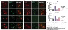
Mouse anti Human MBP antibody, clone 12 (MCA409S) used to detect myelin im mouse samples by immunofluorescence.
Image caption:
CHPG enhances numbers of differentiated human oligodendrocytes derived from iPSCs. A Schematic illustration of human iPSC-derived oligodendrocyte culture. B–C CHPG (30 μM final concentration) alone, or CHPG together with mGluR5 antagonist MPEP (20 μM final concentration), or BDNF (10 ng/ml final concentration) was added during pre-OL stage (day 35–40, panel B) or immature-to-mature OL stage (day 50–55, panel C).
From: Huang Y, Geywitz C, Bandaru A, Glass IA; Birth Defects Research Laboratory; Schirmer L, Nobuta H, Dreyfus CF.
The mGluR5 agonist CHPG enhances human oligodendrocyte differentiation.
Acta Neuropathol Commun. 2025 Oct 3;13(1):210.
doi: 10.1186/s40478-025-02124-7.
This image is from an open access article distributed under terms of a Creative Commons Attribution License.

Mouse anti Human MBP antibody, clone 12 (MCA409S) used to detect myelin im mouse samples by immunofluorescence.
Image caption:
CHPG enhances numbers of differentiated human primary oligodendrocytes. A Schematic illustration of the derivation of human mixed glial culture from primary brain tissues. B Human brain cell culture containing neurons, astrocytes, and OPCs were treated with CHPG (30μM final concentration) alone, or CHPG together with mGluR5 antagonist MPEP (20 μM final concentration), or with BDNF (10ng/ml final concentration) for 3 days in Glial medium (see Methods). C The frequency of primary processes was counted for every O4+ cell and expressed as percent of total cells exhibiting 1–3, 4–6 or > 6 processes. D The proportion of mature OL, defined by the percentage of MBP+O4+ double positive cells relative to all O4+ cells, was quantified. CHPG treatment significantly increased the proportion of mature OL, which was reversed by MPEP (N = 3 independent experiments). Effects of BDNF did not differ significantly from those of CHPG. Scale bar = 25μm. Data are expressed as mean ± SEM
From: Huang Y, Geywitz C, Bandaru A, Glass IA; Birth Defects Research Laboratory; Schirmer L, Nobuta H, Dreyfus CF.
The mGluR5 agonist CHPG enhances human oligodendrocyte differentiation.
Acta Neuropathol Commun. 2025 Oct 3;13(1):210.
doi: 10.1186/s40478-025-02124-7.
This image is from an open access article distributed under terms of a Creative Commons Attribution License.
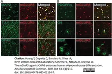
Mouse anti Human MBP antibody, clone 12 (MCA409S) used to detect myelin im mouse samples by immunofluorescence.
Image caption:
mGluR5 expression in human brain cells. A mGluR5 antibody and cell type specific antibodies were used to immunofluorescently determine mGluR5 expression. MAP2+ and TUJ1+ neurons express mGluR5 (solid arrowheads). GFAP+ astrocytes are negative for mGluR5 (empty arrowheads). B In the OL lineage cells, only PDGFRα+ OPC and O4+ immature cells express mGluR5 (solid arrowheads), but not MBP+ mature OLs. Scale bar = 40 μm
From: Huang Y, Geywitz C, Bandaru A, Glass IA; Birth Defects Research Laboratory; Schirmer L, Nobuta H, Dreyfus CF.
The mGluR5 agonist CHPG enhances human oligodendrocyte differentiation.
Acta Neuropathol Commun. 2025 Oct 3;13(1):210.
doi: 10.1186/s40478-025-02124-7.
This image is from an open access article distributed under terms of a Creative Commons Attribution License.
Filter by Application:
IF WB P C IC Reset| Rat anti MBP antibody, clone 12 recognizes myelin basic protein from a wide range of species. Rat anti MBP antibody, clone 12 reacts weakly with peptides ending in the Phe 91 where the 91-92 Phe-Phe bond is broken. Synthetic peptide 82-99 reacts very well with Rat anti MBP antibody, clone 12, as does intact MBP. Further epitope analysis indicates binding to a region defined by amino acids 82-87 (DENPVV). Rat anti MBP antibody, clone 12 has been reported as being suitable for use in western blotting (Glynn et al. 1987). |
- Target Species
- Bovine
- Species Cross-Reactivity
-
Target Species Cross Reactivity Mouse Rabbit Mammals Expected from Sequence Rat Guinea Pig Sheep Human Chicken Pig - N.B. Antibody reactivity and working conditions may vary between species.
- Product Form
- Tissue Culture Supernatant - liquid
- Buffer Solution
- 0.1M TRIS
- Preservative Stabilisers
0.1% Sodium Azide - Immunogen
- Bovine MBP.
- Fusion Partners
- Spleen cells from an immunized outbred rat were fused with cells of the mouse NS0 myeloma cell line.
- Regulatory
- For research purposes only
- Guarantee
- 12 months from date of despatch
This product is shipped at ambient temperature. It is recommended to aliquot and store at -20°C on receipt. When thawed, aliquot the sample as needed. Keep aliquots at 2-8°C for short term use (up to 4 weeks) and store the remaining aliquots at -20°C.
Avoid repeated freezing and thawing as this may denature the antibody. Storage in frost-free freezers is not recommended.
Avoid repeated freezing and thawing as this may denature the antibody. Storage in frost-free freezers is not recommended.
This product has been reported to work in the following applications. This information is derived from testing within our laboratories, peer-reviewed publications or personal communications from the originators. Please refer to references indicated for further information. For general protocol recommendations, please visit the antibody protocols page.
| Application Name | Verified | Min Dilution | Max Dilution |
|---|---|---|---|
| ELISA |  |
||
| Immunofluorescence | |
||
| Radioimmunoassays | |
||
| Western Blotting | |
Where this product has not been tested for use in a particular technique this does not necessarily exclude its use in such procedures. Suggested working dilutions are given as a guide only. It is recommended that the user titrates the product for use in their own system using appropriate negative/positive controls.
References for MBP antibody
-
Groome, N.P. et al. (1986) Region-specific immunoassays for human myelin basic protein.
J Neuroimmunol. 12 (4): 253-64. -
Glynn, P. et al. (1987) Basic protein dissociating from myelin membranes at physiological ionic strength and pH is cleaved into three major fragments.
J Neurochem. 48 (3): 752-9. -
Hruby, S. et al. (1987) Monoclonal antibodies reactive with myelin basic protein.
Mol Immunol. 24 (12): 1359-64. -
Relvas, J.B. et al. (2001) Expression of dominant-negative and chimeric subunits reveals an essential role for beta1 integrin during myelination.
Curr Biol. 11: 1039-43. -
Massa, P.T. et al. (2002) Critical role for protein tyrosine phosphatase SHP-1 in controlling infection of central nervous system glia and demyelination by Theiler's murine encephalomyelitis virus.
J Virol. 76:8335-46. -
Massa, P.T. et al. (2004) Dysmyelination and reduced myelin basic protein gene expression by oligodendrocytes of SHP-1-deficient mice.
J Neurosci Res. 77: 15-25. -
Homchaudhuri L et al. (2009) Influence of membrane surface charge and post-translational modifications to myelin basic protein on its ability to tether the Fyn-SH3 domain to a membrane in vitro.
Biochemistry. 48 (11): 2385-93. -
Relucio, J. et al. (2009) Laminin alters fyn regulatory mechanisms and promotes oligodendrocyte development.
J Neurosci. 29: 11794-806.
View The Latest Product References
-
Savvaki, M. et al. (2010) The expression of TAG-1 in glial cells is sufficient for the formation of the juxtaparanodal complex and the phenotypic rescue of tag-1 homozygous mutants in the CNS.
J Neurosci. 30: 13943-54. -
Pohl, H.B. et al. (2011) Genetically induced adult oligodendrocyte cell death is associated with poor myelin clearance, reduced remyelination, and axonal damage.
J Neurosci. 31 (3): 1069-80. -
Laursen, L.S. et al. (2011) Translation of myelin basic protein mRNA in oligodendrocytes is regulated by integrin activation and hnRNP-K.
J Cell Biol. 192: 797-811. -
Monk, K.R. et al. (2011) Gpr126 is essential for peripheral nerve development and myelination in mammals.
Development. 138: 2673-80. -
Ioannidou, K. et al. (2012) Time-lapse imaging of the dynamics of CNS glial-axonal interactions in vitro and ex vivo.
PLoS One. 7: e30775. -
Horn, M. et al. (2012) Myelin is dependent on the Charcot-Marie-Tooth Type 4H disease culprit protein FRABIN/FGD4 in Schwann cells.
Brain. 135 (Pt 12): 3567-83. -
di Petna, A. et al. (2013) Oxidative stress and proinflammatory cytokines contribute to demyelination and axonal damage in a cerebellar culture model of neuroinflammation.
PLoS One. 8 (2): e54722. -
Müller, C. et al. (2015) SncRNA715 Inhibits Schwann Cell Myelin Basic Protein Synthesis.
PLoS One. 10 (8): e0136900. -
Brügger V et al. (2015) HDAC1/2-Dependent P0 Expression Maintains Paranodal and Nodal Integrity Independently of Myelin Stability through Interactions with Neurofascins.
PLoS Biol. 13 (9): e1002258. -
Natrajan, M.S. et al. (2015) Retinoid X receptor activation reverses age-related deficiencies in myelin debris phagocytosis and remyelination.
Brain. 138(Pt 12):3581-97 -
Rittchen S et al. (2015) Myelin repair in vivo is increased by targeting oligodendrocyte precursor cells with nanoparticles encapsulating leukaemia inhibitory factor (LIF).
Biomaterials. 56: 78-85. -
Friess, M. et al. (2016) Intracellular ion signaling influences myelin basic protein synthesis in oligodendrocyte precursor cells.
Cell Calcium. 60 (5): 322-30. -
Fernandes, A.R. & Chari, D.M. (2016) Part II: Functional delivery of a neurotherapeutic gene to neural stem cells using minicircle DNA and nanoparticles: Translational advantages for regenerative neurology.
J Control Release. 238: 300-10. -
Crawford, A.H. et al. (2016) Pre-Existing Mature Oligodendrocytes Do Not Contribute to Remyelination following Toxin-Induced Spinal Cord Demyelination.
Am J Pathol. 186 (3): 511-6. -
Lim, J.L. et al. (2016) Protandim Protects Oligodendrocytes against an Oxidative Insult.
Antioxidants (Basel). 5 (3): pii: E30. -
Isoda, M. et al. (2016) Robust production of human neural cells by establishing neuroepithelial-like stem cells from peripheral blood mononuclear cell-derived feeder-free iPSCs under xeno-free conditions.
Neurosci Res. 110: 18-28. -
Fernandes, A.R. & Chari, D.M. (2016) Part I: Minicircle vector technology limits DNA size restrictions on ex vivo gene delivery using nanoparticle vectors: Overcoming a translational barrier in neural stem cell therapy.
J Control Release. 238: 289-99. -
Moreno, B. et al. (2017) Methylthioadenosine promotes remyelination by inducing oligodendrocyte differentiation
Mult Scler Demyel Disord. 2, 3. -
Qin, J. et al. (2017) GD1a Overcomes Inhibition of Myelination by Fibronectin via Activation of Protein Kinase A: Implications for Multiple Sclerosis.
J Neurosci. 37 (41): 9925-38. -
Aranmolate, A. et al. (2017) Myelination is delayed during postnatal brain development in the mdx mouse model of Duchenne muscular dystrophy.
BMC Neurosci. 18 (1): 63. -
Yu, Q. et al. (2017) Strain differences in cuprizone induced demyelination.
Cell Biosci. 7: 59. -
Grigoletto, J. et al. (2017) Higher levels of myelin phospholipids in brains of neuronal α-Synuclein transgenic mice precede myelin loss.
Acta Neuropathol Commun. 5 (1): 37. -
Dillenburg, A. et al. (2018) Activin receptors regulate the oligodendrocyte lineage in health and disease.
Acta Neuropathol. 135 (6): 887-906. -
Sekizar, S. & Williams, A. (2019) Ex Vivo Slice Cultures to Study Myelination, Demyelination, and Remyelination in Mouse Brain and Spinal Cord.
Methods Mol Biol. 1936: 169-83. -
Nocita, E. et al. (2019) EGFR/ErbB Inhibition Promotes OPC Maturation up to Axon Engagement by Co-Regulating PIP2 and MBP.
Cells. 8 (8): 844. -
Martinez-rachadell, L. et al. (2019) Cell-specific expression of insulin/insulin-like growth factor-I receptor hybrids in the mouse brain.
Growth Horm IGF Res. 45: 25-30. -
Swire, M. et al. (2019) Endothelin signalling mediates experience-dependent myelination in the CNS.
Elife. 8:e49493 -
Vogel, J.K. et al. (2020) Sox9 overexpression exerts multiple stage-dependent effects on mouse spinal cord development.
Glia. 68 (5): 932-46. -
Vanheel, H. et al. (2020) Duodenal acidification induces gastric relaxation and alters epithelial barrier function by a mast cell independent mechanism.
Sci Rep. 10 (1): 17448. -
Kerman, B.E. et al. (2020) Motoneuron expression profiling identifies an association between an axonal splice variant of HDGF-related protein 3 and peripheral myelination.
J Biol Chem. 295 (34): 12233-46. -
Klein, B. et al. (2020) DCX+ neuronal progenitors contribute to new oligodendrocytes during remyelination in the hippocampus.
Sci Rep. 10 (1): 20095. -
Yetiş, Ç. et al. (2020) Myelin detection in fluorescence microscopy images using machine learning.
J Neurosci Methods. 346: 108946. -
Werkman, I.L. et al. (2020) Transcriptional heterogeneity between primary adult grey and white matter astrocytes underlie differences in modulation of in vitro myelination.
J Neuroinflammation. 17 (1): 373. -
Melero-Jerez, C. et al. (2021) Myeloid-derived suppressor cells support remyelination in a murine model of multiple sclerosis by promoting oligodendrocyte precursor cell survival, proliferation, and differentiation.
Glia. 69 (4): 905-24. -
Holloway, R.K. et al. (2021) Microglial inflammasome activation drives developmental white matter injury.
Glia. 69 (5): 1268-80. -
Schultz, V. et al. (2021) Zika Virus Infection Leads to Demyelination and Axonal Injury in Mature CNS Cultures.
Viruses. 13 (1): 91. -
Meireles, A.M. et al. (2018) The Lysosomal Transcription Factor TFEB Represses Myelination Downstream of the Rag-Ragulator Complex.
Dev Cell. 47 (3): 319-330.e5. -
Lloyd, A.F. et al. (2019) Central nervous system regeneration is driven by microglia necroptosis and repopulation.
Nat Neurosci. 22 (7): 1046-52. -
Yamazaki, R. et al. (2021) Macroscopic detection of demyelinated lesions in mouse PNS with neutral red dye.
Sci Rep. 11 (1): 16906. -
Alhajlah, S. et al. (2021) Overexpression of Reticulon 3 Enhances CNS Axon Regeneration and Functional Recovery after Traumatic Injury.
Cells. 10 (8): 2015. -
Swire, M. et al. (2021) Oligodendrocyte HCN2 Channels Regulate Myelin Sheath Length.
J Neurosci. 41 (38): 7954-64. -
Niu, J. et al. (2021) Oligodendroglial ring finger protein Rnf43 is an essential injury-specific regulator of oligodendrocyte maturation.
Neuron. 109 (19): 3104-18.e6. -
Moyon, S. et al. (2021) TET1-mediated DNA hydroxymethylation regulates adult remyelination in mice.
Nat Commun. 12 (1): 3359. -
Bechler, M.E. (2019) A Neuron-Free Microfiber Assay to Assess Myelin Sheath Formation.
Methods Mol Biol. 1936: 97-110. -
Sekizar, S. & Williams, A. (2019) Ex Vivo. Slice Cultures to Study Myelination, Demyelination, and Remyelination in Mouse Brain and Spinal Cord.
Methods Mol Biol. 1936: 169-83. -
Li, S. et al. (2019) Induction of immunological tolerance to myelinogenic glial-restricted progenitor allografts.
Brain. 142 (11): 3456-72. -
Berghoff, S.A. et al. (2021) Microglia facilitate repair of demyelinated lesions via post-squalene sterol synthesis.
Nat Neurosci. 24 (1): 47-60. -
Kalafatakis, I. et al. (2021) The beneficial role of the synthetic microneurotrophin BNN20 in a focal demyelination model.
J Neurosci Res. 99 (5): 1474-95. -
Grigoletto, J. et al. (2022) Loss of Corticostriatal Mu-Opioid Receptors in α-Synuclein Transgenic Mouse Brains.
Life (Basel). 12 (1): 63. -
Poggi, G. et al. (2022) Effects of chronic social stress on oligodendrocyte proliferation-maturation and myelin status in prefrontal cortex and amygdala in adult mice
Neurobiol Stress. 18: 100451. -
McNamara, N.B. et al. (2023) Microglia regulate central nervous system myelin growth and integrity.
Nature. 613 (7942): 120-9. -
Aber, E.R. et al. (2022) Oligodendroglial macroautophagy is essential for myelin sheath turnover to prevent neurodegeneration and death.
Cell Rep. 41 (3): 111480. -
Tanabe, K. et al. (2022) Generation of functional human oligodendrocytes from dermal fibroblasts by direct lineage conversion.
Development. 149 (20): dev199723. -
Silva, M.E. et al. (2023) DDR1 and Its Ligand, Collagen IV, Are Involved in In Vitro Oligodendrocyte Maturation.
Int J Mol Sci. 24 (2): 1742. -
Freire, M.A.M. et al. (2023) Astrocytosis, Inflammation, Axonal Damage and Myelin Impairment in the Internal Capsule following Striatal Ischemic Injury
Cells. 12 (3): 457. -
Holloway, R.K. et al. (2023) Localized microglia dysregulation impairs central nervous system myelination in development.
Acta Neuropathol Commun. 11 (1): 49. -
Rocha, D.N. et al. (2023) It takes two to remyelinate: A bioengineered platform to study astrocyte-oligodendrocyte crosstalk and potential therapeutic targets in remyelination.
Biomater Adv. 151: 213429. -
Kaplanis, S.I. et al. (2023) Nicotinamide enhances myelin production after demyelination through reduction of astrogliosis and microgliosis.
Front Cell Neurosci. 17: 1201317. -
Ma, D. et al. (2023) Human iPSC-derived endothelial cells promote CNS remyelination via BDNF and mTORC1 pathway
Glia. 72(1):133-55. -
Dominicis, A. et al. (2023) N-Acetylaspartate Drives Oligodendroglial Differentiation via Histone Deacetylase Activation.
Cells. 12 (14): 1861. -
Molina-Gonzalez, I. et al. (2023) Astrocyte-oligodendrocyte interaction regulates central nervous system regeneration.
Nat Commun. 14 (1): 3372. -
Ktena, N. et al. (2022) Autophagic degradation of CNS myelin maintains axon integrity.
Cell Stress. 6 (12): 93-107. -
Cai, Y. et al. (2024) Embryonic origins of forebrain oligodendrocytes revisited by combinatorial genetic fate mapping.
bioRχiv. 23 Jan [Epub ahead of print]. -
Seo, T. et al. (2023) MARCH family E3 ubiquitin ligases selectively target and degrade cadherin family proteins
bioRχiv 10 Aug [Epub ahead of print]. -
Gigliotta, A. et al. (2024) Genetic background modulates the effect of glucocorticoids on proliferation, differentiation and myelin formation of oligodendrocyte lineage cells.
Eur J Neurosci. Feb 22 [Epub ahead of print]. -
Jarjour, A.A. et al. (2020) The formation of paranodal spirals at the ends of CNS myelin sheaths requires the planar polarity protein Vangl2.
Glia. 68 (9): 1840-1858. -
Odell, E. et al. (2024) Minimum Effective Dose of Clemastine in a Mouse Model of Preterm White Matter Injury.
bioRχiv. Mar 01 [Epub ahead of print]. -
Aberle, T. et al. (2024) Sox10 Activity and the Timing of Schwann Cell Differentiation Are Controlled by a Tle4-Dependent Negative Feedback Loop.
Int J Mol Sci. ;25 (10): 5234. -
Bagheri, H. et al. (2024) Myelin basic protein mRNA levels affect myelin sheath dimensions, architecture, plasticity, and density of resident glial cells.
Glia. 72 (10): 1893-1914. -
Jörg, L.M. et al. (2024) Transcription Factors Sox8 and Sox10 Contribute with Different Importance to the Maintenance of Mature Oligodendrocytes.
Int J Mol Sci. 25 (16): 8754. -
Dorrier, C.E. et al. (2021) CNS fibroblasts form a fibrotic scar in response to immune cell infiltration.
Nat Neurosci. 24 (2): 234-44. -
Jang, G. et al. (2024) Visceral adiposity is associated with iron deposition and myelin loss in the brains of aged mice.
Neurochem Int. 179: 105833. -
Xie, Q. et al. (2025) Immune cell response after intracerebral hemorrhage in piglets and the treatment effects of deferoxamine and minocycline.
Exp Neurol. : 115354. 23 Jun [Epub ahead of print]. -
Huang, Y. et al. (2025) The mGluR5 agonist CHPG enhances human oligodendrocyte differentiation.
Acta Neuropathol Commun. 13 (1): 210. -
Ktena, N. et al. (2025) Disruption of Oligodendroglial Autophagy Leads to Myelin Morphological Deficits, Neuronal Apoptosis, and Cognitive Decline in Aged Mice.
Glia. 73 (7): 1383-1397.
- Synonyms
- Myelin Basic Protein
- RRID
- AB_325004
- UniProt
- P02687
- Entrez Gene
- MBP
- GO Terms
- GO:0005886 plasma membrane
- GO:0005515 protein binding
- GO:0019911 structural constituent of myelin sheath
- GO:0043209 myelin sheath
View more products with MBP specificity
Please Note: All Products are "FOR RESEARCH PURPOSES ONLY"
View all Anti-Bovine ProductsAlways be the first to know.
When we launch new products and resources to help you achieve more in the lab.
Yes, sign me up