MitoPT™ JC-1, TMRE and TMRM Kits

- On This Page
- Overview
- MitoPT™ JC-1, TMRE and TMRM kits
MitoPT™ JC-1, TMRE & TMRM Kits Overview

- Choice – MitoPT™ JC-1, TMRE & TMRM Kits
- Quick & easy staining method clearly differentiates non-apoptotic & apoptotic cells
- MitoPT™ TMRE & TMRM Kits - ideal for single cell multi-parametric analyses
- Suitable for flow cytometry, fluorescence microscopy or a plate reader
An early indication of apoptosis involves a collapse in the electrochemical gradient across the mitochondrial membrane.
This is mediated by the opening of permeability transition (PT) pores and is known as the mitochondrial PT event. It is measured by the change in the mitochondrial membrane potential.
Loss of mitochondrial membrane potential can be detected by three fluorescent cationic dyes:
- 5,5’,6,6’-tetrachloro-1,1’,3,3’-tetraethylbenzimidazolocarbocyanine iodide (JC-1)
- Tetramethylrhodamine ethyl ester (TMRE) and
- Tetramethylrhodamine methyl ester (TMRM)
MitoPT™ JC-1, TMRE and TMRM Kits
| Description | Target | Format | Clone | Applications | Citations | Code |
|---|

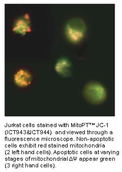
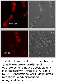
These dyes are lipophilic and have a delocalized positive charge, which enables them to enter cells and the negatively-charged healthy mitochondria. In non-apoptotic mitochondria, the dyes aggregate and will brightly fluoresce.
As apoptosis occurs and the mitochondrial membrane potential collapses, the dyes disperse throughout the cell and the fluorescence either diminishes (MitoPT™ TMRE and TMRM kits) or changes color (MitoPT™ JC-1 kit).
This alteration in fluorescence allows an easy distinction between non-apoptotic and apoptotic cells which can be read with a flow cytometer, fluorescence microscope or fluorescence plate reader.
| MitoPT™ Kit | Non-apoptotic cells | Apoptotic cells |
|---|---|---|
| JC-1 | Red fluorescence | Green fluorescence |
| TMRE | Orange fluorescence | Weak orange fluorescence |
| TMRM | Orange fluorescence | Weak orange fluorescence |
View the other sections in our quality apoptosis antibodies and kits product range:
Reference:
- Antonsson, B., et al. (2000) Biochem J. 345:271-278
- Cossarizza, A., et al. (1993) Biochem. Biophys. Res. Commun. 197:40-45
- Desagher, S., et al. (1999) J. Cell Biol. 144:891-901
- Kroemer, G. and J.C. Reed. (2000). Nature Med. 6(5): 513-519
- Luo, X., et al. (1998) Cell 94:481-490
- Reers, M., et al. (1991) Biochemistry 30:4480-4486
- Smiley, S. T., et al. (1991) Proc. Natl. Acad. Sci. 88:3671-3675
- Wlodkowic, D., J. Skommer, and J. Pelkonen. (2006). Leukemia Res. 30:1187-1192.


