Legionnaires’ Disease

- On This Page
- Legionella
- Legionnaires’ disease
- Legionella life cycle
- Antibodies to legionella
- Our legionella antibody range
- References
 Legionnaires’ disease, a severe pneumonia, is caused through infection of lung tissue by Legionella spp. bacteria. It was first observed in the summer of 1976, when 221 attendees of the American Legion convention displayed an unusual lung infection; 34 cases were fatal.
A large investigation by the US Centers for Disease Control and Prevention (CDC) identified the pathogen by the end of 1976, a newly discovered rod-shaped, gram-negative bacterium, named Legionella pneumophila. Subsequently it appeared that the bacterium had been first isolated in the late 1940s, but never characterized. Additionally, Legionella was also shown to be responsible for a series of flu-like diseases in 1968 that had been termed Pontiac fever (Fraser et al. 1977).
Legionella
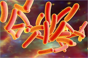
Legionella are gram-negative, rod-shaped gamma-proteobacteria that are widely distributed in freshwater and moist soil, meaning that they can populate any structures that contain water as well as survive in biofilms. It was also the first bacterium found to use protozoan hosts, aquatic amoebae, to multiply. Protozoans provide an intracellular replication pathway for the generation of infective Legionella vesicles; cyst forming protozoans provide enhanced environmental protection. This characteristic provided the clue that Legionella may use human lung macrophages for replication (Brenner et al. 1979).
Legionnaires’ Disease
Humans are primarily infected by inhaling Legionella-containing aerosols from water sources as diverse as showers and air-conditioning systems. Although rare, human-to-human transmission has been found (Correia et al. 2016). While anyone can be infected with Legionella, people with underlying medical conditions like diabetes or cancer, smokers, and males over 50 years old, are more likely to catch Legionnaires’ disease. There is also a seasonal bias, summer and early autumn are peak infection times.
Legionnaires’ disease is also increasing, in Europe cases rose from 4921 in 2011 to 11,343 in 2018; in the US from 2301 in 2005 to 7104 in 2018. Fatality rates vary from 2.2-10.3% but can reach 48% in hospital settings (Nisar et al. 2020). Of the 65 species described so far, Legionella pneumophila is responsible for 80-90% of cases, and within this strain serogroup 1 causes 90% of infections (Yu et al. 2002).
Legionella Life Cycle
Legionella manage to survive in a variety of phagocytic hosts, many species of amoeba, and mammalian cell lines. This is facilitated by a biphasic life cycle that has a noninfective replication stage and a virulent transmissive phase. In the former, genes for growth and division are activated and those that control infectivity are downregulated. Once bacterial density has increased, the reverse happens and bacteria leave the host in search of new infection targets (Oliva et al. 2018).
One of the key regulators that controls this switch is the RNA-binding protein, carbon storage regulator A (CsrA), a global, posttranscriptional regulator of gene expression that supports the replicative stage. Once a certain bacterial density has been reached, CsrA is bound by small noncoding RNAs that abrogate its function, leading to the virulent phase. CsrA also regulates the expression of at least 40 defective in organelle trafficking/intracellular multiplication (Dot/Icm) Type IVb secretion system (T4SS) effector proteins (Sahr et al. 2017). The T4SS works in conjunction with the type 2 secretion system (T2SS) in L. pneumophila to support infection, maintenance, and replication in the host.
After the initial binding of the bacteria by the host cell, the T2SS is critical for successful infection and maintenance of L. pneumophila in the host. It deploys at least 25 effector proteins, mostly enzymes; among them a phospholipase C and chitinase that have been shown to promote bacterial persistence in lung tissues (Cianciotto 2009).
To maintain and replicate inside host cells, Legionella create an intracellular environment, the Legionella-containing vacuole (LCV), that supplies them with nutrients while avoiding lysosomal degradation. The T4SS secretes over 300 proteins that interfere with host cell functions to ensure bacterial survival (Schroeder 2017, Qin et al. 2017). Some of their functions are:
- Interfering with endocytic maturation and preventing fusion with lysosomes (Horwitz 1983)
- Stopping vacuolar acidification by blocking host vacuolar ATPase (Xu et al. 2010)
- Redirecting the LCV away from the canonical endocytic pathway (Hoffmann et al. 2014)
- Depleting transport and fusion factors from endosomes, rendering them fusion incompetent (Gaspar and Machner 2014)
- Recruiting ER-derived vesicles to the LCV membrane (Robinson and Roy 2006)
The recruitment of the ER vesicles is achieved via ubiquitination of host cell reticulon 4 (Rtn4), leading to tubule rearrangements as well as modifications in Rtn4 associated with the replication compartment (Kotewicz et al. 2017). A further example of L. pneumophila’s ability to hijack host cell processes is the manipulation of the cell-death pathways, implemented via the SidF T4SS effector. SidF binds the proapoptotic BNIP3 and Bcl-rambo, and neutralizes their functions, preventing apoptosis of the infected host cells (Banga et al. 2007). Conversely, it has been suggested that L. pneumophila’s VipD protein can mediate caspase-3 activation and thus induce apoptosis (Zhu et al. 2013).
Antibodies to Legionella
While a large body of data has been accumulated on Legionella spp., much remains to be elucidated about these bacteria. To aid with basic Legionella spp detection, Bio-Rad offers a polyclonal antibody, suitable for immunofluorescence. Rabbit Anti-Legionella SPP, OBT0943, will detect serogroups 1-12 and is available in purified, biotin, and HRP conjugated formats for use in ELISA, frozen sections, and immunoblots.
Bio-Rad supplies secondary antibodies certified to work in immunofluorescence detection. Additionally, if you are new to immunofluorescence staining have a look at the 10 Tips for Selecting and Using Fluorophores in IF Experiments.
Our Legionella Antibody Range
| Description | Target | Format | Clone | Applications | Citations | Code |
|---|
References
- Banga S et al. (2007). Legionella pneumophila inhibits macrophage apoptosis by targeting pro-death members of the Bcl2 protein family. PNAS. (12), 5,121-26.
- Brenner DJ et al. (1979). Classification of the Legionnaires’ disease bacterium: Legionella pneumophila, genus novum, species nova, of the family Legionellaceae, familia nova. Ann. Intern. Med. 90(4), 656-658.
- Cianciotto NP (2009). Many substrates and functions of type II secretion: lessons learned from Legionella pneumophila. Future Microbiol. 4(7), 797-805.
- Correia AM et al. (2016). Probable person-to-person transmission of Legionnaires’ disease. N. Engl. J. Med. 374(5), 497-498.
- Fraser DW et al. (1977). Legionnaires' disease: description of an epidemic of pneumonia. N. Engl. J. Med. 297(22), 1,189-1,197.
- Gaspar AH and Machner MP (2014). VipD is a Rab5-activated phospholipase A1 that protects Legionella pneumophila from endosomal fusion. PNAS. 111(12), 4,560-4,565.
- Hoffmann C et al. (2014). Functional analysis of novel Rab GTPases identified in the proteome of purified Legionella-containing vacuoles from macrophages. Cell. Microbiol. 16(7), 1,034-1,052.
- Horwitz MA (1983). The Legionnaires’ disease bacterium (Legionella pneumophila) inhibits phagosome-lysosome fusion in human monocytes. J. Exp. Med. 158(6), 2,108-2,126.
- Kotewicz KM et al. (2017). A single Legionella effector catalyzes a multistep ubiquitination pathway to rearrange tubular endoplasmic reticulum for replication. Cell Host Microbe. 21(2), 169-181
- Nisar MA et al. (2020). Legionella pneumophila and Protozoan Hosts: Implications for the control of hospital and potable water systems. Pathogens. 9(4), 286.
- Oliva G et al. (2018). The life cycle of Legionella pneumophila: Cellular differentiation is linked to virulence and metabolism. Front. Cell. Infect. Microbiol. 8, 3.
- Qin T et al. (2017). Distribution of secretion systems in the genus Legionella and its correlation with pathogenicity. Front. Microbiol. 8, 388.
- Robinson CG and Roy CR (2006). Attachment and fusion of endoplasmic reticulum with vacuoles containing Legionella pneumophila. Cell. Microbiol. 8(5), 793-805.
- Sahr T et al. (2017). The Legionella pneumophila genome evolved to accommodate multiple regulatory mechanisms controlled by the CsrA-system. PLOS Genet. 13(2), e1006629.
- Schroeder GN (2017). The toolbox for uncovering the functions of LegionellaDot/Icm type IVb secretion system effectors: current state and future directions. Front. Cell. Infect. Microbiol. 7, 528.
- Xu L et al. (2010). Inhibition of host vacuolar H+-ATPase activity by a Legionella pneumophila effector. PLOS Pathog. 6(3), 1000822.
- Yu VL et al. (2002). Distribution of Legionella species and serogroups isolated by culture in patients with sporadic community-acquired legionellosis: an international collaborative survey. J. Infect. Dis. 186(1), 127-128.
- Zhu W et al. (2013). Induction of caspase 3 activation by multiple Legionella pneumophila Dot/Icm substrates. Cell.Microbiol. 15(11), 1,783-1,795.


