NogoA antibody
Rabbit anti Human NogoA (N-Terminal)
- Product Type
- Polyclonal Antibody
- Isotype
- Polyclonal IgG
- Specificity
- NogoA
- Region
- (N-TERMINAL)


Filter by Application:
C IF Reset| Rabbit anti Human NOGOA antibody recognizes human Reticulon-4 (RTN4), also known as NogoA, neurite outgrowth inhibitor, Foocen, Neuroendocrine-specific protein, Neuroendocrine-specific protein C homolog, Reticulon-5 or RTN-x. NogoA is a 1192 amino acid multi pass transmembrane protein associated with the endoplasmic reticulum, a member of a family of integral membrane proteins termed reticulons. Six isoforms of NogoA can be generated by alternative splicing, the canonical isoform 1 is predominantly expressed in the brain and testis with weaker expression in the heart and skeletal muscle. Reticulons are involved in various neurodegenerative diseases such as Amyotrophic lateral sclerosis,and multiple sclerosis (Chiurchiù et al. 2014). Reticulon proteins have been demonstrated to regulate many cellular processes and interact with multiple proteins and receptors such as BACE. NogoA was initially identified as a myelin-associated neurite outgrowth inhibitor (Niederöst et al. 2002). NogoA is highly expressed in oligodendrocytes in the white matter of the CNS (Kuhlmann et al. 2008). Blocking NogoA activity with antibodies or other factors results in improved long distance axonal regeneration and functional recovery in experimental CNS lesion models (Schwab 2004). NOGOA has a predicted molecular weight of 130 kDa however, despite its predicted molecular weight, NogoA typically migrates at ~180 kDa in an SDS-PAGE. Rabbit anti human NOGOA antibody is expected to recognize all isoforms of NogoA. |
- Target Species
- Human
- Species Cross-Reactivity
-
Target Species Cross Reactivity Mouse Rat - N.B. Antibody reactivity and working conditions may vary between species.
- Product Form
- Purified IgG - liquid
- Antiserum Preparation
- Antisera to human NogoA were raised by repeated immunisation of rabbits with highly purified antigen. Purified IgG prepared from whole serum by affinity chromatography.
- Buffer Solution
- Phosphate buffered saline
- Preservative Stabilisers
- 0.02% Sodium Azide (NaN3)
- Immunogen
- Synthetic peptide sequence corresponding to a 23 amino acid sequence from near the carboxy terminus of Human NogoA
- Approx. Protein Concentrations
- IgG concentration 1.0mg/ml
- Regulatory
- For research purposes only
- Guarantee
- 12 months from date of despatch
This product is shipped at ambient temperature. It is recommended to aliquot and store at -20°C on receipt. When thawed, aliquot the sample as needed. Keep aliquots at 2-8°C for short term use (up to 4 weeks) and store the remaining aliquots at -20°C.
Avoid repeated freezing and thawing as this may denature the antibody. Storage in frost-free freezers is not recommended.
Avoid repeated freezing and thawing as this may denature the antibody. Storage in frost-free freezers is not recommended.
This product has been reported to work in the following applications. This information is derived from testing within our laboratories, peer-reviewed publications or personal communications from the originators. Please refer to references indicated for further information. For general protocol recommendations, please visit the antibody protocols page.
| Application Name | Verified | Min Dilution | Max Dilution |
|---|---|---|---|
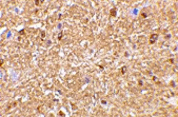
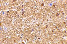
| Immunohistology - Paraffin 1 |  |
2.5ug/ml | |
| Western Blotting | |
0.5 | 1.0ug/ml |
- 1This product requires antigen retrieval using heat treatment prior to staining of paraffin sections.Sodium citrate buffer pH 6.0 is recommended for this purpose.
Where this product has not been tested for use in a particular technique this does not necessarily exclude its use in such procedures. Suggested working dilutions are given as a guide only. It is recommended that the user titrates the product for use in their own system using appropriate negative/positive controls.
- Histology Positive Control Tissue
- Mouse brain
- Western Blotting
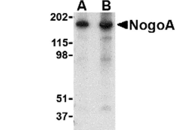
- AHP1799 detects a band of approximately 180 kDa in Mouse Brain tissue lysate.
| Description | Product Code | Applications | Pack Size | List Price | Your Price | Quantity | |
|---|---|---|---|---|---|---|---|
| Antigen Retrieval Buffer, pH8.0 | BUF025A | P | 500 ml | Log in | |||
| List Price | Your Price | ||||||
| Log in | |||||||
| Description | Antigen Retrieval Buffer, pH8.0 | ||||||
| TidyBlot Western Blot Detection Reagent:HRP | STAR209P | WB * | 0.5 ml | Log in | |||
| List Price | Your Price | ||||||
| Log in | |||||||
| Description | TidyBlot Western Blot Detection Reagent:HRP | ||||||
References for NogoA antibody
-
Dann, A. et al. (2011) Cytosolic RIG-I-like helicases act as negative regulators of sterile inflammation in the CNS.
Nat Neurosci. 15: 98-106. -
Gerondopoulos, A. et al. (2014) Rab18 and a Rab18 GEF complex are required for normal ER structure.
J Cell Biol. 205 (5): 707-20.
Further Reading
-
Chen, M.S. et al. (2000) Nogo-A is a myelin-associated neurite outgrowth inhibitor and an antigen for monoclonal antibody IN-1.
Nature. 403 (6768): 434-9. -
Dupuis, L. et al. (2002) Nogo provides a molecular marker for diagnosis of amyotrophic lateral sclerosis.
Neurobiol Dis. 10 (3): 358-65. -
Yan, R. et al. (2006) Reticulon proteins: emerging players in neurodegenerative diseases.
Cell Mol Life Sci. 63 (7-8): 877-89. -
Schweigreiter, R. & Bandtlow, C.E. (2006) Nogo in the injured spinal cord.
J Neurotrauma. 23 (3-4): 384-96.
- RRID
- AB_10612769
- UniProt
- Q9NQC3
- Entrez Gene
- RTN4
Request a different product with this specificity
Please Note: All Products are "FOR RESEARCH PURPOSES ONLY"
View all Anti-Human ProductsAlways be the first to know.
When we launch new products and resources to help you achieve more in the lab.
Yes, sign me up