CD8 antibody | CC63
Mouse anti Bovine CD8:RPE
- Product Type
- Monoclonal Antibody
- Clone
- CC63
- Isotype
- IgG2a
- Specificity
- CD8


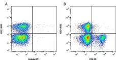
Figure B. FITC conjugated Mouse anti Bovine CD21 antibody, clone CC21 (MCA1424F) and RPE conjugated Mouse anti Bovine CD8 antibody, clone CC63 (MCA837PE). All experiments performed on red cell lysed bovine blood gated on lymphocytes in the presence of 10% bovine serum.
Data acquired on the ZE5 Cell Analyzer.
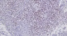

Image courtesy of Dr. J.B. Silvan, Moredun Research Institute, Roslin, UK
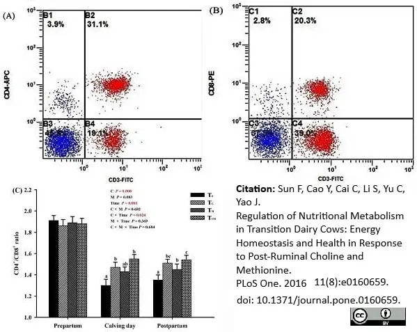
RPE conjugated Mouse anti Bovine CD8 antibody, clone CC63 (MCA837PE) used to detect cytotoxic T-cells in bovine peripheral blood by flow cytometry together with APC conjugated Mouse anti Bovine CD4 antibody, clone CC8 (MCA1653) used for the identification of helper T-cells in the same samples.
Image caption:
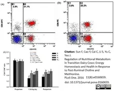
Effects of supplementation with rumen-protected choline (TC), rumen-protected methionine (TM) or both (TCM) on proportions of T lymphocyte subtypes in peripheral blood of transition dairy cows. (A) and (B) present the output images from flow cytometry. (C) displays the effects of each treatment over the three weeks before calving, on calving day, and over the three weeks after calving. a, b, c Values with different superscript letters at the same time point are significantly different (P <0.05).
From: Sun F, Cao Y, Cai C, Li S, Yu C, Yao J (2016)
Regulation of Nutritional Metabolism in Transition Dairy Cows: Energy Homeostasis and Health in Response to Post-Ruminal Choline and Methionine.
PLoS ONE 11(8): e0160659.
This image is from an open access article distributed under terms of a Creative Commons Attribution License.
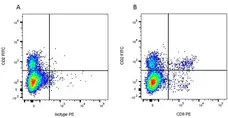
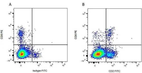
Figure B. FITC conjugated Mouse anti Bovine CD2 antibody, clone CC42 (MCA833F) and RPE conjugated Mouse anti Bovine CD8 antibody, clone CC63 (MCA837PE). All experiments performed on red cell lysed bovine blood gated on mononuclear cells.
Data acquired on the ZE5 Cell Analyzer.
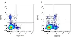
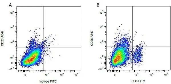
Figure B. Alexa Fluor® 647 conjugated Mouse anti Bovine CD26 antibody, clone CC69 (MCA1652A647) and FITC conjugated Mouse anti Bovine CD8 antibody, clone CC63 (MCA837F). All experiments performed on red cell lysed bovine blood gated on mononuclear cells.
Data acquired on the ZE5 Cell Analyzer.
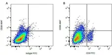
Figure B. RPE conjugated Mouse anti Bovine CD8 antibody, clone CC63 (MCA837PE) and FITC conjugated Mouse anti Bovine CD2 antibody, clone CC42 (MCA833F). All experiments performed on red cell lysed bovine blood gated on mononuclear cells.
Data acquired on the ZE5 Cell Analyzer.
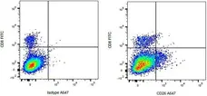
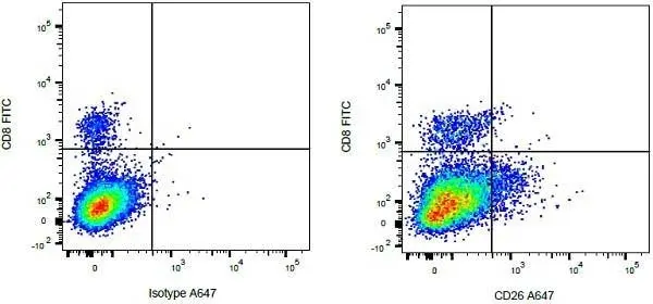
Figure B. FITC conjugated Mouse anti Bovine CD8 antibody, clone CC63 (MCA837F) and Alexa Fluor® 647 conjugated Mouse anti Bovine CD26 antibody, clone CC69 (MCA1652A647). All experiments performed on red cell lysed bovine blood gated on mononuclear cells.
Data acquired on the ZE5 Cell Analyzer.
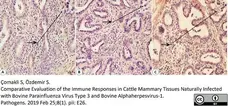
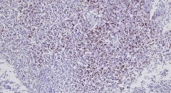
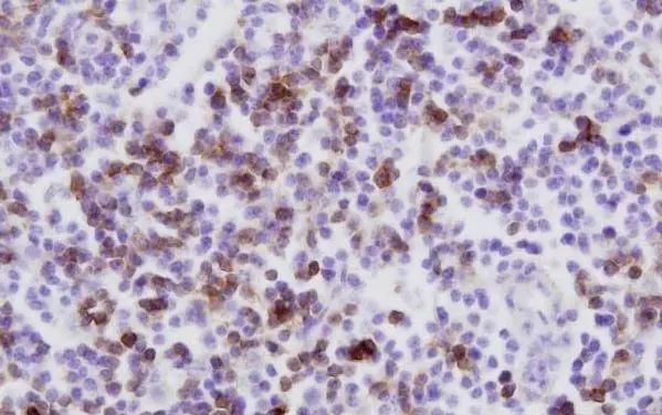
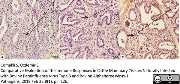
Mouse anti Bovine CD8 antibody, clone CC63 (MCA837GA) used for the identification of CD8 expressing lymphocytes in bovine tissue sections by immunohistochemistry on formalin fixed, paraffin sections.
Image caption:
Immunohistochemical staining of CD8 + T cells of the Bovine mammary tissue using paraffin-embedded sections (A–C): (A) The intermediate immunoreactivity of CD8 + T lymphocytes (arrow) in the interalveolar connective tissue with the monoclonal antibody specific for CD8+ T cells in Bovine mammary tissue infected with BPIV-3. (B) The low immunoreactivity of T lymphocytes (arrow) in the interalveolar connective tissue with the monoclonal antibody specific for CD8+ T cells in Bovine mammary tissue infected with BoHV-1. (C) The high reactivity distributional of T lymphocytes in the interalveolar connective tissue with the monoclonal antibody specific for CD8+ T cells (arrow) in Bovine mammary tissue infected with BoHV-1 and BPIV-3. IHC. Bar, 20μm.
From: Çomakli S, Özdemir S.
Comparative Evaluation of the Immune Responses in Cattle Mammary Tissues Naturally Infected with Bovine Parainfluenza Virus Type 3 and Bovine Alphaherpesvirus-1.
Pathogens. 2019; 8 (1). pii: E26.
doi: 10.3390/pathogens8010026.
This image is from an open access article distributed under the terms of a Creative Commons Attribution License.
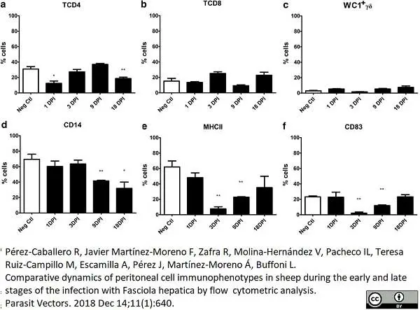
FITC conjugated Mouse anti Bovine CD8 antibody, clone CC63 (MCA837F) used for the evaluation of CD8 expression on ovine peritoneal cells by flow cytometry.
Image caption:
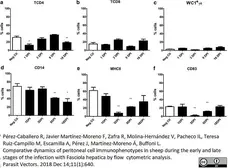
Percentage of cell populations in the peritoneal fluid during the early stages of infection. The specific cell populations are shown as follows: (a) TCD4; (b) TCD8; (c) WC1+γδ; (d) CD14; (e) MHCII; (f) CD83. Cell subsets of uninfected (Neg. Ctl) and infected sheep (Group 1) are shown in columns. Mann-Whitney U-test was used to compare data from Groups 1 and 3. Values represent the mean ± standard deviation (SD). Asterisks indicate different levels of significance between groups: *P ≤ 0.05, **P ≤ 0.01
From: Pérez-Caballero R, Javier Martínez-Moreno F, Zafra R, Molina-Hernández V, Pacheco IL, Teresa Ruiz-Campillo M, Escamilla A, Pérez J, Martnez-Moreno Á, Buffoni L.
Comparative dynamics of peritoneal cell immunophenotypes in sheep during the early and late stages of the infection with Fasciola hepatica by flow cytometric analysis.
Parasit Vectors. 2018 11(1):640.
doi: 10.1186/s13071-018-3250-5.
This image is from an open access article distributed under the terms of a Creative Commons Attribution License.
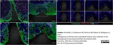
Mouse anti Bovine CD8 antibody, clone CC63 used for the identification of CD8 expressing cells in the bovine nasopharynx by immunofluorescence.
Image caption:
Nasopharyngeal primary infection sites of vaccinated cattle are infiltrated by CD8+/CD3- (presumptive NK) cells at 24 hpi.
Multichannel immunofluorescent technique. A). FMDV VP1 (red) protein within cytokeratin+ cells (green) in epithelial crypt of the nasopharyngeal mucosa of non-vaccinated steer at 24 hpi (animal number 3). CD8+ (aqua)/ CD3+ (purple) double-positive CTLs are present in the subepithelial compartment amongst larger populations of CD8-/CD3+ T-lymphocytes. 20x magnification, scale bar 50μm. B) 40x magnification of region identified in (A), demonstrating consistent co-localization of CD8 (aqua) with CD3 (purple). Scale bars 25μm. B) Select channels of image shown in (B). CD3+ cells (purple) include single-positive (T-lymphocytes) or CD8+/CD3+ double-positive (CTLs). CD8 (aqua) is exclusively detected in combination with CD3 (purple). Scale bars 25 μm. D) FMDV VP1 (red) protein co-localize with cytokeratin (green) in a focal surface erosion within follicle-associated epithelium of nasopharyngeal mucosa of vaccinated steer at 24 hpi (animal number 11). A distinct population of cells defined as presumptive NK-cells based on a CD8+ (aqua)/CD3- (purple) phenotype is present in submucosal and epithelial compartments surrounding the focus of infection. A smaller population of CTLs (CD8+/CD3+) is present amongst abundant non-CTL T-lymphocytes (CD8-/CD3+). 20x magnification, scale bar 50μm. E) Higher magnification of region identified in showing CD8+/CD3- cells (aqua) representing presumptive NK cells in close proximity of FMDV infected focus. 40x magnification, scale bars 25 μm F) Individual channels of image shown in (E) demonstrating inconsistent co-localization of CD8 (aqua) and CD3 (purple). Scale bar 25 μm.
From: Stenfeldt C, Eschbaumer M, Pacheco JM, Rekant SI, Rodriguez LL, Arzt J (2015)
Pathogenesis of Primary Foot-and-Mouth Disease Virus Infection in the Nasopharynx of Vaccinated and Non-Vaccinated Cattle.
PLoS ONE 10(11): e0143666.
This image is from an open access article distributed under terms of a Creative Commons Attribution License.
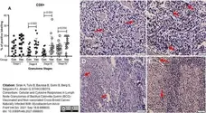
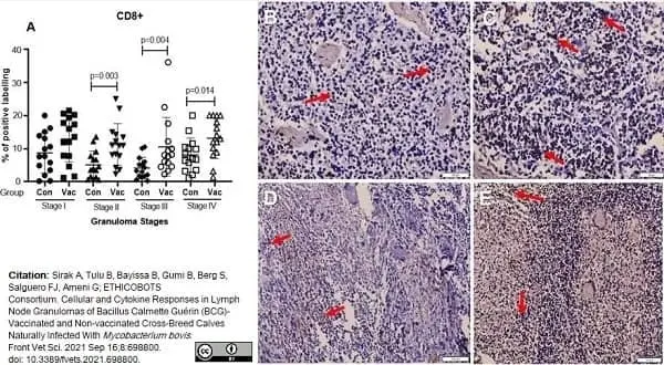
Mouse anti Bovine CD8 antibody, clone CC63 (MCA837GA) used to evaluate CD8 expression in bovine granulomatous lesions by immunohistochemistry on formalin fixed, paraffin embedded tissue sections.
Image caption:
Immunolabelling for CD8+ cells (ABC IHC stain, DAB brown chromogen). (A) Comparison of CD8+ cell distribution in different granuloma stages of BCG vaccinated (Vac) and non-vaccinated (Con) calves. (B) Stage I granuloma from non-vaccinated calf showing very few stained CD8+ cells (arrows). (C) Stage I granuloma from BCG vaccinated calf showing stained CD8+ cells (arrows). (D) Stage III granuloma from non-vaccinated calf showing few stained CD8+ cells (arrows). (E) Stage III granuloma from vaccinated calf showing abundance of CD8+ cells (arrows). Bar equals 50μm in (B,C) and 100μm in (D,E).
From: Sirak A, Tulu B, Bayissa B, Gumi B, Berg S, Salguero FJ, Ameni G; ETHICOBOTS Consortium.
Cellular and Cytokine Responses in Lymph Node Granulomas of Bacillus Calmette Guérin (BCG)-Vaccinated and Non-vaccinated Cross-Breed Calves Naturally Infected With Mycobacterium bovis.
Front Vet Sci. 2021 Sep 16;8:698800.
doi: 10.3389/fvets.2021.698800.
This image is from an open access article distributed under terms of a Creative Commons Attribution License.
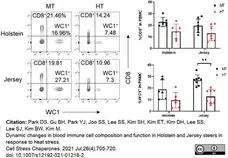
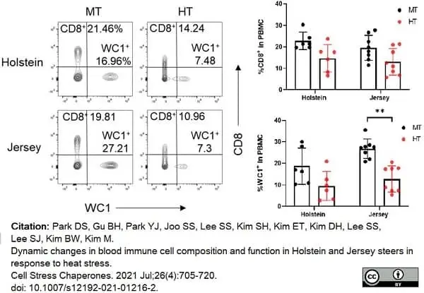
Alexa Fluor® 647 conjugated Mouse anti Bovine CD8 antibody, clone CC63 (MCA837A647) used to identify T cell populations in bovime blood samples by flow cytometry.
Image caption:
Changes in T and B lymphocytes among PBMCs in Holstein and Jersey steers subjected to heat stress. Flow cytometry analysis to identify lymphocytes subset population. Heat stress caused reduction in lymphocyte populations in the PBMCs of both Holstein and Jersey steers. The lymphocytes comprised CD4+ T cells. MT indicates moderate THI condition and HT indicates high THI condition. * = P <0.05, ** = P <0.01
From: Park DS, Gu BH, Park YJ, Joo SS, Lee SS, Kim SH, Kim ET, Kim DH, Lee SS, Lee SJ, Kim BW, Kim M.
Dynamic changes in blood immune cell composition and function in Holstein and Jersey steers in response to heat stress.
Cell Stress Chaperones. 2021 Jul;26(4):705-20.
doi: 10.1007/s12192-021-01216-2.
This image is from an open access article distributed under terms of a Creative Commons Attribution License.
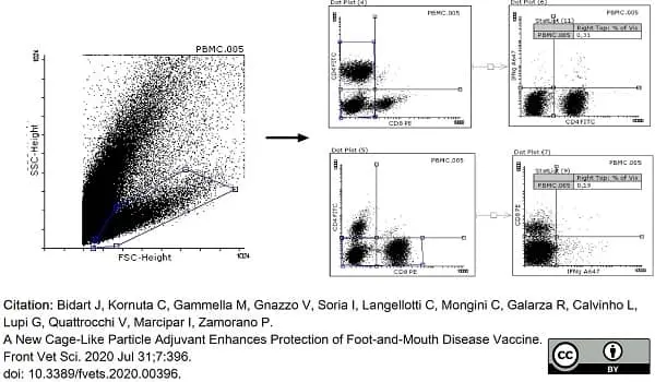
R-Phycoerythrin conjugated Mouse anti Bovine CD8 antibody, clone CC63 (MCA837PE) used to evaluate CD8 expression on bovine lymphocytes by flow cytometry.
Image caption:
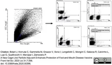
Representative dot plots, from bovine PBMCs, used for selecting the lymphocyte region based on side sideward scatter (SSC) on the y-axis and forward side scatter (FSC) on the x-axis. Then, we selected the CD8 region based on fluorescence anti-CD8 stain on the y-axis and CD4 region based on fluorescence anti-CD4 stain on the x-axis. PBMCs incubated for 18 h with iFMDV are shown.
From: Bidart J, Kornuta C, Gammella M, Gnazzo V, Soria I, Langellotti C, Mongini C, Galarza R, Calvinho L, Lupi G, Quattrocchi V, Marcipar I, Zamorano P.
A New Cage-Like Particle Adjuvant Enhances Protection of Foot-and-Mouth Disease Vaccine.
Front Vet Sci. 2020 Jul 31;7:396.
doi: 10.3389/fvets.2020.00396.
This image is from an open access article distributed under terms of a Creative Commons Attribution License.
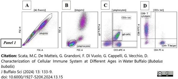
Fitc conjugated Mouse anti Bovine CD8 antibody, clone CC63 (MCA837F) used to label lymphocyts from buffalo blood by flow cytometry.
Image caption:
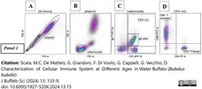
Gating strategy used in this study: (A) A dot plot FSC-A vs. FSC-H on All events was used to exclude doublets, a morphological gate was drawn to highlight single cells (singlets); (B) a dot plot FSC-A vs. SSC-Aon singlets was used to identify leukocyte populations: in Panel 1 a morphological gate was drawn to highlight lymphocytes (B), (C) a dot plot CD3APC-A vs. γδ TCRPC7-A on lymphocytes was used to identify total and γδ T lymphocytes; (D) a dot plot CD4PE-A vs. CD8FITC-Aon CD3+ tot was used to identify the CD4+ helper and CD8+ cytotoxic subsets.
From: Scatà, M. C.De Matteis, G. Grandoni, F. Di Vuolo, G. .Cappelli, G. Vecchio, D. (2024).
Characterization of Cellular Immune System at Different Ages in Water Buffalo (Bubalus bubalis).
Journal of Buffalo Science, 13, 133–139.
doi: 10.6000/1927-520X.2024.13.15
This image is from an open access article distributed under terms of a Creative Commons Attribution License.
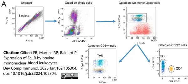
Alexa Fluor 700 conjugated Mouse anti Bovine CD8 antibody, clone CC63 (MCA837A700) used to label T lymphocytes in bovine blood by flow cytometry.
Image caption:
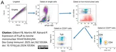
Expression of the FcμR by blood T lymphocytes. A) Gating strategy to distinguish viable CD3pos T cells.
From: Gilbert FB, Martins RP, Rainard P.
Expression of FcμR by bovine mononuclear blood leukocytes.
Dev Comp Immunol. 2025 Jan;162:105304.
doi: 10.1016/j.dci.2024.105304.
This image is from an open access article distributed under terms of a Creative Commons Attribution License.
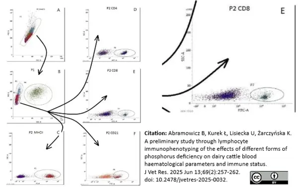
Alexa Fluor® 700 conjugated Mouse anti Bovine CD8 antibody, clone CC63 (MCA1653A647) used for gating of bovine cell populations by flow cytometry. [ Enlargement on right].
Image caption:
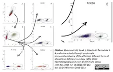
Gating strategy for immunophenotyping of bovine peripheral blood lymphocytes. A – single-cell gating; B – lymphocyte gating; C–F – fluorescence cytograms of positive cells.
From: Abramowicz B, Kurek Ł, Lisiecka U, Żarczyńska K.
A preliminary study through lymphocyte immunophenotyping of the effects of different forms of phosphorus deficiency on dairy cattle blood haematological parameters and immune status.
J Vet Res. 2025 Jun 13;69(2):257-262.
doi: 10.2478/jvetres-2025-0032.
This image is from an open access article distributed under terms of a Creative Commons Attribution License.

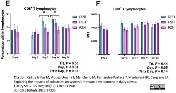
RPE conjugated Mouse anti Bovine CD8 antibody, clone CC63 (MCA837PE) used for the evaluation of CD8+ cells in bovine samples.
Image caption:
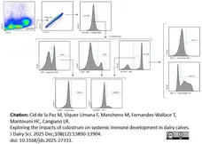
Changes in the proportion of CD8+ T lymphocytes (E) were evaluated in colostrum-deprived calves (DPR) and calves fed fresh colostrum (FSH) or frozen colostrum (FZN). Changes in the receptor expression based on median fluorescence intensity (MFI) of the coreceptor CD8 (F) in colostrum-deprived calves, fresh colostrum calves, and frozen colostrum calves from d 0 before treatment to d 28. Trt = treatment. Asterisks indicate a treatment-by-day interaction at P ≤0.05; pound signs indicate a treatment-by-day interaction at P ≤0.10. Values are mean ± SE; error bars represent SEM.
From: Cid de la Paz M, Viquez-Umana F, Mancheno M, Fernandez-Wallace T, Mantovani HC, Cangiano LR.
Exploring the impacts of colostrum on systemic immune development in dairy calves.
J Dairy Sci. 2025 Dec;108(12):13890-13904.
doi: 10.3168/jds.2025-27333.
This image is from an open access article distributed under terms of a Creative Commons Attribution License.
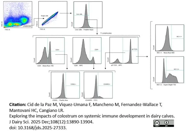
RPE conjugated Mouse anti Bovine CD8 antibody, clone CC63 (MCA837PE) used in the gating strategy for bovine lymphocytes by flow cytometry.
Image caption:
Gating strategy for T lymphocytes. Lymphocytes were identified by a scatter plot, followed by single cells, live cells, and then CD4+ Lymphocytes, CD8+ Lymphocytes, CD4+ Lymphocytes, γδ T Lymphocytes, WC1.1+ γδ T Lymphocytes, WC1.2+ γδ T Lymphocytes, CD62L, and CD45RO. SSC = side scatter; FSC = forward scatter; A = signal area; H = signal height.
From: Cid de la Paz M, Viquez-Umana F, Mancheno M, Fernandez-Wallace T, Mantovani HC, Cangiano LR.
Exploring the impacts of colostrum on systemic immune development in dairy calves.
J Dairy Sci. 2025 Dec;108(12):13890-13904.
doi: 10.3168/jds.2025-27333.
This image is from an open access article distributed under terms of a Creative Commons Attribution License.
Filter by Application:
F C P IF Reset| Mouse anti Bovine CD8 antibody, clone CC63 reacts with the bovine CD8 antigen expressed by a subset of T lymphocytes. The antibody precipitates molecules of ~34 kDa and ~38 kDa under reducing conditions. Clone CC63 has been reported as being suitable for use on formalin dichromate (FD5) fixed paraffin embedded tissue with amplification and antigen retrieval techniques (Gutierrez et al. 1999). |
- Target Species
- Bovine
- Species Cross-Reactivity
-
Target Species Cross Reactivity Sheep Goat - N.B. Antibody reactivity and working conditions may vary between species.
- Product Form
- Purified IgG conjugated to R. Phycoerythrin (RPE) - lyophilized
- Reconstitution
- Reconstitute with 1 ml distilled water
- Preparation
- Purified IgG prepared by affinity chromatography on Protein A from tissue culture supernatant
- Buffer Solution
- Phosphate buffered saline
- Preservative Stabilisers
0.09% Sodium Azide 1% Bovine Serum Albumin 5% Sucrose - Fusion Partners
- Spleen cells from an immunized mouse were fused with cells of the mouse NS1 myeloma cell line.
- Max Ex/Em
-
Fluorophore Excitation Max (nm) Emission Max (nm) RPE 488nm laser 496 578 - Regulatory
- For research purposes only
- Guarantee
- 12 months from date of despatch
This product is shipped at ambient temperature.
Prior to reconstitution store at +4oC. Following reconstitution store at +4oC.
This product should be stored undiluted.
DO NOT FREEZE. This product is photosensitive and should be protected from light. Should this product contain a precipitate we recommend microcentrifugation before use.
Prior to reconstitution store at +4oC. Following reconstitution store at +4oC.
This product should be stored undiluted.
DO NOT FREEZE. This product is photosensitive and should be protected from light. Should this product contain a precipitate we recommend microcentrifugation before use.
This product has been reported to work in the following applications. This information is derived from testing within our laboratories, peer-reviewed publications or personal communications from the originators. Please refer to references indicated for further information. For general protocol recommendations, please visit the antibody protocols page.
| Application Name | Verified | Min Dilution | Max Dilution |
|---|---|---|---|
| Flow Cytometry |  |
Neat |
Where this antibody has not been tested for use in a particular technique this does not necessarily exclude its use in such procedures. Suggested working dilutions are given as a guide only. It is recommended that the user titrates the antibody for use in their own system using appropriate negative/positive controls.
- Flow Cytometry
- Use 10ul of the suggested working dilution to label 106 cells in 100ul.
How to Use the Spectraviewer
Watch the Tool Tutorial Video ▸- Start by selecting the application you are interested in, with the option to select an instrument from the drop down menu or create a customized instrument
- Select the fluorophores or fluorescent proteins you want to include in your panel to check compatibility
- Select the lasers and filters you wish to include
- Select combined or multi-laser view to visualize the spectra
| Description | Product Code | Applications | Pack Size | List Price | Your Price | Quantity | |
|---|---|---|---|---|---|---|---|
| Mouse IgG2a Negative Control:RPE | MCA929PE | F | 100 Tests | Log in | |||
| List Price | Your Price | ||||||
| Log in | |||||||
| Description | Mouse IgG2a Negative Control:RPE | ||||||
References for CD8 antibody
-
MacHugh, N.D. & Sopp P (1991) Individual antigens of cattle. Bovine CD8 (BoCD8).
Vet Immunol Immunopathol. 27 (1-3): 65-9. -
Gutierrez, M. et al. (1999) The detection of CD2+, CD4+, CD8+, and WC1+ T lymphocytes, B cells and macrophages in fixed and paraffin embedded bovine tissue using a range of antigen recovery and signal amplification techniques.
Vet Immunol Immunopathol. 71 (3-4): 321-34. -
Winkler, M.T. et al. (1999) Bovine herpesvirus 1 can infect CD4(+) T lymphocytes and induce programmed cell death during acute infection of cattle.
J Virol. 73 (10): 8657-68. -
Winkler, M.T. et al. (2000) Persistence and reactivation of bovine herpesvirus 1 in the tonsils of latently infected calves.
J Virol. 74 (11): 5337-46. -
Twizere, J.C. et al. (2000) Discordance between bovine leukemia virus tax immortalization in vitro and oncogenicity in vivo.
J Virol. 74 (21): 9895-902. -
Harris, J. et al. (2002) Expression of caveolin by bovine lymphocytes and antigen-presenting cells.
Immunology. 105: 190-5. -
Toman, M. et al. (2003) Immunological characteristics of cale with Mycobacterium avium subsp. paratuberculosis infection
Vet. Med. – Czech, 48, 2003: 147–54. -
Vordermeier, H.M. et al. (2004) Cellular immune responses induced in cattle by heterologous prime-boost vaccination using recombinant viruses and bacille Calmette-Guérin.
Immunology. 112: 461-70.
View The Latest Product References
-
Vitale, F. et al. (2006) ESAT-6 peptide recognition by bovine CD8+ lymphocytes of naturally infected cows in herds from southern Italy.
Clin Vaccine Immunol. 13: 530-3. -
Fulton, B.E. Jr. et al. (2006) Dissemination of bovine leukemia virus-infected cells from a newly infected sheep lymph node.
J Virol. 80: 7873-84. -
Liebana, E. et al. (2007) Distribution and activation of T-lymphocyte subsets in tuberculous bovine lymph-node granulomas.
Vet Pathol. 44: 366-72. -
Foulon, E. & Foucras, G. (2008) Two populations of ovine bone marrow-derived dendritic cells can be generated with recombinant GM-CSF and separated on CD11b expression.
J Immunol Methods. 339 (1): 1-10. -
Sidders, B. et al. (2008) Screening of highly expressed mycobacterial genes identifies Rv3615c as a useful differential diagnostic antigen for the Mycobacterium tuberculosis complex.
Infect Immun. 76: 3932-9. -
Lynch, E.M. et al. (2010) Effect of abrupt weaning at housing on leukocyte distribution, functional activity of neutrophils, and acute phase protein response of beef calves.
BMC Vet Res. 6: 39. -
Coad, M. et al. (2010) Repeat tuberculin skin testing leads to desensitisation in naturally infected tuberculous cattle which is associated with elevated interleukin-10 and decreased interleukin-1 beta responses.
Vet Res. 41: 14. -
Constantinoiu, C.C. et al. (2010) Local immune response against larvae of Rhipicephalus (Boophilus) microplus in Bos taurus indicus and Bos taurus taurus cattle.
Int J Parasitol. 40: 865-75. -
La Manna, M.P. et al. (2011) Expansion of intracellular IFN-γ positive lymphocytes during Mycoplasma agalactiae infection in sheep.
Res Vet Sci. 91 (3): e64-7. -
Sanchez, J. et al. (2011) Microscopical and immunological features of tuberculoid granulomata and cavitary pulmonary tuberculosis in naturally infected goats.
J Comp Pathol. 145 (2-3): 107-17. -
Lacroux, C. et al. (2012) Prionemia and leukocyte-platelet-associated infectivity in sheep transmissible spongiform encephalopathy models.
J Virol. 86 (4): 2056-66. -
Brodzki, P. et al. (2014) Phenotyping of leukocytes and granulocyte and monocyte phagocytic activity in the peripheral blood and uterus of cows with endometritis.
Theriogenology. 82 (3): 403-10. -
Silva, A.P. et al. (2015) Encapsulated Brucella ovis Lacking a Putative ATP-Binding Cassette Transporter (&Detla;abcBA) Protects against Wild Type Brucella ovis in Rams.
PLoS One. 10 (8): e0136865. -
Leite FL et al. (2015) ZAP-70, CTLA-4 and proximal T cell receptor signaling in cows infected with Mycobacterium avium subsp. paratuberculosis.
Vet Immunol Immunopathol. 167 (1-2): 15-21. -
Stenfeldt, C. et al. (2015) Pathogenesis of Primary Foot-and-Mouth Disease Virus Infection in the Nasopharynx of Vaccinated and Non-Vaccinated Cattle.
PLoS One. 10 (11): e0143666. -
Romero-Palomo, F. et al. (2017) Immunopathologic Changes in the Thymus of Calves Pre-infected with BVDV and Challenged with BHV-1.
Transbound Emerg Dis. 64 (2): 574-84. -
Schmidt, N. et al. (2018) Decreased STEC shedding by cattle following passive and active vaccination based on recombinant Escherichia coli Shiga toxoids.
Vet Res. 49 (1): 28. -
Pérez-caballero, R. et al. (2018) Comparative dynamics of peritoneal cell immunophenotypes in sheep during the early and late stages of the infection with Fasciola hepatica by flow cytometric analysis.
Parasit Vectors. 11 (1): 640. -
Benedictus, L. et al. (2019) Immunization of young heifers with staphylococcal immune evasion proteins before natural exposure to Staphylococcus aureus induces a humoral immune response in serum and milk.
BMC Vet Res. 15 (1): 15. -
Nakajima, N. et al. (2019) Effects of direct exposure to cold weather under grazing in winter on the physiological, immunological, and behavioral conditions of Japanese Black beef cattle in central Japan.
Anim Sci J. 90 (8): 1033-41. -
de Araújo, F.F. et al. (2019) Distinct immune response profile during Rhipicephalus (Boophilus) microplus. infestations of guzerat dairy herd according to the maternal lineage ancestry (mitochondrial DNA).
Vet Parasitol. 273: 36-44. -
Kolar, Q.K. et al. (2020) Anatomical distribution of respiratory tract leukocyte cell subsets in neonatal calves.
Vet Immunol Immunopathol. 227: 110090. -
Risalde, M.A. et al. (2020) BVDV permissiveness and lack of expression of co-stimulatory molecules on PBMCs from calves pre-infected with BVDV.
Comp Immunol Microbiol Infect Dis. 68: 101388. -
Bidart, J. et al. (2020) A New Cage-Like Particle Adjuvant Enhances Protection of Foot-and-Mouth Disease Vaccine.
Front Vet Sci. 7: 396. -
Brodzki, P. et al. (2020) Selected leukocyte subpopulations in peripheral blood and uterine washings in cows before and after intrauterine administration of cefapirin and methisoprinol.
Anim Sci J. 91 (1): e13306. -
Bloomer, S.A. et al. (2020) Aging results in accumulation of M1 and M2 hepatic macrophages and a differential response to gadolinium chloride.
Histochem Cell Biol. 153 (1): 37-48. -
Gondaira, S. et al. (2020) Immunosuppression in Cows following Intramammary Infusion of Mycoplasma bovis.
Infect Immun. 88 (3) :e00521-19. -
Damani-Yokota, P. et al. (2021) Transcriptional programming and gene regulation in WC1+ γδ T cell subpopulations.
Mol Immunol. 142: 50-62. -
Sirak, A. et al. (2021) Cellular and Cytokine Responses in Lymph Node Granulomas of Bacillus Calmette Guérin (BCG)-Vaccinated and Non-vaccinated Cross-Breed Calves Naturally Infected With Mycobacterium bovis.
Front Vet Sci. 8: 698800. -
Colombatti, M.O. et al. (2021) Evaluation of a virulent strain of Mycobacterium avium subsp. paratuberculosis used as a heat-killed vaccine.
Vaccine. 39 (51): 7401-12. -
Park, D.S. et al. (2021) Dynamic changes in blood immune cell composition and function in Holstein and Jersey steers in response to heat stress.
Cell Stress Chaperones. 26 (4): 705-20. -
Nashiruddullah, N. et al. (2021) Dermal Response to Experimental Orfvirus (ORFV) Infection in Goats, Mice and Rabbit
Indian J Anim Res. 56 (8): B-4266 1003-9. -
Kato-Mori, Y. et al. (2021) Characterization of a variant CD4 molecule in Japanese Black cattle.
Vet Immunol Immunopathol. 232: 110167. -
Casaro, S. et al. (2022) Flow cytometry panels for immunophenotyping dairy cattle peripheral blood leukocytes
VetImmunol Immunopathol. 248: 110417. -
Elsayed, M.S.A.E. et al. (2022) Real-time PCR using atpE, conventional PCR targeting different regions of difference, and flow cytometry for confirmation of Mycobacterium bovis. in buffaloes and cattle from the Delta area of Egypt.
BMC Microbiol. 22 (1): 154. -
Korbonits, L. et al. (2022) Mycobacterium avium subsp. paratuberculosis Infected Cows Reveal Divergent Immune Response in Bovine Peripheral Blood Derived Lymphocyte Proteome.
Metabolites. 12 (10): 924. -
Tucker, N. et al. (2023) Bovine blood and milk T-cell subsets in distinct states of activation and differentiation during subclinical Staphylococcus aureus mastitis.
J Reprod Immunol. 156: 103826. -
Özbek, M. & Bayraktaroğlu, A.G. (2019) Developmental study on the ileal Peyer's patches of sheep, and cytokeratin-18 as a possible marker for M cells in follicle associated epithelium.
Acta Histochem. 121 (3): 311-22. -
Benedictus, L. et al. (2019) Immunization of young heifers with staphylococcal immune evasion proteins before natural exposure to Staphylococcus aureus induces a humoral immune response in serum and milk.
BMC Vet Res. 15 (1): 15. -
Yang, L. et al. (2018) Association of the expression of Th cytokines with peripheral CD4 and CD8 lymphocyte subsets after vaccination with FMD vaccine in Holstein young sires.
Res Vet Sci. 119: 79-84. -
Andrés, S. et al. (2024) Essential oil supplementation in milk replacers: short- and long-term impacts on feed efficiency, the faecal microbiota and the plasma metabolome in dairy calves.
J Dev Orig Health Dis. : 1-11. -
Seemann, L. et al. (2024) Dietary L-carnitine supplementation modifies blood parameters of mid-lactating dairy cows during standardized lipopolysaccharide-induced inflammation.
Front Immunol. 15: 1390137. -
Hong, S. et al. (2024) Impact of an Injectable Trace Mineral Supplement on the Immune Response and Outcome of Mannheimia haemolytica Infection in Feedlot Cattle.
Biol Trace Elem Res. Jun 10 [Epub ahead of print]. -
Müller, C.B.M. et al. (2024) Interactions between rumen epithelium-associated microbiota and host immunological and metabolic adaptations in response to different milk replacer feeding intensities in dairy calves
Animal Nutrition. 7 Sept [Epub ahead of print]. -
Brodzki, P. et al. (2024) The influence of probiotic administration on selected leukocyte subpopulations and the serum amyloid A concentration in the peripheral blood of dairy cows during different lactation periods
Journal of Veterinary Research. 09 Oct [Epub ahead of print]. -
Scatà, C.M. et al. (2024) Characterization of Cellular Immune System at Different Ages in Water Buffalo (Bubalus bubalis)
Journal of Buffalo Science. 13: 133-139. -
Gilbert, F.B. et al. (2025) Expression of FcμR by bovine mononuclear blood leukocytes.
Dev Comp Immunol. 162: 105304. -
Díaz-Otero, F. et al. (2025) Comparative longitudinal analysis of T lymphocyte subpopulations in calves vaccinated with different doses of BCG-Phipps or with culture filtrate protein extract of Mycobacterium bovis in a natural transmission setting.
BMC Vet Res. 21 (1): 78. -
Abramowicz, B. et al. (2025) A preliminary study through lymphocyte immunophenotyping of the effects of different forms of phosphorus deficiency on dairy cattle blood haematological parameters and immune status.
J Vet Res. 69 (2): 257-62. -
Cid de la Paz, M. et al. (2025) Exploring the impacts of colostrum on systemic immune development in dairy calves.
J Dairy Sci. 108 (12): 13890-13904.
- RRID
- AB_2075540
- UniProt
- P31783
- Entrez Gene
- CD8A
- GO Terms
- GO:0016021 integral to membrane
MCA837PE
If you cannot find the batch/lot you are looking for please contact our technical support team for assistance.
View more products with CD8 specificity
Please Note: All Products are "FOR RESEARCH PURPOSES ONLY"
View all Anti-Bovine ProductsAlways be the first to know.
When we launch new products and resources to help you achieve more in the lab.
Yes, sign me up