F4/80 antibody | Cl:A3-1
Rat anti Mouse F4/80
- Product Type
- Monoclonal Antibody
- Clone
- Cl:A3-1
- Isotype
- IgG2b
- Specificity
- F4/80
| Product Code | Applications | Pack Size | List Price | Your Price | Qty | ||||||||||
|---|---|---|---|---|---|---|---|---|---|---|---|---|---|---|---|
|
|||||||||||||||
|
|||||||||||||||
|
|||||||||||||||
|
|||||||||||||||


Rat anti Mouse F4/80 antibody, clone A3-1 (MCA497G) used for the detection of macrophages in mouse lung and pancreas by immunohistochemistery on formalin fixed, paraffin tissue sections.
Image caption:
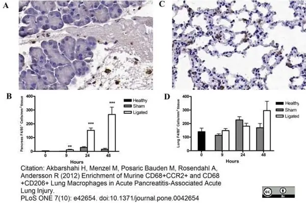
Changes in pancreatic and lung F4/80+ macrophage number during acute pancreatitis. Representative photomicrographs of pancreas (A) and lung (C) sections stained for F4/80 as indicator of macrophage infiltration (brown) 24 h after pancreatitis induction (original magnification, ×20). Quantification of F4/80+ cells showed a significantly increased enrichment in the pancreas from 9 h following pancreatitis compared to sham controls (B). No significant difference was observed in the number of lung F4/80+ cells between pancreatitis and sham control groups (D). Bars show mean ± SEM, n = 10 per group. **P<0.01, ***P<0.001, by two-tailed Student t-test.
From: Akbarshahi H, Menzel M, Posaric Bauden M, Rosendahl A, Andersson R (2012)
Enrichment of Murine CD68+CCR2+ and CD68+CD206+ Lung Macrophages in Acute Pancreatitis-Associated Acute Lung Injury.
PLoS ONE 7(10): e42654.
This image is from an open access article distributed under the terms of the Creative Commons Attribution License.


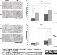
Rat anti Mouse F4/80 antibody, clone A3-1 (MCA497G) used for the identification of macrophages in injured soleus muscle by immunohistochemistry of formalin fixed, paraffin embedded tissue sections.
Image caption:
PCNA and F4/80 immunohistochemical expression after muscular injury. WT: wild type, KO: knockout. (A) Immunohistochemistry of PCNA. (Scale bar = 50μm), (B) Quantitative analysis of PCNA-positive cells, (C) Immunohistochemistry of F4/80. (Scale bar = 50μm), (D) Quantitative analysis of F4/80-positive cells. (B and D) Both PCNA and F4/80 expression in p21KO mice at 3 days after injury were the highest (p < 0.05).
From: Chinzei N, Hayashi S, Ueha T, Fujishiro T, Kanzaki N, Hashimoto S, et al. (2015)
P21 Deficiency Delays Regeneration of Skeletal Muscular Tissue.
PLoS ONE 10(5): e0125765.
This image is from an open access article distributed under the terms of the Creative Commons Attribution License.
Rat anti Mouse F4/80 antibody, clone A3-1 (MCA497R) used for the detection of infiltrating macrophages in tumor tissue by immunofluorescence.
Image caption:
Presence of inflammatory cells in tumor tissue. Macrophage and neutrophil infiltration are unaffected by loss of serglycin in RIP1-Tag2 tumors. Tumor sections from 15w RTposSGwt and RTposSGko mice were immunostained from the neutrophil and macrophage markers Gr-1 (a) and F4/80 (b) respectively. For Gr-1, the number of positive cells/mm2 was calculated (c) and there was no difference in infiltration of neutrophils between the two groups. For F4/80, the area of positive staining was measured (d) and although there was a slight trend to decreased macrophage infiltration in serglycin deficient animals, this was not significant. Each data point in represents an individual animal. Statistical analysis was performed using a two-tailed Mann-Whitney test. Error bars represent mean ± SEM.
From: Hamilton A, Basic V, Andersson S, Abrink M, Ringvall M (2015)
Loss of Serglycin Promotes Primary Tumor Growth and Vessel Functionality in the RIP1-Tag2 Mouse Model for Spontaneous Insulinoma Formation.
PLoS ONE 10(5): e0126688.
This image is from an open access article distributed under the terms of the Creative Commons Attribution License.

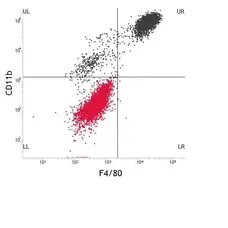
Rat anti Mouse F4/80 antibody, clone A3-1 (MCA497GA) used for the detection of macrophage processes in mouse kidney tissue sections by immunofluorescence.
Image caption:
Dual immunofluorescence staining showing the association of F4/80+ cells with deposited C3. Shown is a representative immunofluorescence micrograph of a Crry−/−C3−/− kidney 7 days after transplantation into a wildtype recipient. Considerable deposition of C3 (green) is evident in the basolateral aspects of tubules, along with closely approximated F4/80+ cellular processes (red).
From: Chaves LD, Bao L, Wang Y, Chang A, Haas M, Quigg RJ (2014)
Loss of CD11b Exacerbates Murine Complement-Mediated Tubulointerstitial Nephritis.
PLoS ONE 9(3): e92051.
This image is from an open access article distributed under the terms of the Creative Commons Attribution License.

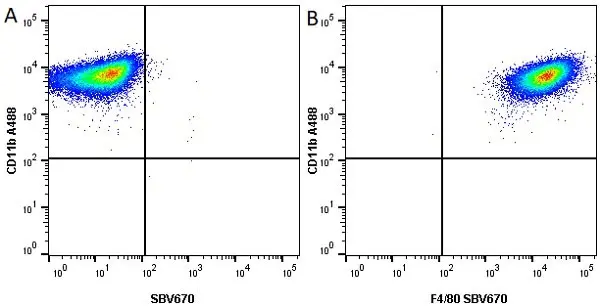
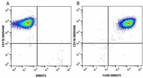
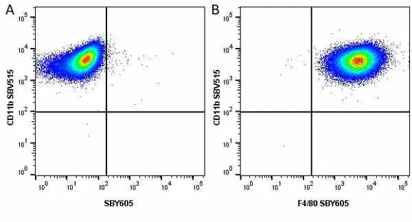
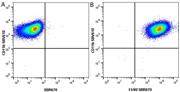
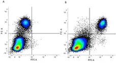
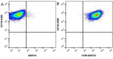
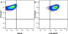
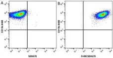
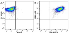
FITC-conjugated Rat anti Mouse F4/80 antibody, clone A3-1 (MCA497F) used for the detection of F4/80 expressing cells in peritoneal cell exudates by flow cytometry.
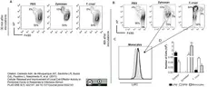
Image caption:
Identification of resident peritoneal Mø subsets. PC from C57BL/6 were harvested and stained with fluorochrome-labeled antibodies directed against F4/80, CD19, CD11c and IAb for flow cytometry analysis. (A) Doublet cells were excluded according to forward scatter profiles (FSC-A and FSC-H). Subsequently, (B) CD19 high cells and (C) CD11c high cells were also excluded, and (D) F4/80+ cells were selected. (E) F4/80 and IAb expression defined three populations: LPM (F4/80highIAb-neg), SPM (F4/80lowIAb-high) and granulocytes (F4/80lowIAb-neg). These three subpopulations were purified by cell sorting on a FACS Vantage, and their morphology was evaluated ex vivo from cytospin slides (F), or after in vitro culture in chamber slides for 12 h (G). Slides were stained with hematoxylin and eosin (H&E) and analyzed by optical microscopy (40×).
From: Cassado AdA, de Albuquerque JAT, Sardinha LR, Buzzo CdL, Faustino L, Nascimento R, et al. (2011)
Cellular Renewal and Improvement of Local Cell Effector Activity in Peritoneal Cavity in Response to Infectious Stimuli.
PLoS ONE 6(7): e22141.
This image is from an open access article distributed under the terms of the Creative Commons Attribution License.
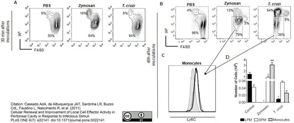
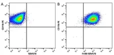
FITC conjugated Rat anti Mouse F4/80 antibody, clone A3-1 (MCA497F) used for the detection of F4/80 expressing cells in peritoneal cell exudates by flow cytometry.
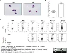
Image caption:
Zymosan and T. cruzi injection alters the Mø compartment of PerC. C57BL/6 mice were injected i.p. with zymosan (1 mg/mouse) or T. cruzi (106 parasites/mouse) and at 30 min (A) or 48 h (B) after stimulation, PC from naive and injected mice were harvested and stained as described in M&M. Sequential gates were made as shown in Fig. S1. Plots show the frequencies of each subpopulation. (C) F4/80lowMHCIIint cells present within PerC 48 h after injections were evaluated according the expression of Ly6C. Gray lines represent FMO [23], [25], the black lines show F4/80lowMHCIIint cells from zymosan- (hairline) or T. cruzi-(bold line)-exposed PerC. (D) Total numbers of SPM, LPM and monocytes 48 h after zymosan or T. cruzi exposure (within Mø gate) are shown in panel. Data are representative of more than 3 independent experiments.
From: Cassado AdA, de Albuquerque JAT, Sardinha LR, Buzzo CdL, Faustino L, Nascimento R, et al. (2011)
Cellular Renewal and Improvement of Local Cell Effector Activity in Peritoneal Cavity in Response to Infectious Stimuli.
PLoS ONE 6(7): e22141.
This image is from an open access article distributed under the terms of the Creative Commons Attribution License.
FITC-conjugated Rat anti Mouse F4/80 antibody, clone A3-1 (MCA497F) used for the detection of F4/80 expressing cells in peritoneal cell exudates by flow cytometry.
Image caption:
SPM are more responsive than LPM and monocytes to infectious stimuli. C57BL/6 mice were injected i.p. with zymosan (1 mg/mouse) or T. cruzi (106 parasites/mouse) and PCs were harvested 30 min or 48 h after stimulation. (A) SPM and LPM from C57BL/6 zymosan-exposed mice were FACS-sorted 30 min after injection and the presence of internalized zymosan particles was observed by optical microscopy. Slides were made with 105 cells from purified Mø subsets, stained with H&E and analyzed by optical microscopy (40×). (B) Numbers represent the mean ±SD of internalized zymosan particles per cell in each Mø subset. *** p<0.001 when compared to the LPM group. (C) PC from control or 48 h-exposed mice were cultured for 6 h in the presence of brefeldin A with or without LPS (1 μg/ml) plus rIFN-γ (5 ng/ml). Titles above plots indicate in vivo - in vitro stimulations. Values inside gates represent the frequencies of IL-12-producing cells in each subpopulation. Data are representative of more than 3 independent experiments.
From: Cassado AdA, de Albuquerque JAT, Sardinha LR, Buzzo CdL, Faustino L, Nascimento R, et al. (2011)
Cellular Renewal and Improvement of Local Cell Effector Activity in Peritoneal Cavity in Response to Infectious Stimuli.
PLoS ONE 6(7): e22141.
This image is from an open access article distributed under the terms of the Creative Commons Attribution License.
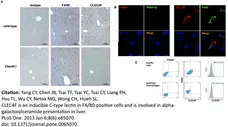
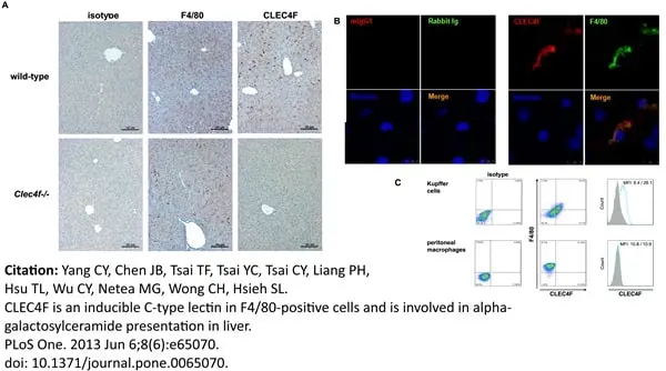
Rat anti Mouse F4/80 antibody, clone A3-1 (MCA497GA) used for the evaluation of F4/80 expression on isolated Kuppfer and peritoneal cells by flow cytometry.
Image caption:
CLEC4F is co-expressed with F4/80 on liver Kupffer cells.
(A) CLEC4F and F4/80 immunohistochemistry of parafilm-embedded liver sections from wild-type and Clec4f−/− mice. (B) Double immunofluorescence of CLEC4F and F4/80 in wild-type livers was performed. Nuclei were counterstained with Hoechst 33342. Signals were determined by confocal microscope (magnification 10×63). (C) Coexpression of CLEC4F and F4/80 on Kupffer cells, but not peritoneal macrophages. Cells were double stained with Alexa Fluor 647-conjugated anti-F4/80 and PE-conjugated anti-CLEC4F mAb. Alexa Fluor 647-conjugated rat IgG2b and PE-conjugated mIgG1 were used as isotype controls.
From: Yang C-Y, Chen J-B, Tsai T-F, Tsai Y-C, Tsai C-Y, Liang P-H, et al. (2013)
CLEC4F Is an Inducible C-Type Lectin in F4/80-Positive Cells and Is Involved in Alpha-Galactosylceramide Presentation in Liver.
PLoS ONE 8(6): e65070.
This image is from an open access article distributed under the terms of the Creative Commons Attribution License.
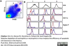
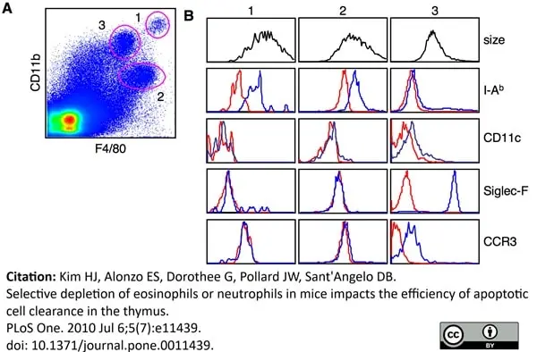
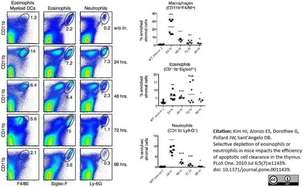
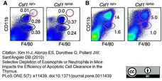
Rat anti Mouse F4/80 antibody, clone A3-1 (MCA497R) used for the evaluation of F4/80 expression on thymic myeloid cells by flow cytometry.
Image caption:
Heterogeneous phenotype of thymic resident myeloid cells. (A) Thymic stromal cells were enriched by collagenase/dispase digestion followed by percoll density gradient centrifugation. Cells were then stained with anti-CD11b and anti-F4/80 and analyzed by FACS. Red circles mark the three different cell populations discussed in the text. In this experiment, populations #1, #2 and #3 represented 0.02%, 0.43% and 0.61% of the analyzed cells, respectively. While the absolute cell number varied as a result of the preparation, the percentage of each cell type relative to each other was consistent over many experiments. (B) Size and surface marker expression (blue lines) of each population was determined by FACS. Populations were identified as in (A) and stained with the antibodies as indicated. Isotype controls (red lines) were used to correct differences in the autofluorescence of each cell population. Data are representative of more than ten experiments (a and b).
From: Kim H-J, Alonzo ES, Dorothee G, Pollard JW, Sant'Angelo DB (2010)
Selective Depletion of Eosinophils or Neutrophils in Mice Impacts the Efficiency of Apoptotic Cell Clearance in the Thymus.
PLoS ONE 5(7): e11439.
This image is from an open access article distributed under the terms of the Creative Commons Attribution License.
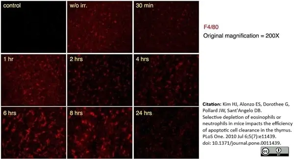
Rat anti Mouse F4/80 antibody, clone A3-1 (MCA497GA) used for the evaluation of F4/80 expression on thymic myeloid cells following irradiation by immunofluorescence.
Image caption:
Time course analysis of F4/80 positive cells following irradiation.
Thymuses from wild type B6 mice were harvested and frozen post-irradiation at the indicated times (1 hour through 24 hours). Sections were stained with anti-F4/80 (red), which identifies macrophages and eosinophils. Panel labeled "control" was a section stained with secondary antibody only. Panel labeled “w/o irr.” was a section from a nonirradiated mouse. Original magnification was 200X.
From: Kim H-J, Alonzo ES, Dorothee G, Pollard JW, Sant'Angelo DB (2010)
Selective Depletion of Eosinophils or Neutrophils in Mice Impacts the Efficiency of Apoptotic Cell Clearance in the Thymus.
PLoS ONE 5(7): e11439.
This image is from an open access article distributed under the terms of the Creative Commons Attribution License.
Rat anti Mouse F4/80 antibody, clone A3-1 (MCA497R) used for the evaluation of F4/80 expression on thymic myeloid cells following irradiation by flow cytometry.
Image caption:
Influx of innate cells rapidly declines twenty-four hours after irradiation. Thymuses were harvested from mice at the indicated times after irradiation. Stromal cells were enriched as described and stained with anti-CD11b, -F4/80, SiglecF and -Ly-6G. Numbers in the plots are the percentage of cells within the electronic gate. N = 6 and the data show the results from two independent experiments. The mean is shown by a horizontal bar and statistics were calculated with 2-tailed, nonpaired Mann-Whitney test. * = <0.01; ** = <0.001; *** = <0.0001..
From: Kim H-J, Alonzo ES, Dorothee G, Pollard JW, Sant'Angelo DB (2010)
Selective Depletion of Eosinophils or Neutrophils in Mice Impacts the Efficiency of Apoptotic Cell Clearance in the Thymus.
PLoS ONE 5(7): e11439.
This image is from an open access article distributed under the terms of the Creative Commons Attribution License.
Rat anti Mouse F4/80 antibody, clone A3-1 (MCA497R) used for the evaluation of F4/80 expression on spleen and bone marrow cells by flow cytometry.
Image caption:
FACS analyses of the innate cells in the spleen and bone marrow from WT and Csf op/op mice. Cells from the spleen and bone marrow from WT and Csfop/op mice were stained for CD11b and F4/80. Eosinophils and neutrophils were identified by staining cells for CD11b and SiglecF or CD11b and Gr-1, respectively. Percent of cells in each area is indicated in the Figure.
From: Kim H-J, Alonzo ES, Dorothee G, Pollard JW, Sant'Angelo DB (2010)
Selective Depletion of Eosinophils or Neutrophils in Mice Impacts the Efficiency of Apoptotic Cell Clearance in the Thymus.
PLoS ONE 5(7): e11439.
This image is from an open access article distributed under the terms of the Creative Commons Attribution License.
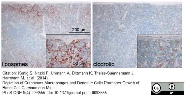
Rat anti Mouse F4/80 antibody, clone A3-1 (MCA497R) used for the identification of macrophages in mouse spleen using immunohistochemistry on formalin fixed, paraffin embedded tissue sections.
Image caption:
Depletion of F4/80-expressing cells in spleens derived from clodrolip-treated Ptchflox/floxERT2+/− mice. Immunohistochemical analysis using an anti-F4/80 antibody of paraffin-embedded spleens of Ptchflox/floxERT2+/− mice treated with empty liposomes or clodrolip.
From: König S, Nitzki F, Uhmann A, Dittmann K, Theiss-Suennemann J, Herrmann M, et al. (2014)
Depletion of Cutaneous Macrophages and Dendritic Cells Promotes Growth of Basal Cell Carcinoma in Mice.
PLoS ONE 9(4): e93555.
This image is from an open access article distributed under the terms of the Creative Commons Attribution License.
Rat anti Mouse F4/80 antibody, clone A3-1 (MCA497GA) used for the identification of infiltrating macrophages in tumor tissue by immunofluorescence.
Image caption:
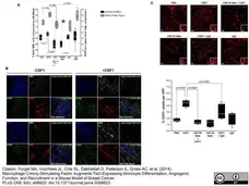
CSF1 has no effect on tumor growth but increases percent tumor TEMs and augments angiogenesis. (A) After two weeks of treatment, tumors were removed, homogenized and immunostained with antibodies specific for F4/80 and Tie2 to identify total F4/80+ cells and F4/80+/Tie2+ cells (Tie2-expressing macrophages, TEMs). While there was a marked increase in total F4/80+ macrophages with CSF1 treatment, the percent of F4/80+/Tie2+ TEMs was significantly increased in response to CSF1 suggesting a regulatory role for CSF1 in expanding the TEM population. N = 5 mice per group and results represent the mean ± SEM of total F4/80+ and F4/80+/Tie2+ TEMs within the tumors. (B, top and bottom left) PyMT tumors without CSF1 treatment and (top and bottom right) with CSF1 treatment immunostained with CD31 for blood vessels, F4/80 for macrophages, Tie2 for F4/80+/Tie2+ TEMS, and DAPI. Confocal images (using 60× objective (top) and with 3× zoom (bottom) suggest an increase in both F4/80 macrophages and F4/80+/Tie2+ TEMS in the CSF1-treated tumors. Multiply overlap indicates those areas where F4/80 and Tie2 positivity overlap. Individual stains are in Supplementary Figure 3. (C, top) Orthotopically implanted PyMT mammary tumors in wild type C57Bl/6 female mice were allowed to become palpable then intraperitoneally treated with PBS (PBS), CSF1 (100 ng in 100 μls) (CSF1), a neutralizing antibody for the CSF1R (50 mg/kg) 4 hours prior to CSF1 treatment (100 ng in 100 μls) (CSF1R NAb+CSF1), the CSF1R antibody alone (CSF1R NAb), an isotype antibody (50 mg/kg) 4 hours prior to CSF1 (100 ng in 100 μls) treatment (CSF1+IgG), or the isotype antibody alone (IgG) three times per week for two additional weeks. The tumors were immunostained with a CD31-Alexa Flour 546 antibody to recognize endothelial cells that comprise blood vessels. Qualitatively, CSF1 treatment increased the percent of CD31-postitive pixels per high powered field compared to PBS treated tumors, while the neutralizing antibody to CSF1R suppressed the CSF1 effect on angiogenesis. (B, bottom) Quantitatively, the percent of CD31+ pixels per high powered field were quantified as blood vessels (angiogenesis) using Adobe Photoshop histogram analysis. CSF1 treatment significantly increased CD31-positive pixels (angiogenesis) compared to PBS. The neutralizing antibody for CSF1R significantly reduced the ability of CSF1 to up-regulate angiogenesis. N = 5 mice per group and results represent the mean ± SEM of percent CD31-positive pixels per high powered field (HPF).
From: Forget MA, Voorhees JL, Cole SL, Dakhlallah D, Patterson IL, Gross AC, et al. (2014)
Macrophage Colony-Stimulating Factor Augments Tie2-Expressing Monocyte Differentiation, Angiogenic Function, and Recruitment in a Mouse Model of Breast Cancer.
PLoS ONE 9(6): e98623.
This image is from an open access article distributed under the terms of the Creative Commons Attribution License.
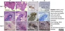
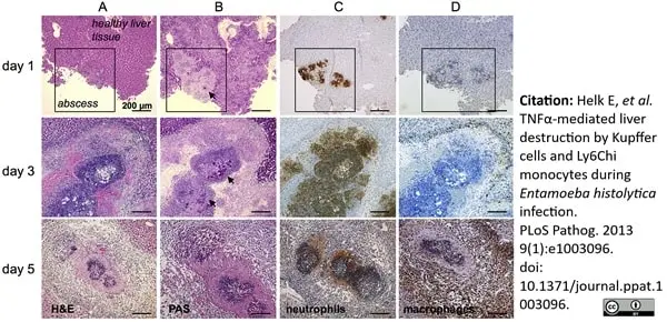
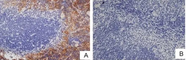
Rat anti Mouse F4/80 antibody, clone A3-1 (MCA497G) used for the identification of infiltrating macrophages in the spleen of Entamoeba histolytica infected mice by immunohistochemistry on formalin fixed, paraffin embedded tissue sections.
Image caption:
Histological and immunohistochemical characterization of cell infiltrates during ALA. (A) H&E staining of mouse liver abscesses (indicated by the square in the top row of images) at the indicated times post-infection with E. histolytica trophozoites. (B) PAS staining shows E. histolytica trophozoites (arrowheads) within the abscess. (C and D) Tissue sections were stained with anti-7/4 (C) and anti-F4/80 (D) antibodies followed by HRP-conjugated secondary antibody to detect neutrophils and macrophages, respectively (brown).
From: Helk E, Bernin H, Ernst T, Ittrich H, Jacobs T, Heeren J, et al. (2013)
TNFα-Mediated Liver Destruction by Kupffer Cells and Ly6Chi Monocytes during Entamoeba histolytica Infection.
PLoS Pathog 9(1): e1003096.
This image is from an open access article distributed under the terms of the Creative Commons Attribution License.
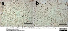
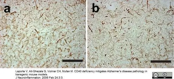
Rat anti Mouse F4/80 antibody, clone A3-1 (MCA497R) used for immunohistochemistry on mouse brain sections
Image caption:
Representative photographs of cortical area in (a) CD40 def. and (b) wild-type mice stained with F4/80 antibody at 8 to 10 weeks old (each bar represents 0.1 mm).
From: Laporte et al.
Journal of Neuroinflammation 2006 3:3.
This image is from an open access article distributed under the terms of the Creative Commons Attribution License.
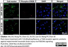
Rat anti Mouse F4/80 antibody, clone A3-1 (MCA497G) used for the detection of microglia in mouse brain by immunofluorescence
Image caption:
Post-injury 7,8-dihydroxyflavone treatment increased brain BDNF levels and promoted CREB activation. D) GFAP (astrocytes), or F4/80 (microglia) is shown in green. Sections were stained with DAPI (blue) to show all nuclei. The scale bar is 50 μm. Ipsi: ipsilateral cortex; Contra: contralateral cortex.
From: Wu C-H, Hung T-H, Chen C-C, Ke C-H, Lee C-Y, et al. (2014)
Post-Injury Treatment with 7,8-Dihydroxyflavone, a TrkB Receptor Agonist, Protects against Experimental Traumatic Brain Injury via PI3K/Akt Signaling.
PLoS ONE 9(11): e113397.
This image is from an open access article distributed under the terms of the Creative Commons Attribution License.
Rat anti Mouse F4/80 antibody, clone A3-1 (MCA497R) used for the detection of infiltrating macrophages in mouse kidney by immunohistochemistry on paraffin embedded material
Image caption:
Reduction in cellular infiltration and inflammatory markers in UUO kidneys exposed to telmisartan or PXS64. Untreated UUO kidneys showed increased F4/80, CD68 and CD45 positively stained cells as compared to the sham operated control animals. PXS64 significantly reduced F4/80 and CD45 positive stained cells (Fig. 6C and E) with a trend to a reduction in CD68 cells, although this was not statistically significant (Fig. 6D). Telmisartan treated kidneys showed a reduction in F4/80 positive cells but no difference in CD45 or CD68 stained cells, suggesting a differential action of PXS64 and telmisartan in modifying cellular infiltration. Results are presented as mean showed (n = 8, *P<0.05 vs. UUO, ** P<0.01 vs. UUO). Magnification x 400.
From: Zhang J, Wong MG, Wong M, Gross S, Chen J, et al. (2015)
A Cationic-Independent Mannose 6-Phosphate Receptor Inhibitor (PXS64) Ameliorates Kidney Fibrosis by Inhibiting Activation of Transforming Growth Factor-β1.
PLoS ONE 10(2): e0116888.
This image is from an open access article distributed under the terms of the Creative Commons Attribution License.
Rat anti Mouse F4/80 antibody, clone A3-1 (MCA497G) used for the identification of macrophages in a murine colitis model by immunohistochemistry on formalin fixed paraffin embedded tissue sections.
Image caption:
Nur77-/- mice show more macrophage and T-cell influx in TNBS-induced colitis. The numbers of T-cells in the mucosa were counted and the influx was calculated by taking the ratio of Healthy / TNBS. Representative photomicrographs of sections stained for T-cells (anti-CD3) are shown (A). mRNA was extracted from colon and qPCR was performed for FoxP3 (B). cDNA levels were normalized for 36B4 housekeeping gene expression. Macrophage surface was quantified in the mucosa and the number of macrophages was counted in the muscle. The ratio of Healthy / TNBS was calculated. Representative photomicrographs of sections stained for macrophages (anti-F4/80) are shown (C). A and C Original magnification x100, D x200. Each symbol represents 1 animal. Data are presented as mean ±SEM; *p<0.05, **p<0.01, ***p<0.001. Scale bars represent 1mm. nr = number, AU = Arbitrary units, FoxP3 = Forkhead box P3 (marker for regulatory T-cells).
From: Hamers AAJ, van Dam L, Teixeira Duarte JM, Vos M, Marinković G, van Tiel CM, et al. (2015)
Deficiency of Nuclear Receptor Nur77 Aggravates Mouse Experimental Colitis by Increased NFκB Activity in Macrophages.
PLoS ONE 10(8): e0133598.
This image is from an open access article distributed under the terms of the Creative Commons Attribution License.
Rat anti MouseF4/80 antibody, clone A3-1 (MCA497R) used for the identification of macrophages in murine kidney by immunofluorescence and immunohistochemistry on formalin fixed, paraffin embedded tissue sections.
Image caption:
Clodronate liposome mediated macrophage depletion in Col4a3KO mice. (A) KO male mice were dosed intraperitonally with CL (KO+CL) or PBSL (KO+PBSL) as control. Animals were entered in the study at the age of 4 weeks and were continually dosed until the age of 8 weeks. Injections were repeated every second day, except for the first two doses, which were injected on consecutive days. (B-D) CL significantly reduced F4/80 positive macrophage infiltrates in KO+CL kidney as compared to KO+PBS or KO mice at the age of 8 weeks. (B, C) Representative images and quantitative assessment of F4/80 stained macrophages in kidney sections from KO+CL and KO+PBSL reveal marked reduction in macrophage infiltrates following CL dosing. Scale bar: 100 μm. (D) Real-time PCR analysis of F4/80 mRNA expression in KO+CL, KO+PBSL, KO, and WT littermates at the age of 8 weeks. Significant reduction in F4/80 mRNA expression following CL dosing was observed. PBSL did not affect endogenous F4/80 mRNA expression as shown by similar F4/80 mRNA expression in KO and KO+PBSL mice. (E) Real-time PCR analysis of CD204 and CD206 mRNA showed significant reduction in the expression of both genes in KO kidney upon CL treatment. (F) Representative images and quantitative assessment of F4/80 and CD206 stained kidney sections from KO+CL and KO+PBSL revealed marked reduction in M1 (F4/80+CD206-) and M2 (F4/80+CD206+) macrophages. Asterisk: F4/80+CD206- M1 macrophages, arrow: F4/80+CD206+ M2 macrophages. Scale bar: 20 μm. Welch corrected ANOVA followed by posthoc pairwise t-test with a Bonferroni correction (C,D,E) and unpaired t-test (F) was used to evaluate differences between groups. (C,D,E,F) n = 5 mice per group. CL: clodronate liposomes; PBSL: PBS liposomes.
From: Kim M, Piaia A, Shenoy N, Kagan D, Gapp B, Kueng B, et al. (2015)
Progression of Alport Kidney Disease in Col4a3 Knock Out Mice Is Independent of Sex or Macrophage Depletion by Clodronate Treatment.
PLoS ONE 10(11): e0141231.
This image is from an open access article distributed under terms of a Creative Commons Attribution License.
Rat anti Mouse F4/80 antibody, clone A3-1 (MCA497R) used for the identification of murine macrophages in the kidney by immunohistochemistry on formalin fixed cryosections.
Image caption:
Immunostainings of macrophages and neutrophils in the kidneys of M-Rac1 FC and KO mice. (A) Representative micrographs of F4/80 immunostaining in the kidneys of Vehicle- or LPS-injected M-Rac1 FC and KO mice. Original magnification x 200. (B) Quantitative analysis of F4/80-positive macrophages. Data are means ± s.e.m. Statistical analysis was performed by two-way ANOVA, n.s. genotype effect, P < 0.01 treatment effect, n.s. interaction effect. **P < 0.01 by Bonferroni's post hoc test. n = 5 per each group. (C) Representative micrographs of Ly-6B.2 (mAb 7/4) immunostaining. (D) Quantitative analysis of Ly-6B.2-positive neutrophils. Statistical analysis was performed by two-way ANOVA, n.s. genotype effect, P < 0.01 treatment effect, n.s. interaction effect. **P < 0.01 by Bonferroni's post hoc test. n = 6 per group.
From: Citation: Nagase M, Kurihara H, Aiba A, Young MJ, Sakai T (2016)
Deletion of Rac1GTPase in the Myeloid Lineage Protects against Inflammation-Mediated Kidney Injury in Mice.
PLoS ONE 11(3): e0150886.
This image is from an open access article distributed under terms of a Creative Commons Attribution License.
Alexa Fluor® 647 conjugated Rat anti Mouse F4/80 antibody, clone A3-1 (MCA497A647) used for the detection of murine Kupffer cells by immunofluorescence
Image caption:
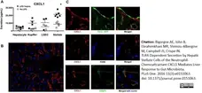
Hepatic stellate cells are the major source of CXCL1, as shown by both quantification of secretion and in situ localization. (A) Quantification of CXCL1 secretion in enriched fractions of hepatocytes, KCs, LSECs and HSCs, freshly isolated and stimulated in vitro with LPS (1 ng/mL LPS, black squares) during 24 hours. Data are representative of three separate experiments with six mice in each group; #P<.05. (B) In-situ localization of CXCL1 in the liver. Immunofluorescent detection for CXCL1 (red) and liver cells nuclei (blue) for nuclei first shows CXCL1 expression in the sinusoids throughout liver parenchyma. (C) Higher resolution shows that CXCL1 (red) is expressed by sub-endothelial cells, which also store retinol droplets in separate compartments, as shown by CRBP1 staining (green). The Cellular Retinol Binding Protein-1 (CRBP-1) is the best marker to detect simultaneously both resting (Glial Fibrillary Acidic Protein, GFAP+) and activated (α-Smooth Muscle Actin, αSMA+) stellate cells in situ. Alexa Fluor-546-CXCL1 (red) staining does not colocalize either with Tie2-GFP in LSECs (green, upper panel), or F4/80 in KCs (blue, middle panel), but with AlexaFluor-488-CRBP1 (green, lower panel), staining both resting and activated HSCs. TOPRO3 was used for nuclei vizualisation.
From: Bigorgne AE, John B, Ebrahimkhani MR, Shimizu-Albergine M, Campbell JS, Crispe IN (2016)
TLR4-Dependent Secretion by Hepatic Stellate Cells of the Neutrophil-Chemoattractant CXCL1 Mediates Liver Response to Gut Microbiota.
PLoS ONE 11(3): e0151063.
This image is from an open access article distributed under the terms of the Creative Commons Attribution License.
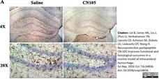
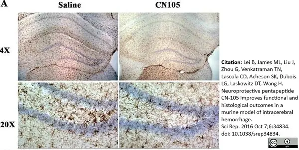
Rat anti Mouse F4/80 antibody, clone A3-1 (MCA497GA) used for the demonstration of microglia in mouse brain by immunohistochemistry on formalin fixed 40μ vibratome sections.
Image caption:
CN-105 reduces brain microglial activation after ICH.
(A) Representative images of F4/80-labelled microglia in ipsilateral hippocampi at 5 days after ICH in mice treated with vehicle or CN-105.
From: Lei B, James ML, Liu J, Zhou G, Venkatraman TN, Lascola CD, Acheson SK, Dubois LG, Laskowitz DT, Wang H.
Neuroprotective pentapeptide CN-105 improves functional and histological outcomes in a murine model of intracerebral hemorrhage.
Sci Rep. 2016 Oct 7;6:34834.
This image is from an open access article distributed under the terms of the Creative Commons Attribution License.
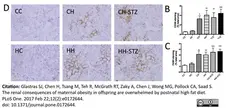
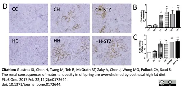
Rat anti Mouse F4/80 antibody, clone A3-1 (MCA497GA) used for the identifcation of macrophages in kidney sections by immunohistochemistry on cryosections.
Image caption:
Renal markers of inflammation at Week 32.
B. Area (%) of CD68 staining, C. Area (%) of F4/80 staining, D. F4/80 representative images at 200x magnification. Results are expressed as mean ± SEM, N = 4–6. *P<0.05, **P<0.01, ***P<0.001, ****P<0.0001 compared to CC.
From: Glastras SJ, Chen H, Tsang M, Teh R, McGrath RT, Zaky A, et al. (2017)
The renal consequences of maternal obesity in offspring are overwhelmed by postnatal high fat diet.
PLoS ONE 12(2): e0172644.
This image is from an open access article distributed under the terms of the Creative Commons Attribution License.
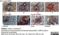
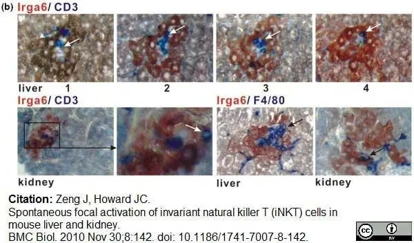
Rat anti Mouse F4/80 antibody, clone A3-1 (MCA497G) used for the identification of macrophages in kidneys and livers of mice by immunohistochemistry on formalin fixed, paraffin embedded tissue sections.
Image caption:
Focal expression of Irga6 in healthy mouse liver and kidney.
T cells and macrophages/dendritic cells are present in the mononuclear cores of the liver and kidney patches. Wax sections of the livers and kidneys from adult C57BL/6 mice were stained for Irga6 (brown/red) and CD3 or F4/80 (blue). For Irga6 and CD3 double staining in liver, consecutive serial sections were analysed (numbered 1, 2, 3, 4). Arrows point to CD3 and F4/80 positive cells. The microscope magnification is 200×; frames show enlargements.
From: Zeng J, Howard JC.
Spontaneous focal activation of invariant natural killer T (iNKT) cells in mouse liver and kidney.
BMC Biol. 2010 Nov 30;8:142.
This image is from an open access article distributed under the terms of the Creative Commons Attribution License.
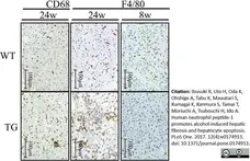
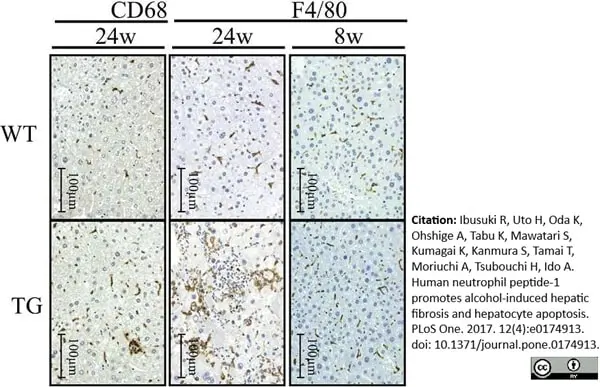
Rat anti Mouse F480, clone A3-1 (MCA497R) used for the demonstration of Kupffer cells in mouse liver by immunohistochemistry on formalin fixed, paraffin embedded tissue sections.
Image caption:
Expression of F4/80 and CD68 in liver tissue assessed by immunostaining using anti-F4/80 or anti-CD68 antibody.
From: Citation: Ibusuki R, Uto H, Oda K, Ohshige A, Tabu K, Mawatari S, et al. (2017)
Human neutrophil peptide-1 promotes alcohol-induced hepatic fibrosis and hepatocyte apoptosis.
PLoS ONE 12(4): e0174913.
This image is from an open access article distributed under the terms of the Creative Commons Attribution License.
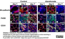
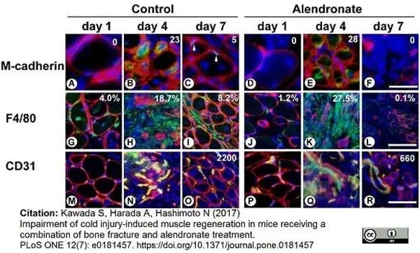
Rat anti Mouse F4/80 antibody, clone A3-1 (MCA497GA) used to identify macrophages in the murine vastus lateralis by immunofluorescence on cryostat sections.
Image caption:
Combination of bone fracture and alendronate treatment causes loss of myogenic cells.
PBS (Control group) or alendronate (Alendronate group) was subcutaneously injected three times at seven-day intervals. The femur was broken and vastus lateralis muscle injured by cold shock on day 14. Then the vastus lateralis muscle was removed on day 1, 4, or 7 after the bone fracture and cold injury. Sections were subjected to immunostaining with antibodies to M-cadherin (green in A-F), F4/80 (green in G-L), CD31 (green in M-R), and laminin α2 (red in A-R). Nuclei were stained with DAPI (blue in A-R). Arrows represent M-cadherin-positive muscle satellite cells in (C). Scale bars, 40 μm in (A-F), 20 μm in (G-R). Images represent a part of each original picture. Numbers represent numbers of M-cadherin-positive muscle satellite cells/myogenic progenitor cells in each original picture (0.17 mm2) in (A-F). Percentages of F4/80-positive cell-occupied area of each original picture (0.68 mm2) are shown in (G-L). Numbers of CD31-positive capillaries per mm2 of each original picture are shown in (O and R).
From: Kawada S, Harada A, Hashimoto N (2017)
Impairment of cold injury-induced muscle regeneration in mice receiving a combination of bone fracture and alendronate treatment.
PLoS ONE 12(7): e0181457.
This image is from an open access article distributed under the terms of the Creative Commons Attribution License.
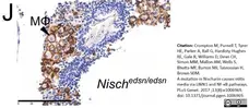
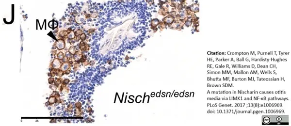
Rat anti Mouse F4/80 antibody, clone A3-1 (MCA497GA) used to identify infiltrating macrophages in mice by immunohistochemistry on formalin fixed, paraffin embedded tissue sections
Image caption:
Middle ear histology indicates chronic otitis media in Nischedsn/edsn mice.
(J) Immunohistochemical staining of a Nischedsn/edsn ear with OM using an F4/80 antibody shows a cellular exudate rich in macrophages (brown). Representative image from four mice scale bar =100μm.
From: Crompton M, Purnell T, Tyrer HE, Parker A, Ball G, Hardisty-Hughes RE, et al. (2017)
A mutation in Nischarin causes otitis media via LIMK1 and NF-κB pathways.
PLoS Genet 13(8): e1006969.
This image is from an open access article distributed under the terms of the Creative Commons Attribution License.
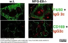
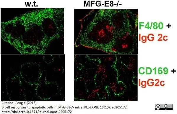
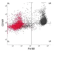
Rat anti Mouse F4/80 antibody, clone A3-1 (MCA497R) used for the identification of macrophages in mouse spleen sections by immunofluorescence.
Image caption:
Localization of IgG2c+ (red) plasma cells in the spleens of 4 month old w.t. and MFG-E8-/- mice was examined by immunofluorescence staining. Red pulp macrophages and Marginal zone macrophages were visualized by anti-F4/80(green, upper) and anti-CD169 (green, lower), respectively. The images are representative of at least 5 mice in each strain.
From: Peng Y (2018)
B cell responses to apoptotic cells in MFG-E8-/- mice.
PLoS ONE 13(10): e0205172.
This image is from an open access article distributed under the terms of the Creative Commons Attribution License.
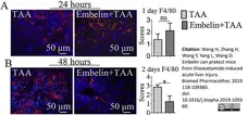
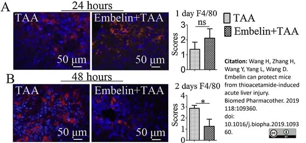
Rat anti Moouse F4/80 antibody, clone A3-1 (MCA497G) used to identify macrophages in mouse liver by immunofluorescence on formalin fixed, paraffin embedded tissue sections.
Image caption:
F4/80 staining. F4/80 activities in livers are examined at 1 and 2 days with immunostaining. (A) 1 day; (B)2 days. For all panels, representative images are shown and arrows denote positive staining. ns p ≥0.05, * p <0.05.
From: Wang H, Zhang H, Wang Y, Yang L, Wang D.
Embelin can protect mice from thioacetamide-induced acute liver injury.
Biomed Pharmacother. 2019 Oct;118:109360.
doi: 10.1016/j.biopha.2019.109360.
This image is from an open access article distributed under the terms of the Creative Commons Attribution License.
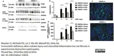
Rat anti Mouse F4/80 antibody, clone A3-1 (MCA497R) used to evaluate F4/80 expression in murine kidney by western blotting and immunofluorescence.
Image caption:
UMOD expression amplifies UUO‐associated interstitial inflammation. Interstitial inflammation is a hallmark feature of kidney fibrosis. Interstitial macrophage density, quantified by kidney F4/80 protein western blotting (n = 5 for sham groups; n = 7 for UUO groups), was significantly lower in the UMOD−/− groups by 41–58% at all time‐points (A, B). Representative day 21 immunofluorescence photomicrographs illustrate a smaller interstitial area expressing the F4/80 macrophage protein (green) in the UMOD−/− kidney (C). Kidney MCP‐1 mRNA levels were significantly lower in the UMOD−/− mice on UUO days 7, 14, and 21 (n = 3 for sham groups; n = 7 for UUO groups) (D). Bar graphs represent means +1SEM; * indicates P value <0.05. Photomicrograph magnification: ×40. Scale bar: 25 μm.
From: Maydan O, McDade PG, Liu Y, Wu XR, Matsell DG, Eddy AA.
Uromodulin deficiency alters tubular injury and interstitial inflammation but not fibrosis in experimental obstructive nephropathy.
Physiol Rep. 2018 Mar;6(6):e13654.
doi:10.14814/phy2.13654.
This image is from an open access article distributed under the terms of the Creative Commons Attribution License.

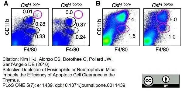
Rat anti Mouse F4/80 antibody, clone A3-1 (MCA497R) used for the evaluation of F4/80 expression on thymic cells by flow cytometry.
Image caption:
Impaired clearance of apoptotic thymocytes in Csf1op/op mice. (A) Thymic stromal cell subpopulations from WT (Csf1op/+) and osteopetrosis (Csf1op/op) mice were stained with the indicated antibodies and analyzed by FACS. Numbers indicate the percentage of cells within the circles. The red circle marks population #1, which is not present in Csf1op/op mice. (B) Comparison of thymic stromal cell subpopulations between WT (Csf1op/+) and osteopetrosis (Csf1op/op) mice 16 hours after irradiation. Thymic stromal cells were enriched and stained for CD11b and F4/80.
From: Kim H-J, Alonzo ES, Dorothee G, Pollard JW, Sant'Angelo DB (2010)
Selective Depletion of Eosinophils or Neutrophils in Mice Impacts the Efficiency of Apoptotic Cell Clearance in the Thymus.
PLoS ONE 5(7): e11439.
This image is from an open access article distributed under the terms of the Creative Commons Attribution License.
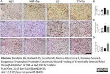
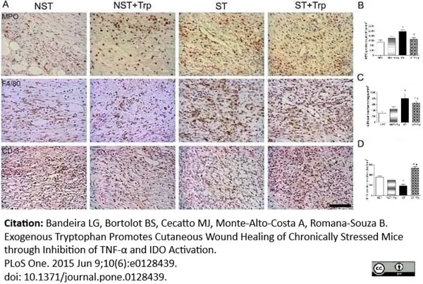
Rat anti Mouse F4/80 antibody, clone A3-1 (MCA497R) used for the identification of macrophages in formalin fixed, paraffin embedded murine skin tissue sections by immunohistochemistry.
Image caption:
L-tryptophan administration reverses stress-induced alterations on inflammatory response and lipid peroxidation. Mice were daily submitted to restraint stress and treated with L-tryptophan (Trp) or vehicle until euthanasia. Two days after the beginning of the stress protocol, two full-thickness excisional lesions (8-mm diameter) were made on the dorsal skin. Five days after wounding, lesions were collected and the number of myeloperoxidase (MPO)-positive neutrophils (B), F4/80-positive macrophages (C) and CD-3 positive T lymphocytes (D) were counted. Representative images (A) of staining for neutrophils, macrophages and T cells on wound area were presented. In addition, the protein levels of monocyte chemotactic protein-1 (MCP-1) (12 kDa) (E) were estimated in wound lysate by western-blotting. Representative images for immunoblotting for MCP-1 (F) were presented. The β-actin (42 kDa) was used as a loading control protein. Vertical black lines show non-adjacent bands from the same blot. To evaluate the oxidative damage in lipids, the levels of lipid peroxides (G) were measured in wound lysate using colorimetric assay. Data (n = 5) are presented as mean ± SEM. #p<0.05 vs. stressed (ST) group. Bar = 50 μm.
From: Bandeira LG, Bortolot BS, Cecatto MJ, Monte-Alto-Costa A, Romana-Souza B (2015)
Exogenous Tryptophan Promotes Cutaneous Wound Healing of Chronically Stressed Mice through Inhibition of TNF-α and IDO Activation.
PLoS ONE 10(6): e0128439.
This image is from an open access article distributed under the terms of the Creative Commons Attribution License.
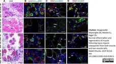
Rat anti Mouse F4/80 antibody, clone 3A-1 (MCA497R) used to identify macrophages in mouse muscle graft sections by immunofluorescence.
Image caption:
Autografted and allografted muscles: morphology and osteopontin expression. Cryosections of EDL muscle grafts at 5 (a, b) or 3 (c, d) days post-surgery. a H&E-stained sections. Arrows indicate regenerating fibres (small fibres with centrally located nuclei). b Sections stained with anti-MyHCemb (green), anti-laminin (red) and DAPI, as used for quantitation of regenerating fibres. c Sections stained with anti-osteopontin (red), anti-desmin (myoblasts; green) and DAPI. In wild-type (WT) EDL muscle grafted into a wild-type host, anti-osteopontin stained both myoblasts (yellow) and non-myoblast mononuclear cells (red). In osteopontin-null (KO) muscle grafted into an osteopontin-null host, no osteopontin was detected. In allografted osteopontin-null muscle in a wild-type host, osteopontin was only detected in non-myoblast mononuclear cells. In allografted wild-type muscle in an osteopontin-null host, osteopontin was only detected in myoblasts. Arrows and arrowheads indicate myoblasts and non-myoblast mononuclear cells, respectively. d Sections stained with anti-osteopontin (red), anti-F4/80 (macrophages; green) and DAPI. In autografted wild-type muscle, anti-osteopontin stained macrophages (*; yellow) and some additional structures (arrows; red). In autografted osteopontin-null muscle, no osteopontin was detected. In allografted osteopontin-null muscle in a wild-type host, osteopontin was only observed in macrophages (*; yellow). In allografted wild-type muscle in an osteopontin-null host, weak osteopontin staining was observed in some non-macrophage structures (arrows), but not in macrophages (†; green). Bars = 50μm
From: Wasgewatte Wijesinghe DK, Mackie EJ, Pagel CN.
Normal inflammation and regeneration of muscle following injury require osteopontin from both muscle and non-muscle cells.
Skelet Muscle. 2019; 9(1):6.
This image is from an open access article distributed under the terms of the Creative Commons Attribution License.
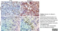
Rat anti Mouse F4/80 antibody, clone A3-1 (MCA497R) used for the identification of macrophages in tumor tissues by immunohistochemistry on formalin fixed cryosections. Comparison is with the distribution of CD11c+ dendritic cells (MCA1369GA) in similar tissue sections.
Image caption:
IHC analysis of Colon-26 tumors untreated and INT230-6-treated tumors ~10 days post dose. Representative images (40× magnification) of IHC staining for F4/80 (macrophages) and CD11c (dendritic cells). In general, the staining appeared more evident in the INT230-6-treated tumors, particularly toward the more necrotic areas.
From: Bender LH, Abbate F, Walters IB.
Intratumoral Administration of a Novel Cytotoxic Formulation with Strong Tissue Dispersive Properties Regresses Tumor Growth and Elicits Systemic Adaptive Immunity in In Vivo Models.
Int J Mol Sci. 2020;2 (12):E4493.
doi:10.3390/ijms21124493
This image is from an open access article distributed under terms of a Creative Commons Attribution License.
Pacific Blue conjugated Rat anti Mouse F4/80 antibody, clone A3-1 (MCA497PB) used for the dendritic cell gating process from mouse white adipose tissue by flow cytometry.
Image caption:
A representative gating scheme of dendritic cell and macrophage populations in the epididymal white adipose tissues. For analysis of dendritic cell and macrophage populations, stromal vascular cells obtained from epididymal white adipose tissues were stained with FITC–conjugated anti-CD206, PE–conjugated anti-CD11c, PerCP–conjugated anti-CD45, PE-Cy7–conjugated anti-F4/80*, APC–conjugated anti-CD64 and APC-Cy7–conjugated anti-MHC class II antibodies.
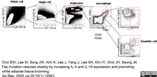
From: Choi EW, Lee M, Song JW, Kim K, Lee J, Yang J, Lee SH, Kim IY, Choi JH, Seong JK.
Fas mutation reduces obesity by increasing IL-4 and IL-10 expression and promoting white adipose tissue browning.
Sci Rep. 2020 Jul 20;10(1):12001.
doi: 10.1038/s41598-020-68971-7.
This image is from an open access article distributed under terms of a Creative Commons Attribution License. * should read "Pacific Blue® conjugated F4/80.

Rat anti Mouse F4/80 antibody, clone A3-1 (MCA497GA) used for the identification of macrophages in murine liver sections by immunohistochemistry on formalin fixed, paraffin embedded material.
Image caption:
Protective effect of clusterin in the pathology of WD-induced NAFLD.(B) Immunostaining for F4/80 in the liver of wild type, wCLU-tg, and CLU-KO mice fed CHOW or WD. *P <0.05; **P <0.01; ***P <0.001. Magnification, × 200.
From: Park JS, Lee WK, Kim HS, Seo JA, Kim DH, Han HC, Min BH.
Clusterin overexpression protects against western diet-induced obesity and NAFLD.
Sci Rep. 2020 Oct 15;10(1):17484.
doi: 10.1038/s41598-020-73927-y.
This image is from an open access article distributed under terms of a Creative Commons Attribution License.
Rat anti Mouse F4/80 antibody, clone A3-1 (MCA497R) used to highlight macrophages in murine kidney by immunohistochemistry on formalin fixed, paraffin embedded tissue sections.
Image caption:
Lithium treatment induces change in M1/M2 phenotype macrophages.
A) Representative areas of macrophage infiltration by F4/80 staining in the cortical area. Arrows denote positive brown staining. Scale bars represent 100 μm. B) Quantitative analysis related to E (n = 4).
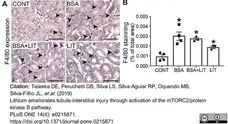
From: Teixeira DE, Peruchetti DB, Silva LS, Silva-Aguiar RP, Oquendo MB, Silva-Filho JL, Takiya CM, Leal-Cardoso JH, Pinheiro AAS, Caruso-Neves C.
Lithium ameliorates tubule-interstitial injury through activation of the mTORC2/protein kinase B pathway.
PLoS One. 2019 Apr 19;14(4):e0215871.
doi: 10.1371/journal.pone.0215871.
This image is from an open access article distributed under terms of a Creative Commons Attribution License.
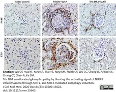
Rat anti Mouse F4/80 antibody, clone A3-1 (MCA497R) used for the demonstration of macrophages in murine kidney by immunohistochemistry on formalin fixed, paraffin embedded tissue sections.
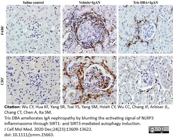
Image caption:
Renal pathology, T cells and macrophages infiltration. F, CD3+ T cells and F4/80+ macrophages in IHC staining; Original magnification, 400×.
From: Wu CY, Hua KF, Yang SR, Tsai YS, Yang SM, Hsieh CY, Wu CC, Chang JF, Arbiser JL, Chang CT, Chen A, Ka SM.
Tris DBA ameliorates IgA nephropathy by blunting the activating signal of NLRP3 inflammasome through SIRT1- and SIRT3-mediated autophagy induction.
J Cell Mol Med. 2020 24(23):13609-22.
doi: 10.1111/jcmm.15663. <.br>This image is from an open access article distributed under terms of a Creative Commons Attribution License.
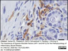
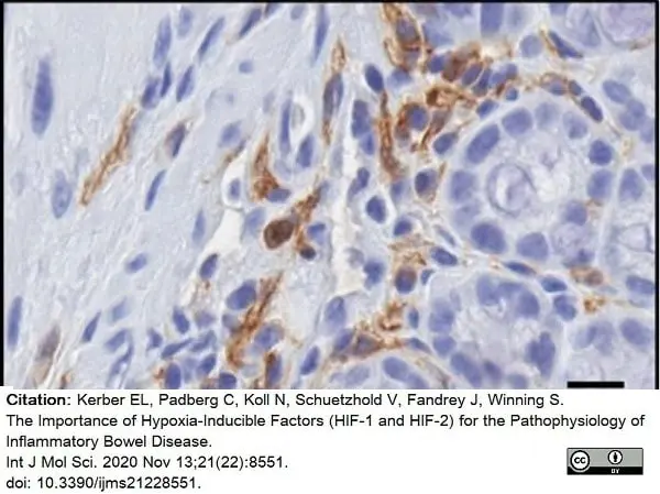
Biotinylated Rat anti Mouse F4/80 antibody, clone A3-1 (MCA497B) used for the demonstration of F4/80 expressing macrophages in mouse tissues by immunohistochemistry on formalin fixed, paraffin embedded tissue sections.
Image caption:
After DSS treatment, increased numbers of F4/80-positive cells were observed in the lamina propria mucosae and tela submucosa in all tissue sections. magnification 400×, scale bar: 50 μm.
From: Kerber EL, Padberg C, Koll N, Schuetzhold V, Fandrey J, Winning S.
The Importance of Hypoxia-Inducible Factors (HIF-1 and HIF-2) for the Pathophysiology of Inflammatory Bowel Disease.
Int J Mol Sci. 2020 Nov 13;21(22):8551.
doi: 10.3390/ijms21228551.
This image is from an open access article distributed under terms of a Creative Commons Attribution License.
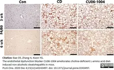
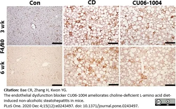
Rat anti mouse F4/80 antibody, clone Cl:A3-1 (MCA497GA) used for the demonstration of macrophages in murine liver by immunohistochemistry on formalin fixed, paraffin embedded tissue sections.
Image caption:
CU06-1004 has anti-inflammatory effects in mice with CDAA diet-induced NASH. Immunohistochemical analysis of F4/80 in liver.
From: Bae C-R, Zhang H, Kwon Y-G (2020)
The endothelial dysfunction blocker CU06-1004 ameliorates choline-deficient L-amino acid diet-induced non-alcoholic steatohepatitis in mice.
PLoS ONE 15(12): e0243497.
doi: 10.1371/journal.pone.0243497
This image is from an open access article distributed under terms of a Creative Commons Attribution License.
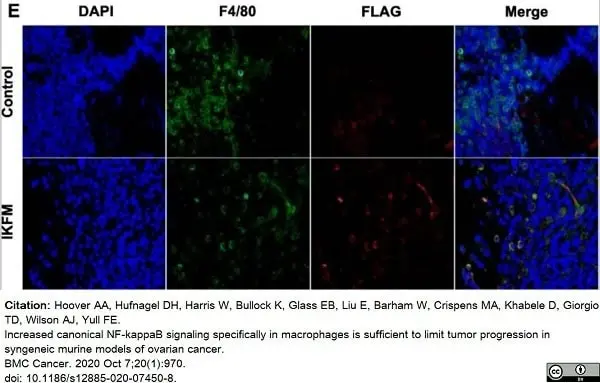
Rat anti Mouse F4/80 antibody, clone A3-1 (MCA497) used to stain macrophages in murine omental tumor tissue by immuofluorescence.
Image caption:
The doxycycline-inducible IKFM model of NF-κB activation in macrophages. E. Representative images from immunofluorescence analysis of TBR5 omental tumor tissue from FVB IKFM mice showing tumor-infiltrating macrophages; nuclei (blue); F4/80 (green); FLAG-IKK2 (red).
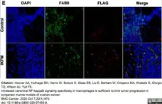
From: Hoover AA, Hufnagel DH, Harris W, Bullock K, Glass EB, Liu E, Barham W, Crispens MA, Khabele D, Giorgio TD, Wilson AJ, Yull FE.
Increased canonical NF-kappaB signaling specifically in macrophages is sufficient to limit tumor progression in syngeneic murine models of ovarian cancer.
BMC Cancer. 2020 Oct 7;20(1):970.
doi: 10.1186/s12885-020-07450-8.
This image is from an open access article distributed under terms of a Creative Commons Attribution License.
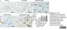
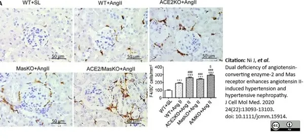
Rat anti Mouse F4/80 antibody, clone A3-1 (MCA497) used to stain macrophages in murine kidney by immunohistochemistry on formalin fixed, paraffin embedded tissue sections.
Image caption:
Double deletion of ACE2 and Mas receptor enhances Ang II‐induced renal inflammation. A, F4/80+ macrophages infiltrating the kidney detected by immunohistochemistry. Data represent the mean ± SEM for groups of 6‐12 mice. *P <.05, ***P <.001 vs. WT + SL; # P <.05, ## P <.01, ### P <.001 vs. WT + Ang II; $ P <.05, $$$ P <.001 vs. ACE2 KO + Ang II and Mas KO + Ang II. Scale bar, 50 μm.
From: Ni J, Yang F, Huang XR, Meng J, Chen J, Bader M, Penninger JM, Fung E, Yu XQ, Lan HY.
Dual deficiency of angiotensin-converting enzyme-2 and Mas receptor enhances angiotensin II-induced hypertension and hypertensive nephropathy.
J Cell Mol Med. 2020 Sep 24;24(22):13093–103.
doi: 10.1111/jcmm.15914.
This image is from an open access article distributed under terms of a Creative Commons Attribution License.

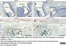
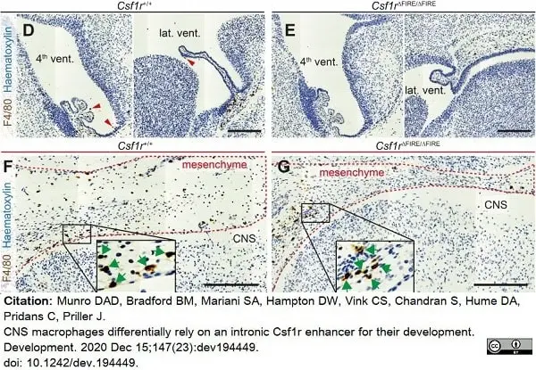
Rat anti Mouse F4/80 antibody, clone A3-1 (MCA497G) used to demonstrate the presence of macrophages in mouse brain by immunohistochemistry on formalin fixed, paraffin embedded tissue sections.
Image caption:
F4/80 staining of macrophages in E12.5 Csf1r+/+ and Csf1rΔFIRE/ΔFIRE embryos. Representative images of E12.5 Csf1r+/+ and Csf1rΔFIRE/ΔFIRE embryonic heads stained for F4/80. (D-E) Anti-F4/80 stained ChP of the lateral (lat.) and fourth (4th) ventricles. (F-G) F4/80+ macrophages (brown) in the cephalic mesenchyme and developing CNS tissue (these images show magnified versions of the red boxed regions in B-C) Inset boxes in F and G show magnified images of cephalic mesenchyme F4/80+ BAMs (green arrows).
From: Munro DAD, Bradford BM, Mariani SA, Hampton DW, Vink CS, Chandran S, Hume DA, Pridans C, Priller J.
CNS macrophages differentially rely on an intronic Csf1r enhancer for their development.
Development. 2020 Dec 15;147(23):dev194449.
doi: 10.1242/dev.194449.
This image is from an open access article distributed under terms of a Creative Commons Attribution License.
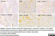
Rat anti Mouse F4/80 antibody, clone A3-1 (MCA497R) used to label macrophages in murine renal tissue by immunohistochemistry on cryostat sections.
Image caption:
SSAO inhibition prevents inflammatory cell infiltrates.
F4/80 Staining. Representative photomicrographs of immunohistochemistry for tubulointerstitial F4/80 stain for activated macrophages and histograms summarising expression. Control IgG1 staining was performed to confirm specific staining (not shown). Data presented as mean ± SEM with (*p<0.05, **p<0.01 vs control, #p<0.05 vs DM) n = 6-8. Bar = 200μm DM, Diabetes Mellitus; SSAOi, Semicarbazide Sensitive Amine Oxidase Inhibitor; Telmi, telmisartan.
From: Wong MY, Saad S, Wong MG, Stangenberg S, Jarolimek W, Schilter H, et al. (2020)
Semicarbazide-sensitive amine oxidase inhibition ameliorates albuminuria and glomerulosclerosis but does not improve tubulointerstitial fibrosis in diabetic nephropathy.
PLoS ONE 15(6): e0234617.
doi: 10.1371/journal.pone.0234617
This image is from an open access article distributed under terms of a Creative Commons Attribution License.
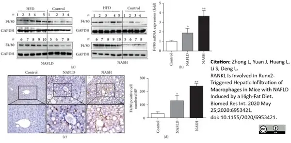
Rat anti Mouse F4/80 antibody, clone A3-1 (MCA497R) used to evaluate F4/80 expression in murine liver by western blotting and immunohistochemistry on formalin fixed, paraffin embedded tissue sections following antigen retrieval.
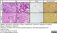
Image caption:
Hepatic infiltration of macrophages accompanies a rise in Runx2 and RANKL during NAFLD development. (a) Western blot detected F4/80 protein expression in liver tissues. (b) Real-time PCR detecting the mRNA expression of F4/80 in liver tissues. (c) Liver sections immunohistochemical staining for F4/80; representative images were amplified 200 and 400 times. (d) Quantification for F4/80+ cells presented as a total number of positive cells/field (×200). (e) The correlation between RANKL protein expression and F4/80-positive cells in each sample in the HFD group.
From: Zhong L, Yuan J, Huang L, Li S, Deng L.
RANKL Is Involved in Runx2-Triggered Hepatic Infiltration of Macrophages in Mice with NAFLD Induced by a High-Fat Diet.
Biomed Res Int. 2020 May 25;2020:6953421.
doi: 10.1155/2020/6953421.
This image is from an open access article distributed under terms of a Creative Commons Attribution License.
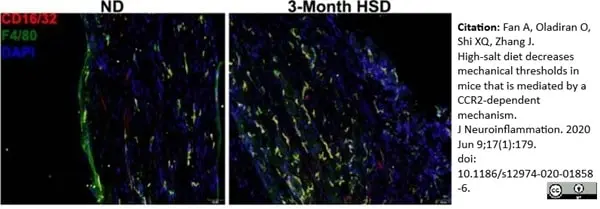
Rat anti Mouse F4/80 antibody, clone A3-1 (MCA497GA) used to label macrophages in murine sciatic nerve cryosections by immunofluorescence.
Image caption:
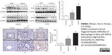
An HSD activates peripheral nerve macrophages.
Longitudinal sections of mouse sciatic nerves were stained with CD16/32 (FcγII/III receptor), F4/80, and DAPI, showing an increased F4/80+ macrophage density in HSD nerves primarily colocalized with CD16/32.
From: Fan A, Oladiran O, Shi XQ, Zhang J.
High-salt diet decreases mechanical thresholds in mice that is mediated by a CCR2-dependent mechanism.
J Neuroinflammation. 2020 Jun 9;17(1):179.
doi: 10.1186/s12974-020-01858-6.
This image is from an open access article distributed under terms of a Creative Commons Attribution License.

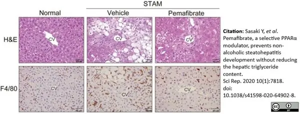
Rat anti Mouse F4/80 antibody, clone A3-1 (MCA497R) used to identify macrophages in murine liver by immunohistochemistry on formalin fixed, paraffin ebmedded tissue sections.
Image caption:
Pemafibrate improves macrovesicular steatosis and F4/80 positive cell accumulation in STAM mice liver. Representative gross morphology of liver, H&E stained and F4/80 stained liver section from normal, vehicle, and pemafibrate treated STAM mice.
From: Sasaki Y, Asahiyama M, Tanaka T, Yamamoto S, Murakami K, Kamiya W, Matsumura Y, Osawa T, Anai M, Fruchart JC, Aburatani H, Sakai J, Kodama T.
Pemafibrate, a selective PPARα modulator, prevents non-alcoholic steatohepatitis development without reducing the hepatic triglyceride content.
Sci Rep. 2020 May 8;10(1):7818.
doi: 10.1038/s41598-020-64902-8.
This image is from an open access article distributed under terms of a Creative Commons Attribution License.
Rat anti Mouse F4/80 antibody, clone A3-1 (MCA497R used to identify macrophages in murine
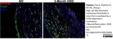
Image caption:
Macrophage marker upregulation in SCs and GERCs during TMEV infection. (a) Under steady-state conditions, F4/80 (green) was weakly expressed in SCs, but not in GERCs. Conversely, the SCs and GERCs migrating to the HCs during the virus infection exhibited strong F4/80 expression (green), indicating the virus-induced activation of SCs and GERCs as macrophages.
From: Hayashi Y, Suzuki H, Nakajima W, Uehara I, Tanimura A, Himeda T, Koike S, Katsuno T, Kitajiri SI, Koyanagi N, Kawaguchi Y, Onomoto K, Kato H, Yoneyama M, Fujita T, Tanaka N.
Cochlear supporting cells function as macrophage-like cells and protect audiosensory receptor hair cells from pathogens.
Sci Rep. 2020 Apr 21;10(1):6740.
doi: 10.1038/s41598-020-63654-9.
This image is from an open access article distributed under terms of a Creative Commons Attribution License.
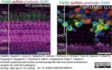
Rat anti Mouse F4/80 antibody, clone A3-1 (MCA497R) used to stain macrophages in formalin fixed paraffin embedded murine liver sections by immunohistochemistry.
Image caption:
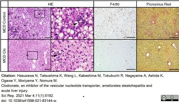
Pharmacologic inhibition of ATP signaling by clodronate protects against methionine- and choline-deficient diet-induced steatohepatitis. Ten-week-old C57BL/6 male mice were fed with normal chow (NC) or a methionine- and choline-deficient (MCD) diet for 4 weeks. The vehicle or 20 mg/kg body weight clodronate (Clo) was administered via daily subcutaneous injection for 4 weeks (n = 5–10 MCD diet-fed, per treatment; n = 3–5 NC-fed, per treatment). (A) Hematoxylin and eosin (HE) staining (arrowheads indicate inflammatory foci), F4/80 immunostaining, and Picrosirius Red staining of paraffin-embedded liver tissue from MCD diet-fed mice treated with the vehicle or clodronate. Scale bars, 100 μm.
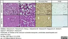
From: Hasuzawa N, Tatsushima K, Wang L, Kabashima M, Tokubuchi R, Nagayama A, Ashida K, Ogawa Y, Moriyama Y, Nomura M.
Clodronate, an inhibitor of the vesicular nucleotide transporter, ameliorates steatohepatitis and acute liver injury.
Sci Rep. 2021 Mar 4;11(1):5192.
doi: 10.1038/s41598-021-83144-w.
This image is from an open access article distributed under terms of a Creative Commons Attribution License.
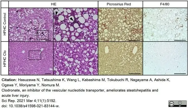
Rat anti Mouse F4/80 antibody, clone A3-1 (MCA497R) used to stain macrophages in formalin fixed paraffin embedded murine liver sections by immunohistochemistry.
Image caption:
Oral administration of clodronate prevents high-fat, high-cholesterol diet-induced steatohepatitis. Ten-week-old C57BL/6 male mice were fed a normal chow (NC) diet or a high-fat, high-cholesterol (HFHC) diet for 24 weeks. The vehicle or clodronate was administered in the drinking water (30 mg/kg body weight/day) for 24 weeks, n = 5 per treatment group. Hematoxylin and eosin (HE) staining (arrowheads indicate inflammatory foci), Picrosirius Red staining, and F4/80 immunostaining of paraffin-embedded mouse liver tissue. Scale bars, 100 μm.
From: Hasuzawa N, Tatsushima K, Wang L, Kabashima M, Tokubuchi R, Nagayama A, Ashida K, Ogawa Y, Moriyama Y, Nomura M.
Clodronate, an inhibitor of the vesicular nucleotide transporter, ameliorates steatohepatitis and acute liver injury.
Sci Rep. 2021 Mar 4;11(1):5192.
doi: 10.1038/s41598-021-83144-w.
This image is from an open access article distributed under terms of a Creative Commons Attribution License.
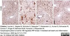
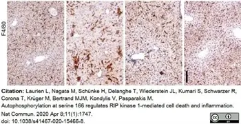
Rat anti Mouse F4/80 antibody, clone A3-1 (MCA497R) used to immunostain macrophages in murine liver by immunohistochemistry on formalin fixed, paraffin embedded tissue sections.
Image caption:
S166 phosphorylation is essential for RIPK1-dependent hepatitis and liver tumorigenesis in NEMOLPC-KO mice.
Representative images of liver sections from 8-week-old mice of the indicated genotypes that were immunostained with the indicated antibody.
Scale bars, 200 μm.
From: Laurien L, Nagata M, Schünke H, Delanghe T, Wiederstein JL, Kumari S, Schwarzer R, Corona T, Krüger M, Bertrand MJM, Kondylis V, Pasparakis M.
Autophosphorylation at serine 166 regulates RIP kinase 1-mediated cell death and inflammation.
Nat Commun. 2020 Apr 8;11(1):1747.
doi: 10.1038/s41467-020-15466-8..
This image is from an open access article distributed under terms of a Creative Commons Attribution License.
Rat anti Mouse F4/80 antibody, clone A3-1 (MCA497R) used to identify macrophages in murine liver by immunohistochemistry on formalin fixed, paraffin embedded tissue sections.
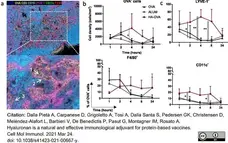
Image caption:
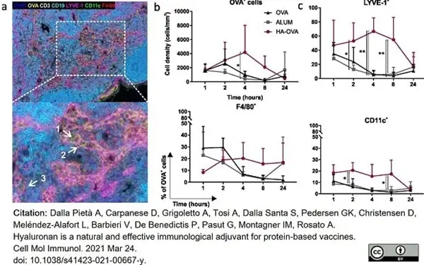
Fluorescence multiplex immunohistochemistry (mIHC) of draining LNs harvested at different time points after i.m. immunization. a Representative seven-color multispectral image of a lymph node collected 4 h after i.m. injection of HA-OVA, scanned at ×20 (upper panel) and ×40 (lower panel) magnification. The white arrows in the ×40 image show examples of an OVA+ DC (1, CD11c), OVA+ lymphatic endothelial cell (2, LYVE-1), and OVA+ macrophage (3, F4/80). b Quantification of OVA+ cell density (cells/mm2) in LNs of mice subjected to i.m. injection with 10 μg of OVA alone, mixed with alum, or conjugated to HA. c Percentage of OVA+ cells among LYVE-1+, CD11c+, and F4/80+ cells. Cumulative data from three independent experiments are presented (n = 3 mice; 6 LNs/group). Multiple t-tests were performed (*P <0.05, **P <0.01, ***P <0.001, ****P <0.0001; P >0.05 if not indicated).
From: Dalla Pietà A, Carpanese D, Grigoletto A, Tosi A, Dalla Santa S, Pedersen GK, Christensen D, Meléndez-Alafort L, Barbieri V, De Benedictis P, Pasut G, Montagner IM, Rosato A.
Hyaluronan is a natural and effective immunological adjuvant for protein-based vaccines.
Cell Mol Immunol. 2021 18(5):1197-1210.
doi: 10.1038/s41423-021-00667-y.
This image is from an open access article distributed under terms of a Creative Commons Attribution License.
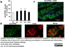
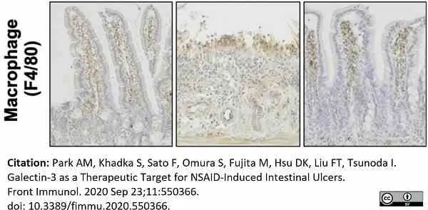
Rat anti Mouse F4/80 antibody, clone A3-1 (MCA497GA) used to label macrophages in murine intestinal tissue sections by immunofluorescence.
Image caption:
Expression of resolvin D1 receptor, lipoxin A4 receptor /formyl peptide receptor 2 (ALX/FPR2) in the small intestine and its dynamics on indomethacin-induced small intestinal damage.
(b) Relative amount of normalized ALX/FPR2 protein expressed as a percentage of untreated control group (0 h group). *p <0.05 vs. 0 h group. N = 4. (c) Representative image of immunofluorescent staining for localization analysis of ALX/FPR2 in damaged small intestinal mucosa. DAPI: 4’,6-diamidino-2-phenylindole. Bars in histological images: 50 μm. (d) Expression of ALX/FPR2 and its colocalization with a marker of mature macrophages F4/80. These are enlarged views of the part indicated by white squares in image.
From: Kuzumoto T, Tanigawa T, Higashimori A, Kitamura H, Nadatani Y, Otani K, et al. (2021)
Protective role of resolvin D1, a pro-resolving lipid mediator, in nonsteroidal anti-inflammatory drug-induced small intestinal damage.
PLoS ONE 16(5): e0250862.
doi: 10.1371/journal.pone.0250862
This image is from an open access article distributed under terms of a Creative Commons Attribution License.
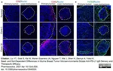
Rat anti Mouse F4/80 antibody, clone A3-1 (MCA497R) used to identify macrophages in murine tumor tissues.
Image caption:
Evaluation of immune microenvironment in parental-, clone-1-, clone-16-derived primary tumor and liver metastases. Immunofluorescence imaging of (a) CD8, (c) CD45, and (e) F480 in both primary and liver metastases. Scale bar, 100 μm.
From: Liu YT, Goel S, Kai M, Moran Guerrero JA, Nguyen T, Mai J, Shen H, Ziemys A, Yokoi K.
Seed- and Soil-Dependent Differences in Murine Breast Tumor Microenvironments Dictate Anti-PD-L1 IgG Delivery and Therapeutic Efficacy.
Pharmaceutics. 2021 Apr 10;13(4):530.
doi: 10.3390/pharmaceutics13040530.
This image is from an open access article distributed under terms of a Creative Commons Attribution License.
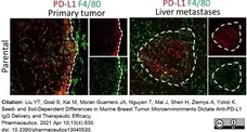
Rat anti Mouse F4/80 antibody, clone A3-1 (MCA497R) used to identify macrophages in murine tumor tissues.
Image caption:
Verification of immune maker that express PD-L1 by immunohistochemical imaging of primary tumor and liver metastases. Immunofluorescence imaging of PD-L1 expression (red) and F4/80 (green) in parental- primary tumors and liver metastases. Scale bar, 100μm
From: Liu YT, Goel S, Kai M, Moran Guerrero JA, Nguyen T, Mai J, Shen H, Ziemys A, Yokoi K.
Seed- and Soil-Dependent Differences in Murine Breast Tumor Microenvironments Dictate Anti-PD-L1 IgG Delivery and Therapeutic Efficacy.
Pharmaceutics. 2021 Apr 10;13(4):530.
doi: 10.3390/pharmaceutics13040530.
This image is from an open access article distributed under terms of a Creative Commons Attribution License.
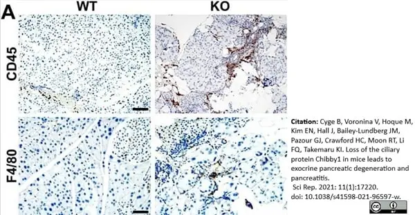
Rat anti Mouse F4/80 antibody, clone A3-1 (MCA497R) used to label macrophages in murine pancreas by immunohistochemistry on formalin fixed, paraffin embedded tissue sections.
Image caption:
Fibrosis and chronic inflammation in the Cby1-KO pancreas.
Pancreatic sections from adult mice were immunostained with antibodies for the inflammation markers CD45 (leukocytes) and F4/80 (macrophages). Scale bars: CD45, 100 μm; F4/80, 50 μm.
From: Cyge B, Voronina V, Hoque M, Kim EN, Hall J, Bailey-Lundberg JM, Pazour GJ, Crawford HC, Moon RT, Li FQ, Takemaru KI.
Loss of the ciliary protein Chibby1 in mice leads to exocrine pancreatic degeneration and pancreatitis.
Sci Rep. 2021 Aug 26;11(1):17220.
doi: 10.1038/s41598-021-96597-w.
This image is from an open access article distributed under terms of a Creative Commons Attribution License.
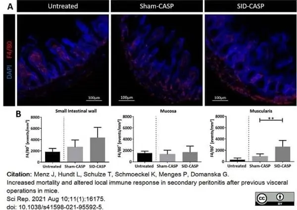
Rat anti Mouse F4/80 antibody, clone A3-1 (MCA497R) used to label macrophages in murine intestine by immunofluorescence.
Image caption:
Representative immunofluorescence images of macrophages (F4/80+) in the small intestinal wall. C57BL/6 N mice underwent CASP following SID or a sham operation. (A) Aboral 1 cm of ileum was cryofixed and sections were stained for infiltrating macrophages using antibody against F4/80-antigen labelled with Alexa-647 dye and DAPI for staining of cell nuclei. Photos were taken from at least 6 mice per group. (Magnification 20x) (B) Fluorescence was assigned to the mucosal region and outer layers of muscularis and serosa (see materials and methods for detail). F4/80 positive events in (B1) whole cross section of small intestine, (B2) mucosa and (B3) muscularis were compared between operated groups. N = 6 to 8 (technical failures have been excluded); Mann–Whitney U-Test; **p <0.01.
From: Menz J, Hundt L, Schulze T, Schmoeckel K, Menges P, Domanska G.
Increased mortality and altered local immune response in secondary peritonitis after previous visceral operations in mice.
Sci Rep. 2021 Aug 10;11(1):16175.
doi: 10.1038/s41598-021-95592-5.
This image is from an open access article distributed under terms of a Creative Commons Attribution License.
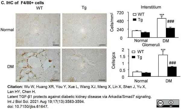
Rat anti Mouse F4/80 antibody, clone A3-1 (MCA497) used to detect F4/80 expressing macrophages in murine renal tissue by immunohistochemistry on formalin fixed, paraffin embedded sections.
Image caption:
Latent TGF-β1 attenuates renal fibrosis and inflammation in streptozotocin-induced type 1 diabetic mice. Mice were euthanized 16 weeks after diabetes induction, and renal tissues were collected. (C) Immunohistochemistry of F4/80-positive cells showing macrophage infiltration in glomeruli and tubulointerstitium. DM, diabetes mellitus. WT, latent TGF-β1 wild-type mice. Tg, latent TGF-β1 transgenic mice. Data represent the means ± SEM for groups of six animals. Scale bar: 50 μm. *P <0.05, **P <0.01, ***P <0.001 versus normal mice; #P <0.05, ##P <0.01, ###P <0.001 versus WT DM mice.
From: Wu W, Huang XR, You Y, Xue L, Wang XJ, Meng X, Lin X, Shen J, Yu X, Lan HY, Chen H.
Latent TGF-β1 protects against diabetic kidney disease via Arkadia/Smad7 signaling.
Int J Biol Sci. 2021 Aug 19;17(13):3583-94.
doi: 10.7150/ijbs.61647.
This image is from an open access article distributed under terms of a Creative Commons Attribution License.
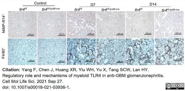
Rat anti Mouse F4/80 antibody, clone A3-1 (MCA497R) used to label macrophages in murine kidneys by immunohistochemistry on formalin fixed, paraffin embedded tissue sections.
Image caption:
Deficiency of myeloid TLR4 inhibits macrophage-dominant renal infiltrates in experimental anti-GBM GN. A Representative images of neutrophil (NIMP-R14+) and macrophage (F4/80+) accumulation in kidney sections (magnification × 40).
From: Yang F, Chen J, Huang XR, Yiu WH, Yu X, Tang SCW, Lan HY.
Regulatory role and mechanisms of myeloid TLR4 in anti-GBM glomerulonephritis.
Cell Mol Life Sci. 2021 Sep 27 [Epub ahead of print].
doi: 10.1007/s00018-021-03936-1. .
This image is from an open access article distributed under terms of a Creative Commons Attribution License.
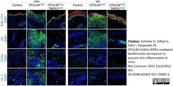
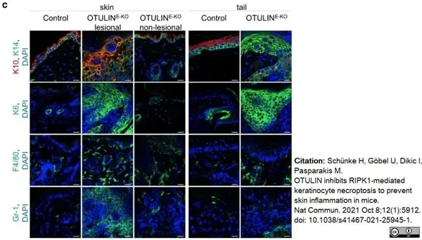
Rat anti Mouse F4/80 antibody, clone A3-1 (MCA497R) used to detect macrophages in mouse skin by immunofluorescence.
Image caption:
Representative images of skin sections from 3-week-old mice of the indicated genotypes, immunostained with anti-K10, anti-K14 (n = 5), anti-K6 (n = 5), anti-F4/80 (n = 3) and anti-Gr-1 (n = 3) antibodies and counterstained with DAPI (DNA stain). Scale bars: K10, K14, K6 = 20 μm; F4/80, Gr-1 = 30 μm.
From: Schünke H, Göbel U, Dikic I, Pasparakis M.
OTULIN inhibits RIPK1-mediated keratinocyte necroptosis to prevent skin inflammation in mice.
Nat Commun. 2021 Oct 8;12(1):5912.
doi: 10.1038/s41467-021-25945-1.
This image is from an open access article distributed under terms of a Creative Commons Attribution License.
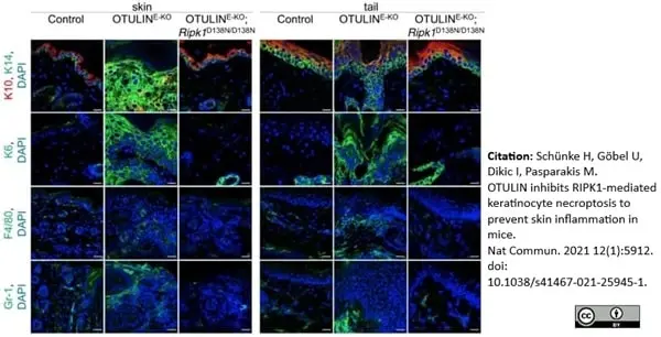
Rat anti Mouse F4/80 antibody, clone A3-1 (MCA497R) used to detect macrophages in mouse skin by immunofluorescence.
Image caption:
Representative images of skin sections from 3-week-old mice of the indicated genotypes, immunostained with anti-K10, anti-K14 (n = 4), anti-K6 (n = 3), anti-F4/80 (n = 3) and anti-Gr-1 (n = 3) antibodies and counterstained with DAPI (DNA stain). Scale bars: K10, K14, K6 = 20 μm; F4/80, Gr-1 = 30 μm.
From: Schünke H, Göbel U, Dikic I, Pasparakis M.
OTULIN inhibits RIPK1-mediated keratinocyte necroptosis to prevent skin inflammation in mice.
Nat Commun. 2021 Oct 8;12(1):5912.
doi: 10.1038/s41467-021-25945-1.
This image is from an open access article distributed under terms of a Creative Commons Attribution License.

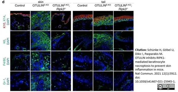
Rat anti Mouse F4/80 antibody, clone A3-1 (MCA497R) used to detect macrophages in mouse skin by immunofluorescence.
Image caption:
Representative images from skin sections from 3-week-old mice of the indicated genotypes, immunostained with anti-K10, anti-K14 (n = 5), anti-K6 (n = 5), anti-F4/80 (n = 5) and anti-Gr-1 (n = 5) antibodies and counterstained with DAPI (DNA stain). Scale bars: K10, K14, K6 = 20 μm; F4/80, Gr-1 = 30 μm.
From: Schünke H, Göbel U, Dikic I, Pasparakis M.
OTULIN inhibits RIPK1-mediated keratinocyte necroptosis to prevent skin inflammation in mice.
Nat Commun. 2021 Oct 8;12(1):5912.
doi: 10.1038/s41467-021-25945-1.
This image is from an open access article distributed under terms of a Creative Commons Attribution License.

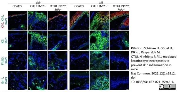
Rat anti Mouse F4/80 antibody, clone A3-1 (MCA497R) used to detect macrophages in mouse skin by immunofluorescence.
Image caption:
Representative images from skin sections from 3-week-old mice of the indicated genotypes, immunostained with anti-K10, anti-K14 (n = 4), anti-K6 (n = 3), anti-F4/80 (n = 3) and anti-Gr-1 (n = 3) antibodies and counterstained with DAPI (DNA stain). Scale bars: K10, K14, K6 = 20 μm; F4/80, Gr-1 = 30 μm.
From: Schünke H, Göbel U, Dikic I, Pasparakis M.
OTULIN inhibits RIPK1-mediated keratinocyte necroptosis to prevent skin inflammation in mice.
Nat Commun. 2021 Oct 8;12(1):5912.
doi: 10.1038/s41467-021-25945-1.
This image is from an open access article distributed under terms of a Creative Commons Attribution License.

Rat anti Mouse F4/80 antibody, clone A3-1 (MCA497R) used to label macrophages in murine kidney by immunohistochemistry on formalin fixed, paraffin embedded tissue sections.
Image caption:
Circulating FGF-2-induced tubulo-interstitial changes in HIV-Tg26 mice and mimic those seen in childhood HIVAN.
Representative immunohistochemistry staining for PCNA+, vimentin+ (both brown), α-smooth muscle actin+ (α-SMA+) and F4/80+ macrophages (both red) in renal sections harvested from WT and HIV-Tg26 mice 14 days after the injections of rAd-LacZ (control) or rAd-FGF-2 vectors (n=3-4 mice per group). Scale bar: 20 μ m.
From: Das JR, Jerebtsova M, Tang P, Li J, Yu J, Ray PE.
Circulating fibroblast growth factor-2 precipitates HIV nephropathy in mice.
Dis Model Mech. 2021 Jul 1;14(7):dmm048980.
doi: 10.1242/dmm.048980.
This image is from an open access article distributed under terms of a Creative Commons Attribution License.
Insert meta-description and applications
Image caption:
Insert image caption
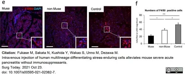
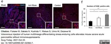
From: Fukase M, Sakata N, Kushida Y, Wakao S, Unno M, Dezawa M. Intravenous injection of human multilineage-differentiating stress-enduring cells alleviates mouse severe acute pancreatitis without immunosuppressants. Surg Today. 2021 Oct 23. doi: 10.1007/s00595-021-02382-7.
This image is from an open access article distributed under terms of a Creative Commons Attribution License.
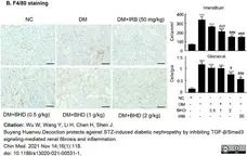
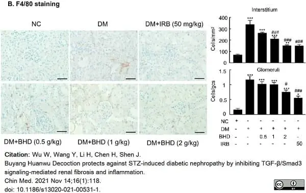
Rat anti Mouse F4/80 antibody, clone A3-1 (MCA497R) used to identify macrophages in murine renal tissue by immunohistochemistry on formalin fixed, paraffin embedded sections.
Image caption:
BHD reduces renal fibrosis and inflammation in STZ-induced diabetic mice. B Immunohistochemistry of F4/80-positive cells showing macrophage infiltration in glomeruli and tubulointerstitium. DM, diabetes mellitus. Irbesartan (IRB) was taken as a positive control. Data represent the means ± SEM for groups of six animals. Scale bar: 50μm. *P <0.05, **P <0.01, ***P <0.001 versus normal mice. #P <0.05, ##P <0.01, ###P <0.001 versus DM mice.
From: Wu W, Wang Y, Li H, Chen H, Shen J.
Buyang Huanwu Decoction protects against STZ-induced diabetic nephropathy by inhibiting TGF-β/Smad3 signaling-mediated renal fibrosis and inflammation.
Chin Med. 2021 Nov 14;16(1):118.
doi: 10.1186/s13020-021-00531-1.
This image is from an open access article distributed under terms of a Creative Commons Attribution License.
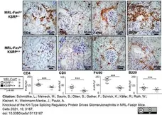
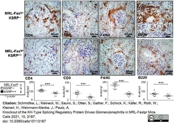
Rat anti mouse F4/80 antibody, clone A3-1 (MCA497G) used to identify macrophages in murine renal tissue by immunohistochemistry on formalin fixed, paraffin embedded sections.
Image caption:
Frequencies of intra-renal CD4, CD8, F4/80 and B220 positive cells in the kidneys of 19-week-old female MRL-FaslprKSRP−/− mice compared to MRL-FaslprKSRP+/+ mice Immunostaining was used to analyze intra-renal frequency of CD4, CD8, F4/80 and B220 positive cells in the kidneys of female 19-week-old MRL-FaslprKSRP+/+ and MRL-FaslprKSRP−/− mice. Data presented are the mean + SEM of the score with 8 to 12 animals per genotype (*** p <0.001 compared to WT, t-test). Representative image of immunostaining with 40× magnification.
From: Schmidtke, L.; Meineck, M.; Saurin, S.; Otten, S.; Gather, F.; Schrick, K.; Käfer, R.; Roth, W.; Kleinert, H.; Weinmann-Menke, J.; Pautz, A.
Knockout of the KH-Type Splicing Regulatory Protein Drives Glomerulonephritis in MRL-Faslpr Mice.
Cells 2021, 10, 3167.
10.3390/cells10113167
This image is from an open access article distributed under terms of a Creative Commons Attribution License.
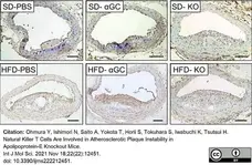
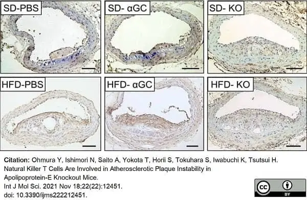
Rat anti Mouse F4/80 antibody, clone A3-1 (MCA497R) used to label macrophages in atherosclerotic plaques by immunohistochemistry on formalin fixed cryosections.
Image caption:
Representative photomicrographs of anti-F4/80 immunohistochemistry of cross-sections at the brachiocephalic artery from 6 groups of SD-PBS (n = 4), SD-αGC (n = 6), SD-KO (n = 3), HFD-PBS (n = 18), HFD-αGC (n = 16), and HFD-KO (n = 7) mice. Scale bar = 100 μm.
From: Ohmura Y, Ishimori N, Saito A, Yokota T, Horii S, Tokuhara S, Iwabuchi K, Tsutsui H.
Natural Killer T Cells Are Involved in Atherosclerotic Plaque Instability in Apolipoprotein-E Knockout Mice.
Int J Mol Sci. 2021 Nov 18;22(22):12451.
doi: 10.3390/ijms222212451.
This image is from an open access article distributed under terms of a Creative Commons Attribution License.
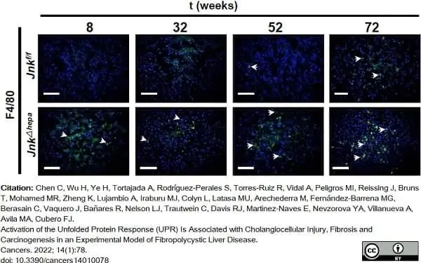
Rat anti mouse F4/80 antibody, clone A3-1 (MCA497R) used to visualize macrophages in murine hepatic tissues by immunofluorescence on cryosections.
Image caption:
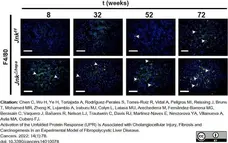
Representative IF microphotographs of 8 to 72 week-old Jnkf/f and JnkΔhepa mice for F4/80. Scale bar, 200 μm.
From: Chen C, Wu H, Ye H, Tortajada A, Rodríguez-Perales S, Torres-Ruiz R, Vidal A, Peligros MI, Reissing J, Bruns T, Mohamed MR, Zheng K, Lujambio A, Iraburu MJ, Colyn L, Latasa MU, Arechederra M, Fernández-Barrena MG, Berasain C, Vaquero J, Bañares R, Nelson LJ, Trautwein C, Davis RJ, Martinez-Naves E, Nevzorova YA, Villanueva A, Avila MA, Cubero FJ.
Activation of the Unfolded Protein Response (UPR) Is Associated with Cholangiocellular Injury, Fibrosis and Carcinogenesis in an Experimental Model of Fibropolycystic Liver Disease.
Cancers. 2022; 14(1):78.
doi: 10.3390/cancers14010078
This image is from an open access article distributed under terms of a Creative Commons Attribution License.
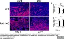
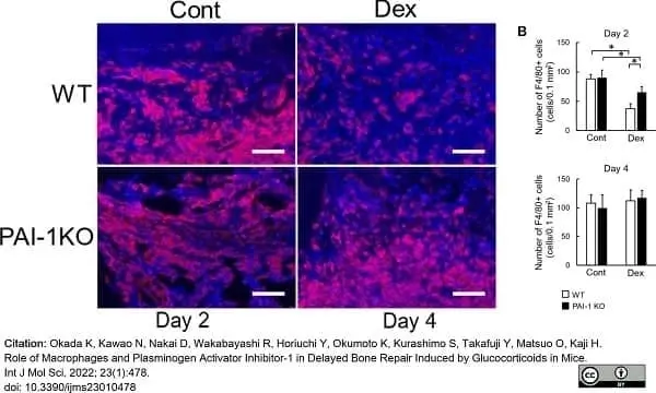
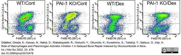
Rat anti Mouse F4/80 antibody, clone A3-1 (MCA497R) used to label macrophages in murine bone marrow by immunofluorescence on formalin fixed, paraffin embedded tissue sections.
Image caption:
Effects of PAI-1 deficiency on Dex-induced decreases in macrophage number after femoral bone injury.
(A) Microphotographs of F4/80-positive cells at damaged sites 2 days after femoral bone injury in control and Dex-treated WT and PAI-1 KO mice. Scale bars indicate 50μm. (B) Number of F4/80-positive cells at damaged sites 2 and 4 days after femoral bone injury in control and Dex-treated WT and PAI-1 KO mice. Data represent the mean ± SEM: n = 5 mice in each group. * p <0.05.
Cont, control; DAPI, 4′,6-diamidino-2-phenylindole.
From: Okada, K.; Kawao, N.; Nakai, D.; Wakabayashi, R.; Horiuchi, Y.; Okumoto, K.; Kurashimo, S.; Takafuji, Y.; Matsuo, O.; Kaji, H.
Role of Macrophages and Plasminogen Activator Inhibitor-1 in Delayed Bone Repair Induced by Glucocorticoids in Mice
Int. J Mol Sci 2022, 23, 478.
doi: 10.3390/ijms23010478
This image is from an open access article distributed under terms of a Creative Commons Attribution License.
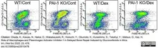
R-phycoerythrin conjugated Rat anti Mouse F4/80 antibody, clone A3-1 (MCA497PE) used to label macrophages in murine bone marrow by Flow cytometry.
Image caption:
Effects of Dex and PAI-1 deficiency on the expression of macrophage-related factors in macrophages derived from the bone marrow of damaged femurs.
Flow cutometric analysis was used to identify F4/80 and CD11b double-positive cells from damaged femurs 2 days after bone injury.
From: Okada, K.; Kawao, N.; Nakai, D.; Wakabayashi, R.; Horiuchi, Y.; Okumoto, K.; Kurashimo, S.; Takafuji, Y.; Matsuo, O.; Kaji, H.
Role of Macrophages and Plasminogen Activator Inhibitor-1 in Delayed Bone Repair Induced by Glucocorticoids in Mice
Int. J Mol Sci 2022, 23, 478.
doi: 10.3390/ijms23010478
This image is from an open access article distributed under terms of a Creative Commons Attribution License.

Rat anti Mouse F4/80 antibody, clone A3-1 (MCA497R) used to label macrophages in murine liver by immunohistochemistry on formalin fixed, paraffin embedded tissue sections and by western blotting on hepatic lysates.
Image caption:
Depletion of Kupffer cells enhances Efavirenz-induced liver injury in vivo. (A–C) Confirmation of macrophage depletion in mice administered with gadolinium chloride (GdCl3): relative mRNA expression levels of F4/80 (RT-PCR), representative images showing F4/80 immunostaining in murine livers (magnification 20×), and representative Western Blot image of F4/80 (each blot lane represents one mouse per condition—saline and GdCl3−) and summary of densitometry data (n = 6).
From: Alegre, F.; Martí-Rodrigo, A.; Polo, M.; Ortiz-Masiá, D.; Bañuls, C.; Pinti, M.; Álvarez, Á.; Apostolova, N.; Esplugues, J.V.; Blas-García, A.
Macrophages Modulate Hepatic Injury Involving NLRP3 Inflammasome: The Example of Efavirenz.
Biomedicines 2022, 10, 109.
doi: 10.3390/biomedicines10010109
This image is from an open access article distributed under terms of a Creative Commons Attribution License.
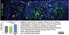
Rat anti Mouse F4/80 antibody, clone A3-1 (MCA497R) used to label macrophages in murine umor sections by immunofluorescence.
Image caption:
MAG-OV+MT stimulates a T lymphocyte-mediated antitumor response. Representative images of EO771 tumor sections examined by terminal immunofluorescent staining and their quantification of signals for macrophages (F4/80). Data are shown as mean ± SD (n = 4). Statistical significance was determined by one-way ANOVA with a Tukey post hoc test.
From: Howard FHN, Al-Janabi H, Patel P, Cox K, Smith E, Vadakekolathu J, Pockley AG, Conner J, Nohl JF, Allwood DA, Collado-Rojas C, Kennerley A, Staniland S, Muthana M.
Nanobugs as Drugs: Bacterial Derived Nanomagnets Enhance Tumor Targeting and Oncolytic Activity of HSV-1 Virus.
Small. 2022 Jan 25:e2104763.
doi: 10.1002/smll.202104763.
This image is from an open access article distributed under terms of a Creative Commons Attribution License.
Alexa Fluor® 647 conjugated Rat anti mouse F4/80 antibody, clone A3-1 (MCA497A647) used to label murine spinal cord macrophages by immunofluorescence.
Image caption:
Immunofluorescence staining for tdTomato (red), CD11b (yellow), F4/80 (cyan), MHCII (blue) and MerTK (green) of spinal cord from mice at peak EAE. A representative picture of three individual mouse samples is shown (n = 3); scale bar, 50 μm (low magnification) and 10 μm (inset).
From: Sarkar T, Patro N, Patro IK.
Perinatal exposure to synergistic multiple stressors lead to cellular and behavioral deficits mimicking Schizophrenia like pathology.
Biol Open. 2022 Feb 2:bio.058870.
doi: 10.1242/bio.058870.
This image is from an open access article distributed under terms of a Creative Commons Attribution License.
Rat anti Mouse F4/80 antibody, clone A3-1 (MCA497R) used to identify infiltrating macrophages in mouse renal tissue by immunohistochemistry on formalin fixed, paraffin embedded tissue sections.
Image caption:
Notch3 overexpression results in severe tubulointerstitial inflammation and fibrosis. F4/80 immunostaining was used for evaluation of infiltrating macrophages, α-smooth muscle actin (αSMA) for fibroblast activation.
From: Djudjaj S, Kavvadas P, Prakoura N, Bülow RD, Migeon T, Placier S, Chadjichristos CE, Boor P, Chatziantoniou C.
Activation of Notch3 in Renal Tubular Cells Leads to Progressive Cystic Kidney Disease. Int J Mol Sci. 2022 Jan 14;23(2):884.
doi: 10.3390/ijms23020884.
This image is from an open access article distributed under terms of a Creative Commons Attribution License.
Rat anti Mouse F4/80 antibody, clone A3-1 (MCA497R) used to label macrophages in murine pulmonary tissue by immunohistochemistry on formalin fixed, paraffin embedded sections.
Image caption:
A–C Representative images (on the left) and quantifications (on the right) showing F4/80+ cells staining positive for A pSTAT3, B PPARγ, C c-MYC immunostainings in alveolar parenchyma and adenoma sections from BRAFV600E and control mice.
Quantifications were performed on at least five different areas of the lung sections in a random way. Data are expressed as mean ± SEM; n = animals per group. *P <0.05; **P <0.01; ***P <0.001, ns = not significant. (ANOVA test with Dunnet post-hoc correction (A, B, C)). Arrows point to selected positive cells for the indicated marker. Insets: magnifications of areas inside dashed squares.
From: Bosso G, Lanuza-Gracia P, Piñeiro-Hermida S, Yilmaz M, Serrano R, Blasco MA.
Early differential responses elicited by BRAFV600E in adult mouse models.
Cell Death Dis. 2022 Feb 10;13(2):142.
doi: 10.1038/s41419-022-04597-z.
This image is from an open access article distributed under terms of a Creative Commons Attribution License.
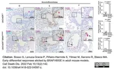
Rat anti Mouse F4/80 antibody, clone A3-1 (MCA497R) used to label macrophages in murine pulmonary tissue by immunohistochemistry on formalin fixed, paraffin embedded sections.
Image caption:
D–F Representative images (top) and quantifications (bottom) showing F4/80+ cells staining positive for D pSTAT3, E PPARγ, F c-MYC double immunostainings in bronchi/bronchiolar parenchyma of lung sections from BRAFV600E and control mice.
Quantifications were performed on at least five different areas of the lung sections in a random way. Data are expressed as mean ± SEM; n = animals per group. *P <0.05; **P <0.01; ***P <0.001, ns = not significant. (ANOVA test with T Student’s test unpaired (D, E, F)). Arrows point to selected positive cells for the indicated marker. Insets: magnifications of areas inside dashed squares.
From: Bosso G, Lanuza-Gracia P, Piñeiro-Hermida S, Yilmaz M, Serrano R, Blasco MA.
Early differential responses elicited by BRAFV600E in adult mouse models.
Cell Death Dis. 2022 Feb 10;13(2):142.
doi: 10.1038/s41419-022-04597-z.
This image is from an open access article distributed under terms of a Creative Commons Attribution License.
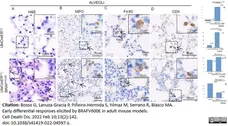
Rat anti Mouse F4/80 antibody, clone A3-1 (MCA497R) used to label macrophages in murine pulmonary tissue by immunohistochemistry on formalin fixed, paraffin embedded sections.
Image caption:
Ubiquitous expression of the oncogenic variant of BRAF results in neutrophils infiltration in alveoli and F4/80-positive cells infiltration in bronchial epithelium as well as in hyperplastic nodules.
Representative images (top) and quantifications (bottom) showing H/E, Myeloperoxidase (MPO), F4/80, and CD4 immunostainings in alveoli (A), (B), (C), (D), from BRAFV600E and control mice. Quantifications in alveoli and bronchi were performed on at least five different areas of the lung sections in a random way. Data are expressed as mean ± SEM; n= animals per group, * P<0.05; ** P<0.01; *** P<0.001, ns=not significant. (T Student’s test unpaired). Arrows point to selected positive cells for the indicated marker. Insets: magnifications of areas inside dashed squares.
From: Bosso G, Lanuza-Gracia P, Piñeiro-Hermida S, Yilmaz M, Serrano R, Blasco MA.
Early differential responses elicited by BRAFV600E in adult mouse models.
Cell Death Dis. 2022 Feb 10;13(2):142.
doi: 10.1038/s41419-022-04597-z.
This image is from an open access article distributed under terms of a Creative Commons Attribution License.
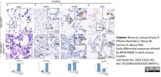
Rat anti Mouse F4/80 antibody, clone A3-1 (MCA497R) used to label macrophages in murine pulmonary tissue by immunohistochemistry on formalin fixed, paraffin embedded sections.
Image caption:
Ubiquitous expression of the oncogenic variant of BRAF results in neutrophils infiltration in alveoli and F4/80-positive cells infiltration in bronchial epithelium as well as in hyperplastic nodules.
Representative images (top) and quantifications (bottom) showing H/E, Myeloperoxidase (MPO), F4/80, and CD4 immunostainings in alveoli (A), (B), (C), (D), from BRAFV600E and control mice. Quantifications in alveoli and bronchi were performed on at least five different areas of the lung sections in a random way. Data are expressed as mean ± SEM; n= animals per group, * P<0.05; ** P<0.01; *** P<0.001, ns=not significant. (T Student’s test unpaired). Arrows point to selected positive cells for the indicated marker. Insets: magnifications of areas inside dashed squares.
From: Bosso G, Lanuza-Gracia P, Piñeiro-Hermida S, Yilmaz M, Serrano R, Blasco MA.
Early differential responses elicited by BRAFV600E in adult mouse models.
Cell Death Dis. 2022 Feb 10;13(2):142.
doi: 10.1038/s41419-022-04597-z.
This image is from an open access article distributed under terms of a Creative Commons Attribution License.

Rat anti Mouse F4/80 antibody, clone A3-1 (MCA497R) used to label macrophages in murine pulmonary tissue by immunohistochemistry on formalin fixed, paraffin embedded sections.
Image caption:
Ubiquitous expression of the oncogenic variant of BRAF results in neutrophils infiltration in bronchi and F4/80-positive cells infiltration in bronchial epithelium as well as in hyperplastic nodules.
Representative images (top) and quantifications (bottom) showing H/E, Myeloperoxidase (MPO), F4/80, and CD4 immunostainings in in bronchi respectively (E) F4/80, (F) MPO and (G) CD4 from BRAFV600E and control mice. Quantifications in alveoli and bronchi were performed on at least five different areas of the lung sections in a random way. Data are expressed as mean ± SEM; n= animals per group, * P<0.05; ** P<0.01; *** P<0.001, ns=not significant. (T Student’s test unpaired). Arrows point to selected positive cells for the indicated marker. Insets: magnifications of areas inside dashed squares.
From: Bosso G, Lanuza-Gracia P, Piñeiro-Hermida S, Yilmaz M, Serrano R, Blasco MA.
Early differential responses elicited by BRAFV600E in adult mouse models.
Cell Death Dis. 2022 Feb 10;13(2):142.
doi: 10.1038/s41419-022-04597-z.
This image is from an open access article distributed under terms of a Creative Commons Attribution License.
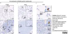
Rat anti Mouse F4/80 antibody, clone A3-1 (MCA497R) used to label macrophages in murine pulmonary tissue by immunohistochemistry on formalin fixed, paraffin embedded sections.
Image caption:
Ubiquitous expression of the oncogenic variant of BRAF results in neutrophils infiltration in bronchi and F4/80-positive cells infiltration in bronchial epithelium as well as in hyperplastic nodules.
Representative images (top) and quantifications (bottom) showing H/E, Myeloperoxidase (MPO), F4/80, and CD4 immunostainings in in bronchi respectively (E) F4/80, (F) MPO and (G) CD4 from BRAFV600E and control mice Arrows point to selected positive cells for the indicated marker. Insets: magnifications of areas inside dashed squares.
From: Bosso G, Lanuza-Gracia P, Piñeiro-Hermida S, Yilmaz M, Serrano R, Blasco MA.
Early differential responses elicited by BRAFV600E in adult mouse models.
Cell Death Dis. 2022 Feb 10;13(2):142.
doi: 10.1038/s41419-022-04597-z.
This image is from an open access article distributed under terms of a Creative Commons Attribution License.


Rat anti Mouse F4/80 antibody, clone A3-1 (MCA497R) used to identify macrophages in murine adipose tissue by immunohistochemistry on formalin fixed, paraffin embedded tissue sections.
Image caption:
Acute endotoxemia affects adipose tissue but does not lead to weight gain in mice under chow diet. Swiss Webster mice were submitted to intraperitoneal administration of 8 consecutive doses of 10 mg/kg LPS, every 3 days. Histological sections of epididymal fat pads after LPS challenges showing adipocyte morphology on hematoxylin and eosin staining and macrophage infiltration (F4/80+).
From: Mendes de Oliveira, E.; Silva, J.C.; Ascar, T.P.; Sandri, S.; Marchi, A.F.; Migliorini, S.; Nakaya, H.T.I.; Fock, R.A.; Campa, A.
Acute Inflammation Is a Predisposing Factor for Weight Gain and Insulin Resistance.
Pharmaceutics 2022, 14, 623.
doi:10.3390/pharmaceutics14030623
This image is from an open access article distributed under terms of a Creative Commons Attribution License.
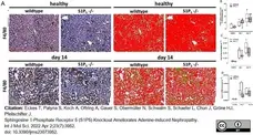
Rat anti Mouse F4/80 antibody, clone A3-1 (MCA497R) used to label macrophages in murine renal tissue by immunohistochemistry on formalin fixed, paraffin embedded tissue sections.
Image caption:
Presence of renal mononuclear phagocytes and expression of inflammasome component in kidney cortex. Expression of markers of mononuclear phagocytes were compared between wild-type (C57BL/6J) and S1P5 knockout mice (S1P5-/-) without (healthy) and with adenine-rich diet for 7 and 14 days. (A) Paraffin-embedded kidney cortex sections were subjected to immunohistochemical staining for F4/80 expression with DAB (brown) and counterstained with Mayer’s hematoxylin solution (blue). Algorithm-based detection of the staining was used to visualize and quantify the area covered by diaminobenzidine (green) and hematoxylin (red). (B) Median DAB area/hematoxylin area ratio was determined for up to 30 single pictures per tissue section, and the medians of all sections per group are shown here. (C,D) Expression levels of Nlrp3 and Adgre1 (also known as Emr1) mRNA were determined by TaqMan® analysis as described in Materials and Methods. The internal standard used was 18s ribosomal RNA. Data are shown as box plots with a line at the median and whiskers from minimum to maximum value; n = 5–9; * p ≤0.05 as indicated; # p ≤0.05; ## p ≤0.01; ### p ≤0.001 compared to the respective healthy controls.
From: Eckes T, Patyna S, Koch A, Oftring A, Gauer S, Obermüller N, Schwalm S, Schaefer L, Chun J, Gröne HJ, Pfeilschifter J.
Sphingosine 1-Phosphate Receptor 5 (S1P5) Knockout Ameliorates Adenine-Induced Nephropathy.
Int J Mol Sci. 2022 Apr 2;23(7):3952.
doi: 10.3390/ijms23073952.
This image is from an open access article distributed under terms of a Creative Commons Attribution License.
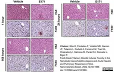
Rat anti mouse F4/80 antibody, clone A3-1 (MCA497R) used to label murine hepatic macrophages following vehicle or E171 administration by immunohistochemistry on formalin fixed, paraffin embedded tissue sections.
Image caption:
Pathological examination of livers of mice treated with E171. Representative images of liver hematoxylin–eosin (HE) and F4/80 immunostaining of mice injected with either vehicle or E171 and euthanized 1, 24, or 168 h after injection. Small microgranulomas were detected at 1 h and 24 h after E171 injection, but not at 168 h, indicative of low-grade transient inflammation due to E171. Scale bars = 50 μm.
From: Sitia G, Fiordaliso F, Violatto MB, Alarcon JF, Talamini L, Corbelli A, Ferreira LM, Tran NL, Chakraborty I, Salmona M, Parak WJ, Diomede L, Bigini P.
Food-Grade Titanium Dioxide Induces Toxicity in the Nematode Caenorhabditis elegans and Acute Hepatic and Pulmonary Responses in Mice.
Nanomaterials (Basel). 2022 May 13;12(10):1669.
doi: 10.3390/nano12101669.
This image is from an open access article distributed under terms of a Creative Commons Attribution License.
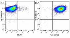
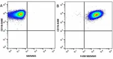

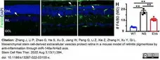
Rat anti Mouse F4/80 antibody, clone A3-1 (MCA497R) used to label macrophages in the mouse retina by immunofluorescence on cryosections.
Image caption:
MSC-EVs inhibit the reactive gliosis and immunoproliferation in the rd10 retina. Retinal sections stained for F4/80 (green). Quantification of the number of F4/80 (M0 macrophage).
From: Zhang J, Li P, Zhao G, He S, Xu D, Jiang W, Peng Q, Li Z, Xie Z, Zhang H, Xu Y, Qi L.
Mesenchymal stem cell-derived extracellular vesicles protect retina in a mouse model of retinitis pigmentosa by anti-inflammation through miR-146a-Nr4a3 axis.
Stem Cell Res Ther. 2022 Aug 3;13(1):394.
doi: 10.1186/s13287-022-03100-x.
This image is from an open access article distributed under terms of a Creative Commons Attribution License.
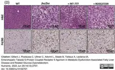
Rat anti Mouse F4/80 antibody, clone A3-1 (MCA497G) used to stain macrophages in murine liver by immunohistochemistry on formalin fixed, paraffin embedded tissue sections.
Image caption:
INT-777, but not RO5527239, partially improves NASH. Representative H&E and F4/80 staining of liver sections (bar size: 100 μm).
From: Gillard J, Picalausa C, Ullmer C, Adorini L, Staels B, Tailleux A, Leclercq IA.
Enterohepatic Takeda G-Protein Coupled Receptor 5 Agonism in Metabolic Dysfunction-Associated Fatty Liver Disease and Related Glucose Dysmetabolism.
Nutrients. 2022 Jun 29;14(13):2707.
doi: 10.3390/nu14132707.
This image is from an open access article distributed under terms of a Creative Commons Attribution License.
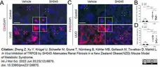
Rat anti Mouse F4/80 antibody, clone A3-1 (MCA497R) used to label macrophages in murine renal tissue by immunofluorescence.
Image caption:
SH045 impact on renal inflammatory cell accumulation after UUO. (A) Representative images of control and UUO-injured kidneys stained with CD4+ T cells (magnification: 400×). Rectangles represent single-cell magnifications. Scale bars are 50 μm. (B) Quantification in renal infiltration of CD4+ T cells (control n = 6, UUO n = 8). (C) Representative images of control and UUO-injured kidneys stained with F4/80+ macrophages (magnification: 400×). Rectangles represent single-cell magnifications. Scale bars are 50 μm. (D) Quantification in renal infiltration of F4/80+ macrophages (control n = 6, UUO n = 8). Data expressed as means ± SD. Two-way ANOVA followed by Sidak’s multiple comparisons post hoc test. * p <0.05, *** p <0.001, and **** p <0.0001 defined as significant. ns, not statistically significant. AU, arbitrary units.
From: Zheng Z, Xu Y, Krügel U, Schaefer M, Grune T, Nürnberg B, Köhler MB, Gollasch M, Tsvetkov D, Markó L.
In Vivo Inhibition of TRPC6 by SH045 Attenuates Renal Fibrosis in a New Zealand Obese (NZO) Mouse Model of Metabolic Syndrome.
Int J Mol Sci. 2022 Jun 20;23(12):6870.
doi: 10.3390/ijms23126870.Insert article citation with space after colon and Hyperlink to PubMed record.
This image is from an open access article distributed under terms of a Creative Commons Attribution License.
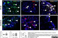
Rat anti Mouse F4/80 antibody, clone A3-1 (MCA497R) used to label CD206+ macrophages in the murine dorsal root ganglia by immunofluorescence.
Image caption:
Macrophage subtypes in DRG of old WT and GLA KO mice. (A–F) Representative photomicrographs of F4/80+ (A,D), CD80+ (B,E), and CD206+ (C,F) macrophages (arrowheads) in DRG of old WT and old GLA KO mice. (G) Quantification of F4/80+ pan-macrophages per mm2 DRG area in old WT (●, n = 5) and old GLA KO mice (◯, n = 5). (H) Quantification of CD80+ M1 macrophages per mm2 DRG area in old WT (●, n = 5) and old GLA KO mice (◯, n = 5). (I) Quantification of CD206+ M2 macrophages per mm2 DRG area in old WT (●, n = 5) and old GLA KO mice (◯, n = 5). Abbreviations: CD = Cluster of Differentiation; DAPI = 4′,6-diamidino-2-phenylindole; DRG = dorsal root ganglia; GLA KO = alpha-galactosidase A knockout; WT = wildtype. Scale bar: 25 μm. ** p <0.01.
From: Spitzel M, Wagner E, Breyer M, Henniger D, Bayin M, Hofmann L, Mauceri D, Sommer C, Üçeyler N.
Dysregulation of Immune Response Mediators and Pain-Related Ion Channels Is Associated with Pain-like Behavior in the GLA KO Mouse Model of Fabry Disease.
Cells. 2022 May 24;11(11):1730.
doi: 10.3390/cells11111730.
This image is from an open access article distributed under terms of a Creative Commons Attribution License.
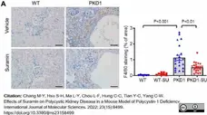
Rat anti mouse F4/80 antibody, clone A3-1 (MCA497R) used to label macrophages in mouse kidney by immunohistochemistry on formalin fixed, paraffin embedded tissue sections.
Image caption:
Suramin treatment inhibited renal inflammation and macrophage infiltration. (A) Immunostaining of kidney sections for F4/80 in different treatment groups as indicated. Quantitative image analysis showed a significant decrease in the percentage of F4/80 positive cells in the suramin-treated Pkd1-miR Tg mice compared with the vehicle-treated controls. Scale bar, 50 μm.
From: Chang, M.-Y.; Hsu, S.-H.; Ma, L.-Y.; Chou, L.-F.; Hung, C.-C.; Tian, Y.-C.; Yang, C.-W.
Effects of Suramin on Polycystic Kidney Disease in a Mouse Model of Polycystin-1 Deficiency.
Int. J. Mol. Sci. 2022, 23, 8499.
doi: 10.3390/ijms23158499
This image is from an open access article distributed under terms of a Creative Commons Attribution License.
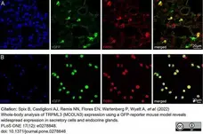
Rat anti Mouse F4/80 antibody, clone A3-1 (MCA497GA used to label pulmonary macrophages by immunofluorescence.
Image caption:
Alveolar macrophages of neonatal and adult express TRPML3.
Immunofluorescence images using F4/80 antibody as macrophage marker (A) in 10 μM lung cryosections or (B) on cells isolated by bronchoalveolar lavage. Images clearly show co-localization of τGFP and F4/80 confirming TRPML3 expression in lung macrophages.
From: Spix B, Castiglioni AJ, Remis NN, Flores EN, Wartenberg P, Wyatt A, Boehm U, Gudermann T, Biel M, García-Añoveros J, Grimm C.
Whole-body analysis of TRPML3 (MCOLN3) expression using a GFP-reporter mouse model reveals widespread expression in secretory cells and endocrine glands.
PLoS One. 2022 Dec 15;17(12):e0278848.
doi: 10.1371/journal.pone.0278848.
This image is from an open access article distributed under terms of a Creative Commons Attribution License.
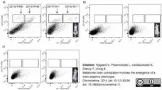
Alexa Fluor 647 conjugated Rat anti mouse F4/80 antibody, clone A3-1 (MCA497A647) used to label macrophages by flow cytometry.
Image caption:
Flow cytometric analysis of brain stroma cells stained with myeloid cell markers CDI lb, F4/80 and Gr-I in: a) metastatic brain; b) healthy brain; c) brain from the metastatic mouse with no metastases in the brain (illustrated by !VIS pictures in the inserts). Numbers indicate percent ages of CD! I b+ stroma cells negative (left quadrant) or positive (left quadrant) for F4/80 or Gr-I.
From: Nygaard V, Prasmickaite L, Vasiliauskaite K, Clancy T, Hovig E.
Melanoma brain colonization involves the emergence of a brain-adaptive phenotype.
Oncoscience. 2014 Jan 10;1(1):82-94.
doi: 10.18632/oncoscience.11.
This image is from an open access article distributed under terms of a Creative Commons Attribution License.
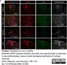
Rat anti Mouse F4/80 antibody, clone A3-1 (MCA497G) used to label macrophages in the spinal column of mice by immunofluorescence.
Image caption:
(K) Longitudinal sections of the spinal cord were made at disease peak and evaluated for Arginase and F4/80 colocalization (n ≥ 4/group). (M−N) Transversal sections of the spinal cord were made at the chronic end phase of the disease where the MBP+ area was evaluated and correct for the number of neurofilament positive axons (n≥ 4/group).
From: Schepers M et al. (2023)
Selective PDE4 subtype inhibition provides new opportunities to intervene in neuroinflammatory versus myelin damaging hallmarks of multiple sclerosis.
Selective PDE4 subtype inhibition provides new opportunities to intervene in neuroinflammatory versus myelin damaging hallmarks of multiple sclerosis.
Brain, Behavior, and Immunity 109. 1-22.
This image is from an open access article distributed under terms of a Creative Commons Attribution License.
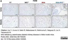
Rat anti mouse F4/80 antibody, clone A3-1 (MCA497G) used to labelmacrophages in mouse kidney by immunohistochemistry on formalin fixed, paraffin embedded tissue sections.
Image caption:
Nrf2 deficiency sensitizes Akita mice to severe renal inflammation. (A–C) Expression of the macrophage marker F4/80 in the kidneys of 18-week-old male WT, Nrf2−/−, Akita and Akita::Nrf2−/− mice. Immunochemistry images of F4/80 (A) and quantitative analysis of the F4/80-positive area within the cortex (B) and medulla (C).
From: Liu Y, Uruno A, Saito R, Matsukawa N, Hishinuma E, Saigusa D, Liu H, Yamamoto M.
Nrf2 deficiency deteriorates diabetic kidney disease in Akita model mice.
Redox Biol. 2022 Dec;58:102525.
doi: 10.1016/j.redox.2022.102525.
This image is from an open access article distributed under terms of a Creative Commons Attribution License.
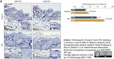
Rat anti Mouse F4/80 antibody (MCA497R) used to label macrophages in murine skin by immunohistochemistry on formalin fixed, paraffin embedded tissue sections.
Image caption:
iASPP deficient keratinocytes induce pro-inflammatory gene expression in vitro and attract macrophages in vivo.
(D) IHC staining of F4/80-positive macrophages in acetone- or TPA-treated skin sections from iASPP WT and KO mice. Scale bar, 50 μm. Histograms below show quantification of the same samples. Values are mean + SD.
From: Al Moussawi K, Chung K, Carroll TM, Osterburg C, Smirnov A, Lotz R, Miller P, Dedeić Z, Zhong S, Oti M, Kouwenhoven EN, Asher R, Goldin R, Tellier M, Murphy S, Zhou H, Dötsch V, Lu X.
Mutant Ras and inflammation-driven skin tumorigenesis is suppressed via a JNK-iASPP-AP1 axis.
Cell Rep. 2022 Oct 18;41(3):111503.
doi: 10.1016/j.celrep.2022.111503.
This image is from an open access article distributed under terms of a Creative Commons Attribution License.
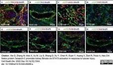
Rat anti Mouse F4/80 antibody, clone A3-1 (MCA497R) used to label macrophages in murine kidney by immunofluorescence.
Image caption:
TNC-expressing cells in fibrotic kidneys were mostly PDGFRβ + NG2 + fibroblasts.
In TNCCreER2-eGFP/+ reporter mice, eGFP positive cells were all PDGFRβ positive, consistent with stromal cells (A). Most of the eGFP-positive cells were also NG2 (a marker of pericyte) positive (B). Some of eGFP positive cells were αSMA positive, while most of αSMA positive myofibroblasts were not eGFP positive (C). These eGFP-positive cells were not positive for FSP1 (D). They were neither CD34 nor CD44 positive progenitors (E, F), and were neither CD68 nor F4/80 positive (G, H). (n = 4, 3 sections for each).
From: Xie Q, Zhang M, Mao X, Xu M, Liu S, Shang D, Xu Y, Chen R, Guan Y, Huang X, Zent R, Pozzi A, Hao CM.
Matrix protein Tenascin-C promotes kidney fibrosis via STAT3 activation in response to tubular injury.
Cell Death Dis. 2022 Dec 15;13(12):1044.
doi: 10.1038/s41419-022-05496-z.
This image is from an open access article distributed under terms of a Creative Commons Attribution License.
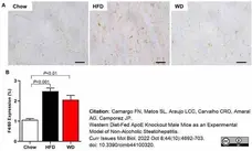
Rat anti Mouse F4/80 antibody, clone A3-1 (MCA497R) used to identify macrophages in mouse liver by immunohistochemistry on formalin fixed, paraffin embedded tissue sections.
Image caption:
ApoE KO Mice fed an HFD or WD displayed increased hepatic inflammation. Representative images of F4/80 expression (scale of 50 μm, objective 40×) (A). Hepatic inflammation represented for F4/80 expression (macrophage activity marker) (B).
From: Camargo FN, Matos SL, Araujo LCC, Carvalho CRO, Amaral AG, Camporez JP.
Western Diet-Fed ApoE Knockout Male Mice as an Experimental Model of Non-Alcoholic Steatohepatitis.
Curr Issues Mol Biol. 2022 Oct 8;44(10):4692-703.
doi: 10.3390/cimb44100320.
This image is from an open access article distributed under terms of a Creative Commons Attribution License.
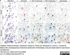
Rat anti Mouse F4/80 antibody, clone A3-1 (MCA497) used to label macroophages in murine lungs by immunohistochemistry on formalin fixed, paraffin embedded tissue sections.
Image caption:
Insert image caption
From: Piñ)eiro-Hermida S, Martínez P, Bosso G, Flores JM, Saraswati S, Connor J, Lemaire R, Blasco MA.
Consequences of telomere dysfunction in fibroblasts, club and basal cells for lung fibrosis development.
Nat Commun. 2022 Oct 6;13(1):5656
doi: 10.1038/s41467-022-32771-6.
This image is from an open access article distributed under terms of a Creative Commons Attribution License.

Rat anti Mouse F4/80 antibody, clone A3-1 (MCA497G) used to identify macrophages in mouse liver spheroids by immunohistochemistry
Image caption:
Presence of cells in MCLS cultures. Immunohistochemistry stains of liver cell specific proteins HNF4α (Hepatocytes), Desmin (HSCs), LYVE1 (LSEC), F4/80 (KCs) at day 4 and 10 of culture.
From: Gawish R, Maier B, Obermayer G, Watzenboeck ML, Gorki AD, Quattrone F, Farhat A, Lakovits K, Hladik A, Korosec A, Alimohammadi A, Mesteri I, Oberndorfer F, Oakley F, Brain J, Boon L, Lang I, Binder CJ, Knapp S.
A neutrophil-B-cell axis impacts tissue damage control in a mouse model of intraabdominal bacterial infection via Cxcr4.
Elife. 2022 Sep 30;11:e78291.
doi: 10.7554/eLife.78291.
This image is from an open access article distributed under terms of a Creative Commons Attribution License.
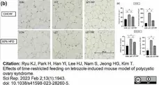
Rat anti Mouse F4/80 antibody, clone A3-1 (MCA497R) used to label macrophages in parametrial fat pads by immunohistochemistry on formalin fixed, paraffin embedded tissue sections.
Image caption:
inflammation markers in parametrial fat tissues.
(b) Representative pictures of F4/80-stained adipose cells of parametrial fat pads are presented. (c) Comparison of the mean adipocyte size and relative expression of F4/80-stained cells in parametrial fat tissues according to the study groups.
From: Ryu KJ, Park H, Han YI, Lee HJ, Nam S, Jeong HG, Kim T.
Effects of time-restricted feeding on letrozole-induced mouse model of polycystic ovary syndrome.
Sci Rep. 2023 Feb 2;13(1):1943.
doi: 10.1038/s41598-023-28260-5.
This image is from an open access article distributed under terms of a Creative Commons Attribution License.
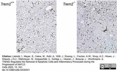
Rat anti Mouse F4/80 antibody, clone A3-1 (MCA497R) used to label macrophages in mouse liver by immunohistochemistry on formalin fixed, paraffin embedded tissue sections.
Image caption:
Hepatic macrophage infiltration after HFD feeding. (a) Representative immunoistochemical images of F4/80 antigen in liver tissue sections from WT- (Trem2+/+) and Trem2-/- - mice fed a HFD for 16 weeks, are shown.
From: Liebold, I.; Meyer, S.; Heine, M.; Kuhl, A.; Witt, J.; Eissing, L.; Fischer, A.W.; Koop, A.C.; Kluwe, J.; Wiesch, J.S.z.; Wehmeyer, M.; Knippschild, U.; Scheja, L.; Heeren, J.; Bosurgi, L.; Worthmann, A.
TREM2 Regulates the Removal of Apoptotic Cells and Inflammatory Processes during the Progression of NAFLD.
Cells 2023, 12, 341.
doi:10.3390/cells12030341
This image is from an open access article distributed under terms of a Creative Commons Attribution License.
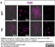
Rat anti Mouse F4/80 antibody, clone A3-1 (MCA497R) used to label macrophages in mouse retina and choroid by immunofluorescence.
Image caption:
Macrophage infiltration and activation in subretina and retina of CAG-HtrA1 Tg mice compared to wild-type control. Representative images of flat mount of retina and choroid separated from CAG-HtrA1 Tg mice (27 months) and wild-type control (29 months). (A) F4/80 (macrophage infiltration)
From: Ahamed W, Yu RMC, Pan Y, Iwata T, Barathi VA, Wey YS, Tun SBB, Qiu B, Tan A, Wang X, Cheung CMG, Wong TY, Yanagi Y.
HTRA1 Regulates Subclinical Inflammation and Activates Proangiogenic Response in the Retina and Choroid.
Int J Mol Sci. 2022 Sep 6;23(18):10206.
doi: 10.3390/ijms231810206.
This image is from an open access article distributed under terms of a Creative Commons Attribution License.

Rat anti Mouse F4/80 antibody, clone A3-1 (MCA497R) used to label macrophages in visceral fat tissues by immunohistochemistry on formalin fixed, paraffin embedded sections.
Image caption:
Visceral fat transplants from aged mice increase macrophage numbers within athersclerotic lesions and fat transplants after transplantation. (d, f) Representative images of (d) Mac2 or (f) F4/80 stained visceral fat (GWAT) sections of young (Y) or aged (A) donors, before (bef) and after (aft) fat transplant (FT) surgery, are shown. The quantification of Mac2 (n = 14‐17/group) or F4/80 (n = 7‐9/group) positive area is shown on the right. (e, g) The change in the percentage of (e) Mac2 or (g) F4/80 positive area after FT, compared to before FT, in young (Y) or aged (A) visceral fat is shown (n = 7 or 14 / group). Results are presented as means ± SEM. Two‐way ANOVA followed by Tukey's post hoc test was used for statistical analysis in (a–c). p values indicate the main effect between Donor‐Y vs. Donor‐A groups. Unpaired two‐tailed Student's t‐test was used for statistical analysis in (d–g).
From: Song J, Farris D, Ariza P, Moorjani S, Varghese M, Blin M, Chen J, Tyrrell D, Zhang M, Singer K, Salmon M, Goldstein DR.
Age-associated adipose tissue inflammation promotes monocyte chemotaxis and enhances atherosclerosis.
Aging Cell. 2023 Feb;22(2):e13783.
doi: 10.1111/acel.13783.
This image is from an open access article distributed under terms of a Creative Commons Attribution License.

Figure B. StarBright UltraViolet 400 conjugated Rat anti Mouse CD11b (MCA711SBUV400) and StarBright UltraViolet 575 conjugated Rat anti Mouse F4/80 (MCA497SBUV575). All experiments performed on J774.2 cells gated on live single cells, in the presence of 10% mouse serum. Data acquired on the ZE5 Cell Analyzer.

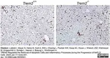
Rat anti Mouse F4/80 antibody, clone A3-1 (MCA497R) used to highlight macrophages in mouse liver by immunohistochemistry on formalin fixed, paraffin embedded tissue sections.
Image caption:
Hepatic macrophage infiltration after HFD feeding. (a) Representative immunoistochemical images of F4/80 antigen in liver tissue sections from WT- (Trem2+/+) and Trem2-/- - mice fed a HFD for 16 weeks, are shown.
From: Liebold I, Meyer S, Heine M, Kuhl A, Witt J, Eissing L, Fischer AW, Koop AC, Kluwe J, Wiesch JSZ, Wehmeyer M, Knippschild U, Scheja L, Heeren J, Bosurgi L, Worthmann A.
TREM2 Regulates the Removal of Apoptotic Cells and Inflammatory Processes during the Progression of NAFLD.
Cells. 2023 Jan 17;12(3):341.
doi: 10.3390/cells12030341.
This image is from an open access article distributed under terms of a Creative Commons Attribution License.
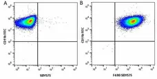
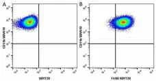
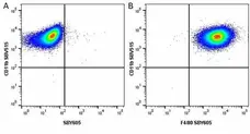


R-Phycoerythrin conjugated Rat anti Mouse F4/80 antibody, clone A3-1 (MCA497PE) used to evaluate F4/80 expression on mouse macrophages by flow cytometry.
Image caption:
Effects of ND on macrophages differentiation. (b) Representative graphs from FACS analysis showing the percent of cells expressing macrophage surface markers, FITC-CD11b and PE-F4/80. (c) Percent of cells expressing CD11b or F4/80. (d) Percent of each cell population based on their surface markers. (e) Effects of ND on the number of surface markers expressed per cell. Results represent fold change in MFI as compared to unstained control from four independent experiments. * p<0.05.
From: Interactions of Carboxylated Nanodiamonds With Mouse Macrophages Cell Line and Primary Cells
Bani-Hani, M. et al.
Int J Orthopaed Res. 6 (1): 30-43.
doi: 10.33140/IJOR.06.01.05.
This image is from an open access article distributed under terms of a Creative Commons Attribution License.
Rat anti Mouse F4/80 antibody, clone A3-1 (MCA497R) used to stain macrophages in murine liver by immunohistochemistry of formalin fixed, paraffin embedded tissue sections.
Image caption:
Liver histopathology and immunohistochemistry. Representative micrographs (H&E, upper panels, immunohistochemical staining, F4/80, middle panels and Sirius Red, lower panels) of the liver from mice injected with bipyramids, plates and rods, compared to vehicle-treated mice sacrificed at 1h after NP injection. Red hatched lines indicate microaggregation of F4/80 positive macrophages; yellow hatched lines indicate areas of hepatic infarcts. The scale bars represent 100 μm.
From: Violatto MB, Sitia G, Talamini L, Morelli A, Tran NL, Zhang Q, Masood A, Pelaz B, Chakraborty I, Cui D, Parak WJ, Salmona M, Bastús NG, Puntes V, Bigini P.
Variations in Biodistribution and Acute Response of Differently Shaped Titania Nanoparticles in Healthy Rodents.
Nanomaterials (Basel). 2023 Mar 25;13(7):1174.
doi: 10.3390/nano13071174. .
This image is from an open access article distributed under terms of a Creative Commons Attribution License.
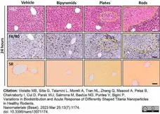
Rat anti Mouse F4/80 antibody, clone A3-1 (MCA497R) used to stain macrophages in murine liver by immunohistochemistry of formalin fixed, paraffin embedded tissue sections.
Image caption:
Liver histopathology and immunohistochemistry. Representative micrographs (H&E, upper panels, immunohistochemical staining, F4/80, middle panels and Sirius Red, lower panels) of the liver from mice injected with bipyramids, plates and rods, compared to vehicle-treated mice sacrificed at 24h after NP injection. Red hatched lines indicate microaggregation of F4/80 positive macrophages; yellow hatched lines indicate areas of hepatic infarcts. The scale bars represent 100 μm.
From: Violatto MB, Sitia G, Talamini L, Morelli A, Tran NL, Zhang Q, Masood A, Pelaz B, Chakraborty I, Cui D, Parak WJ, Salmona M, Bastús NG, Puntes V, Bigini P.
Variations in Biodistribution and Acute Response of Differently Shaped Titania Nanoparticles in Healthy Rodents.
Nanomaterials (Basel). 2023 Mar 25;13(7):1174.
doi: 10.3390/nano13071174. .
This image is from an open access article distributed under terms of a Creative Commons Attribution License.
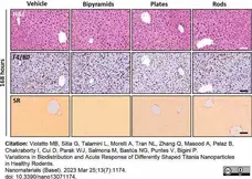
Rat anti Mouse F4/80 antibody, clone A3-1 (MCA497R) used to stain macrophages in murine liver by immunohistochemistry of formalin fixed, paraffin embedded tissue sections.
Image caption:
Liver histopathology and immunohistochemistry. Representative micrographs (H&E, upper panels, immunohistochemical staining, F4/80, middle panels and Sirius Red, lower panels) of the liver from mice injected with bipyramids, plates and rods, compared to vehicle-treated mice sacrificed at 168h after NP injection. Red hatched lines indicate microaggregation of F4/80 positive macrophages; yellow hatched lines indicate areas of hepatic infarcts. The scale bars represent 100 μm.
From: Violatto MB, Sitia G, Talamini L, Morelli A, Tran NL, Zhang Q, Masood A, Pelaz B, Chakraborty I, Cui D, Parak WJ, Salmona M, Bastús NG, Puntes V, Bigini P.
Variations in Biodistribution and Acute Response of Differently Shaped Titania Nanoparticles in Healthy Rodents.
Nanomaterials (Basel). 2023 Mar 25;13(7):1174.
doi: 10.3390/nano13071174. .
This image is from an open access article distributed under terms of a Creative Commons Attribution License.

Rat anti Mouse F4/80 antibody, clone A3-1 (MCA497) used to assess macrophage presence in murine liver by immunohistochemistry on formalin fixed, paraffin embedded tissue sections.
Image caption:
Estradiol reduces hepatic fibrosis and inflammation. Representative images hepatic inflammation represented for F4/80 expression (macrophage activity marker). Black bar represents a scale of 100 μm, 20× objective (A).
From: Araujo, L.C.C.; Cruz, A.G.; Camargo, F.N.; Sucupira, F.G.; Moreira, G.V.; Matos, S.L.; Amaral, A.G.; Murata, G.M.; Carvalho, C.R.O.; Camporez, J.P.
Estradiol Protects Female ApoE KO Mice against Western-Diet-Induced Non-Alcoholic Steatohepatitis.
Int. J. Mol. Sci. 2023, 24, 9845.
doi: 10.3390/ijms24129845
This image is from an open access article distributed under terms of a Creative Commons Attribution License.
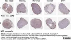
Rat anti Mouse F4/80 antibody, clone A3-1 (MCA497) used to assess macrophage presence in murine liver by immunohistochemistry on formalin fixed, paraffin embedded tissue sections.
Image caption:
Representative images for each staining in Nude and NSG xenografts. E. F4/80 for macrophages.
From: Araujo, L.C.C.; Cruz, A.G.; Camargo, F.N.; Sucupira, F.G.; Moreira, G.V.; Matos, S.L.; Amaral, A.G.; Murata, G.M.; Carvalho, C.R.O.; Camporez, J.P.
Estradiol Protects Female ApoE KO Mice against Western-Diet-Induced Non-Alcoholic Steatohepatitis.
Int. J. Mol. Sci. 2023, 24, 9845.
doi: 10.3390/ijms24129845
This image is from an open access article distributed under terms of a Creative Commons Attribution License.
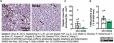
Rat anti Mouse F4/80 antibody, clone A3-1 (MCA497) used to identify macrophages in murine liver by immunohistochemistry on formalin fixed, paraffin embedded tissue sections.
Image caption:
Treatment with SH42 reduces hepatic crown‐like structures, liver collagen content, and plasma alanine transaminase levels in an established NAFLD mode.
E–G Liver sections were stained with F4/80 (E) to quantify (F) hepatic crown‐like structures as indicated by the arrows in Fig 6E and (G) F4/80 positive area.
From: Zhou E, Ge X, Nakashima H, Li R, van der Zande HJP, Liu C, Li Z, Müller C, Bracher F, Mohammed Y, de Boer JF, Kuipers F, Guigas B, Glass CK, Rensen PCN, Giera M, Wang Y.
Inhibition of DHCR24 activates LXRα to ameliorate hepatic steatosis and inflammation.
EMBO Mol Med. 2023 Aug 7;15(8):e16845.
doi: 10.15252/emmm.202216845.
This image is from an open access article distributed under terms of a Creative Commons Attribution License.
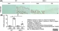
Rat anti Mouse F4/80 antibody (MCA497) used to identify macrophages in murine tissues by immunohistochemistry on formailin fixed, paraffin embedded tissue sections.
Image caption:
The inflammasome is activated in infiltrated macrophages with CD44+ (Experimental 1). (A) Representative images of F4/80 in the WT and ASC−/− mice with or without CG. Bar = 50 μm.
From: Kadoya H, Hirano A, Umeno R, Kajimoto E, Iwakura T, Kondo M, Wada Y, Kidokoro K, Kishi S, Nagasu H, Sasaki T, Taniguchi S, Takahashi M, Kashihara N.
Activation of the inflammasome drives peritoneal deterioration in a mouse model of peritoneal fibrosis.
FASEB J. 2023 Sep;37(9):e23129.
doi: 10.1096/fj.202201777RRR.
This image is from an open access article distributed under terms of a Creative Commons Attribution License.
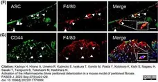
Rat anti Mouse F4/80 antibody (MCA497) used to identify macrophages in murine tissues by immunohistochemistry on formailin fixed, paraffin embedded tissue sections.
Image caption:
The inflammasome is activated in infiltrated macrophages with CD44+ (Experimental 1). (F) Double-immunofluorescence analysis of ASC and F4/80. (G) Double-immunofluorescence analysis of CD44 and F4/80. (H) Flow cytometric evaluation in the mouse peritoneal macrophages with or without CG.
From: Kadoya H, Hirano A, Umeno R, Kajimoto E, Iwakura T, Kondo M, Wada Y, Kidokoro K, Kishi S, Nagasu H, Sasaki T, Taniguchi S, Takahashi M, Kashihara N.
Activation of the inflammasome drives peritoneal deterioration in a mouse model of peritoneal fibrosis.
FASEB J. 2023 Sep;37(9):e23129.
doi: 10.1096/fj.202201777RRR.
This image is from an open access article distributed under terms of a Creative Commons Attribution License.
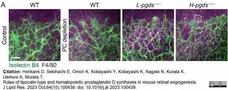
Rat anti Mouse F4/80 antibody, clone A3-1 (MCA497R) used to evaluate macrophage infiltration in mouse retina by immunofluorescence.
Image caption:
The macrophage infiltration in PC depletion-induced pathological angiogenesis. After the PC depletion on P1 WT, L-pgds-/- mice, and H-pgds-/- mice, the P8 retina was excised and used for immunostaining and real-time PCR analysis. (A) Representative pictures of the front of the retinal vessel stained with isolectin B4 (green) and F4/80 (magenta, n = 3-5). Scale bar, 100 μm.
From: Horikami D, Sekihachi E, Omori K, Kobayashi Y, Kobayashi K, Nagata N, Kurata K, Uemura A, Murata T.
Roles of lipocalin-type and hematopoietic prostaglandin D synthases in mouse retinal angiogenesis.
J Lipid Res. 2023 Oct;64(10):100439.
doi: 10.1016/j.jlr.2023.100439.
This image is from an open access article distributed under terms of a Creative Commons Attribution License.
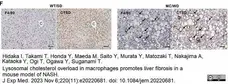
Rat anti Mouse F4/80 antibody, clone A3-1 (MCA497GA) used to label macrophages in murine liver by immunohistochemistry on formalin fixed, paraffin embedded tissue sections.
Image caption:
Cholesterol crystallization and lysosomal stress in CLS-constituting macrophages in NASH livers.
(F) Serial sections of the livers from WT mice fed an SD and MC4R-KO mice fed a WD stained with F4/80 and CTSD antibodies. Arrows, CLS; C, central veins; P, portal veins. Scale bars, 50 μm.
From: Itoh M, Tamura A, Kanai S, Tanaka M, Kanamori Y, Shirakawa I, Ito A, Oka Y, Hidaka I, Takami T, Honda Y, Maeda M, Saito Y, Murata Y, Matozaki T, Nakajima A, Kataoka Y, Ogi T, Ogawa Y, Suganami T.
Lysosomal cholesterol overload in macrophages promotes liver fibrosis in a mouse model of NASH.
J Exp Med. 2023 Nov 6;220(11):e20220681.
doi: 10.1084/jem.20220681.
This image is from an open access article distributed under terms of a Creative Commons Attribution License.
Rat anti Mouse F4/80 antibody, clone A3-1 (MCA497) used to identify macrophages in murine kidney by immunohistochemistry on formalin fixed, paraffin embedded tissue sections.
Image caption:
Deletion of Cx43 in the proximal tubules preserves renal structure and protects against inflammation and fibrosis. Datasets published on Nephroseq were examined for Cx43 expression. Analysis of a published transcriptomic dataset shows that GJA1 expression increases in kidneys from patients with CKD (n = 48 with CKD and n = 5 normal controls: (a) and is positively correlated to proteinuria (b) and declining GFR (c)[. Representative micrographs (d) and quantification of Masson’s Trichrome (tubular dilation) (e), Sirius Red (interstitial fibrosis) (f), F4/80 (macrophage abundance) (g) and FSP1+ (fibroblast number) (h) staining demonstrate that inflammation and fibrosis is significantly ameliorated in UUO-Cx43−/− mice as compared to UUO mice. All groups are n = 5-12 unless otherwise specified, with ANOVA and Tukey post-test used for experimental comparisons except for transcriptomic data where an unpaired t-test with Welch’s correction was used. *P <0.05, **P <0.01, ***P <0.001 and ****P <0.0001
From: Roger E, Chadjichristos CE, Kavvadas P, Price GW, Cliff CL, Hadjadj S, Renciot J, Squires PE, Hills CE.
Connexin-43 hemichannels orchestrate NOD-like receptor protein-3 (NLRP3) inflammasome activation and sterile inflammation in tubular injury.
Cell Commun Signal. 2023 Sep 28;21(1):263.
doi: 10.1186/s12964-023-01245-7.
This image is from an open access article distributed under terms of a Creative Commons Attribution License.
Rat anti Mouse F4/80 antibody, clone A3-1 (MCA497) used to label macrophages in mouse gut by immunohistochemistry on formalin fixed Epon embedded tissue sectionms
Image caption:
Representative images of SI sections from Dars2fl/fl and DARS2IEC-KO mice immunostained against CC3, CC8, CD45 and F4/80. Scale bars, 50 μm (d, e). PAS, Periodic acid- Schiff; ALP, Alkaline Phosphatase; OLFM4, Olfactomedin 4, CC3, Cleaved Caspase 3, CC8, Cleaved Caspase 8.
From: Moschandrea C, Kondylis V, Evangelakos I, Herholz M, Schneider F, Schmidt C, Yang M, Ehret S, Heine M, Jaeckstein MY, Szczepanowska K, Schwarzer R, Baumann L, Bock T, Nikitopoulou E, Brodesser S, Krüger M, Frezza C, Heeren J, Trifunovic A, Pasparakis M.
Mitochondrial dysfunction abrogates dietary lipid processing in enterocytes.
Nature. 2023 Dec 20.
doi: 10.1038/s41586-023-06857-0.
This image is from an open access article distributed under terms of a Creative Commons Attribution License.
Rat anti Mouse F4/80 antibody, clone A3-1 (MCA497) used to identify macrophages in murine renal tissue by immunohistohistochemistry on formalin fixed, paraffin embedded sections.
Image caption:
PAR-1 activation induces pro-fibrotic cytokine expression and potentiates macrophage influx during renal fibrosis.
E: Representative images of F4/80-stained kidney slides of wild-type and PAR-1-deficient (PAR-1−/−) mice 1, 3, 7, and 10 d after UUO and in unobstructed (Sham) control kidneys; scale bars represent 100 μm.
From: IWaasdorp M, de Rooij DM, Florquin S, Duitman J, Spek CA.
Protease-activated receptor-1 contributes to renal injury and interstitial fibrosis during chronic obstructive nephropathy.
J Cell Mol Med. 2019 Feb;23(2):1268-1279.
doi: 10.1111/jcmm.14028.
This image is from an open access article distributed under terms of a Creative Commons Attribution License.
Rat anti Mouse F4/80 antibody, clone A3-1 (MCA497R) used to identify macrophages in murine renal tissues by immunohistochemistry on formalin fixed, paraffin embedded sections.
Image caption:
Petchiether A (PetA) inhibits the expression of proinflammatory cytokines and infiltration of macrophages in the obstructed kidneys of unilateral obstructive (UUO) mice. Mice receiving daily intraperitoneal injection of vehicle or petA (40 mg/kg/d) 4 d before or right after UUO operation were killed 5 d after UUO. A, interleukin-1β (IL-1β), tumour necrosis factor-α (TNF-α), MCP-1 and IL-6 expression were examined by quantitative real-time PCR, as indicated. B, Immunohistochemical staining and (C) semi-quantitative analysis of F4/80+ and IL-1β expression. Data represent the mean ± SEM for 6-8 mice per group. *P <0.05, ***P <0.001 vs the sham group; #P <0.05, ##P <0.01, ###P <0.001 vs the vehicle UUO group. Bar = 50 μmol/L. Magnification ×200. PetA Px, PetA prevention; PetA Tx, PetA treatment
From: You YK, Luo Q, Wu WF, Zhang JJ, Zhu HJ, Lao L, Lan HY, Chen HY, Cheng YX.
Petchiether A attenuates obstructive nephropathy by suppressing TGF-β/Smad3 and NF-κB signalling.
J Cell Mol Med. 2019 Aug;23(8):5576-5587.
doi: 10.1111/jcmm.14454.
This image is from an open access article distributed under terms of a Creative Commons Attribution License.
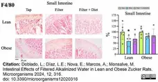
Rat anti Mouse F4/80 antibody, clone A3-1 (MCA497R) used to label macrophages in rat small intestine by immunohistochemistry.
Image caption:
IHC staining of macrophages in tissue sections from the small intestine using antibodies directed against F4/80. Collected data correspond to the positive area divided by the tissue area. The graphs show the % relative to the mean of the control group (tap), mean ±SD. * p <0.05 (t-test). Each data point corresponds to the mean of the values obtained for each rat. Representative images of the groups taken with a 20× objective are included in the left panels.
From: Doblado, L.; Díaz, L.E.; Nova, E.; Marcos, A.; Monsalve, M.
Intestinal Effects of Filtered Alkalinized Water in Lean and Obese Zucker Rats.
Microorganisms 2024, 12, 316.
doi: 10.3390/microorganisms12020316
This image is from an open access article distributed under terms of a Creative Commons Attribution License.
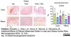
Rat anti Mouse F4/80 antibody, clone A3-1 (MCA497R) used to label macrophages in rat small intestine by immunohistochemistry.
Image caption:
IHC staining of macrophages in tissue sections from the colon using antibodies directed against F4/80. Collected data correspond to the positive area divided by the tissue area. The graphs show the % relative to the mean of the control group (tap), mean ±SD. * p <0.05 (t-test). Each data point corresponds to the mean of the values obtained for each rat. Representative images of the groups taken with a 20× objective are included in the left panels.
From: Doblado, L.; Díaz, L.E.; Nova, E.; Marcos, A.; Monsalve, M.
Intestinal Effects of Filtered Alkalinized Water in Lean and Obese Zucker Rats.
Microorganisms 2024, 12, 316.
doi: 10.3390/microorganisms12020316
This image is from an open access article distributed under terms of a Creative Commons Attribution License.
Rat anti Mouse F4/80 antibody, clone A3-1 (MCA497R) used to identify macrophages in mouse small intestine by immunohistochemistry on formalin fixed paraffin embedded tissue sections..
Image caption:
Indomethacin (Indo)-induced small intestinal ulcers. We treated WT and Gal3KO mice with Indo, and 18 h later, harvested the small intestines (n = 7 per group). We determined the Indo concentration 5 mg/kg body weight by “Human equivalent dose calculation” (42). Indo (FUJIFILM Wako, Osaka, Japan) was solved in a 0.5% NaHCO3 solution and administrated to non-fasted mice by using a stomach tube. (C) Small intestine lesions, 18 h after Indo administration. We stained formalin-fixed paraffin sections with anti-F4/80 antibody (AbD Serotec, Kidlington, United Kingdom) for macrophages. Bar = 50 μm.
From: Park AM, Khadka S, Sato F, Omura S, Fujita M, Hsu DK, Liu FT, Tsunoda I.
Galectin-3 as a Therapeutic Target for NSAID-Induced Intestinal Ulcers.
Front Immunol. 2020 Sep 23;11:550366.
doi: 10.3389/fimmu.2020.550366.
This image is from an open access article distributed under terms of a Creative Commons Attribution License.
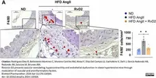
Rat anti Mouse F4/80 antibody, clone A3-1 (MCA497R) used to label macrophages in murine aortic rings by immunohistochemistry on formalin fixed, paraffin embedded tissue sections.
Image caption:
Effects of RvD2 in inflammatory cells infiltration in aorta from mice fed with high fat diet (HFD) plus Angiotensin II (AngII) infusion. (A) Representative immunohistochemistry images and quantification and representative immunofluorescence images of F4/80 positive macrophages infiltration in aortic segments from mice fed with normal diet (ND), HFD AngII or HFD AngII+RvD2. Data represent mean±SEM. *p<0.05 by one-way ANOVA.
From: Rodrigues-Diez R, Ballesteros-Martinez C, Moreno-Carriles RM, Nistal F, Díaz Del Campo LS, Cachofeiro V, Dalli J, García-Redondo AB, Redondo JM, Salaices M, Briones AM.
Resolvin D2 prevents vascular remodeling, hypercontractility and endothelial dysfunction in obese hypertensive mice through modulation of vascular and proinflammatory factors.
Biomed Pharmacother. 2024 Apr 11;174:116564.
doi: 10.1016/j.biopha.2024.116564.
This image is from an open access article distributed under terms of a Creative Commons Attribution License.
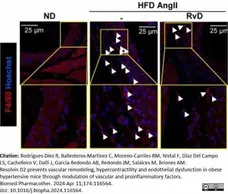
Rat anti Mouse F4/80 antibody, clone A3-1 (MCA497R) used to label macrophages in murine aortic rings by immunofluorescence on formalin fixed, paraffin embedded tissue sections.
Image caption:
Effects of RvD2 in inflammatory cells infiltration in aorta from mice fed with high fat diet (HFD) plus Angiotensin II (AngII) infusion. (A) Representative immunohistochemistry images and quantification and representative immunofluorescence images of F4/80 positive macrophages infiltration in aortic segments from mice fed with normal diet (ND), HFD AngII or HFD AngII+RvD2. Data represent mean±SEM. *p<0.05 by one-way ANOVA.
From: Rodrigues-Diez R, Ballesteros-Martinez C, Moreno-Carriles RM, Nistal F, Díaz Del Campo LS, Cachofeiro V, Dalli J, García-Redondo AB, Redondo JM, Salaices M, Briones AM.
Resolvin D2 prevents vascular remodeling, hypercontractility and endothelial dysfunction in obese hypertensive mice through modulation of vascular and proinflammatory factors.
Biomed Pharmacother. 2024 Apr 11;174:116564.
doi: 10.1016/j.biopha.2024.116564.
This image is from an open access article distributed under terms of a Creative Commons Attribution License.
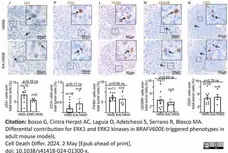
Rat anti Mouse F4/80 antibody, clone A3-1 (MCA497R) used to label macrophages in murine lung by immunohistochemistry on formalin fixed, paraffin embedded tissue sections.
Image caption:
Representative images (top) and quantifications showing (bottom) J p53, K Cleaved Caspase 3 (CC3), L F4/80, M CD45R, N CD3 immunostainings in lung sections from BRAFV600E and Erk, BRAFV600E mice. Quantifications were performed on at least five different lung adenomas from at least five different mice per experimental group. Data are expressed as mean ± SEM; n = tumors per group. *P <0.05; ns not significant. (T-Student’s test unpaired). Arrows point to selected positive cells for the indicated marker. Insets: magnifications of areas inside dashed squares.
From: Bosso G, Cintra Herpst AC, Laguía O, Adetchessi S, Serrano R, Blasco MA.
Differential contribution for ERK1 and ERK2 kinases in BRAFV600E-triggered phenotypes in adult mouse models.
Cell Death Differ. 2024 May 2.
doi: 10.1038/s41418-024-01300-x.
This image is from an open access article distributed under terms of a Creative Commons Attribution License.
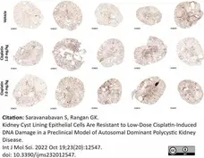
Rat anti Mouse F4/80 antibody, clone A3-1 (MCA497G) used to identify macrophages in murine kidneys by immunohistochemistry on formalin fixed, paraffin embedded sections.
Image caption:
Immunohistochemical staining for F4/80 in Pkd1RC/RC/Atm+/− mice kidney sections at 3 weeks post cisplatin injections (1 mg/kg or 7 mg/kg) or vehicle (7 mg/kg). Scale bar = 1mm.
From: Saravanabavan S, Rangan GK.
Kidney Cyst Lining Epithelial Cells Are Resistant to Low-Dose Cisplatin-Induced DNA Damage in a Preclinical Model of Autosomal Dominant Polycystic Kidney Disease.
Int J Mol Sci. 2022 Oct 19;23(20):12547.
doi: 10.3390/ijms232012547.
This image is from an open access article distributed under terms of a Creative Commons Attribution License.

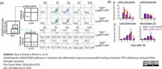
Biotinylated Rat anti Mouse F4/80 antibody, clone A3-1 (MCA497B) used to assess F4/80 levels on murine cell samples by flow cytometry.
Image caption:
Myeloid-specific Ten-Eleven Translocation 2 (Tet2) deletion promotes neutrophil expansion following left anterior descending (LAD) ligation-induced acute myocardial infarction (MI). Data were shown as mean ± SD (*p <0.05; **p <0.005; two-sided unpaired Student's t-test).(D) Flow cytometry analysis on subsets of myeloid cells, including neutrophils, monocytes (Ly6C-high and Ly6C-low) and macrophages/dendritic cells (DCs), in infarct areas. (E) Quantification of the population of Ly6G+ neutrophils, monocytes (Ly6C-high and Ly6C-low) and macrophages/DCs in infarct areas at 0-, 1-, 4-, 7- or 16-day post-MI (n = 3–4).
From: Dou Y, Zhang Y, Rivera L, et al.
Inhibiting the cGAS-STING pathway in myeloid cells effectively improves myocardial healing related to TET2 deficiency-induced DNA damage response.
Clin Transl Med. 2024;14:e1741.
doi: 10.1002/ctm2.1741
This image is from an open access article distributed under terms of a Creative Commons Attribution License.
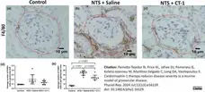
Rat anti Mouse F4/80 antibody, clone A3-1 (MCA497R) used to detect macrophages in murine kidney by immunohistochemistry on formalin fixed, paraffin embedded tissue sections.
Image caption:
Assessment of inflammation following NTS injury and treatment with CT-1. Representative images of glomeruli from control (a), NTS+Saline (b) and NTS+CT-1 (c) treated mice stained for F4/80 21days after NTS injection. Scale bar=10μm. Quantification of (d) the number of F4/80+ cells in the glomerular tuft and (e) the number of F4/80+ cells in the peri-glomerular area (indicated by the dotted line). Individual data points represent average values per mouse and a minimum of 10 glomeruli were assessed per mouse. Healthy controls, n=4; NTS+Saline, n=9; NTS+CT-1, n=8. Data are presented as the mean±SD and analyzed by one-way ANOVA with Holm–Sidak post hoc test
From: Perretta-Tejedor N, Price KL, Jafree DJ, Pomeranz G, Kolatsi-Joannou M, Martínez-Salgado C, Long DA, Vasilopoulou E.
Cardiotrophin-1 therapy reduces disease severity in a murine model of glomerular disease.
Physiol Rep. 2024 Jul;12(13):e16129.
doi: 10.14814/phy2.16129.
This image is from an open access article distributed under terms of a Creative Commons Attribution License.
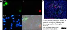
Rat anti Mouse F4/80 antibody, clone A3-1 (MCA497GA) used to label macrophages in axolotls by immunofluorescence.
Image caption:
Macrophages mediate the efficient clearance of senescent cells during salamander limb regeneration.
(B) DiI-liposomes are incorporated specifically by macrophages. Blood cells were extracted from treated animals and stained with antibodies against the specific macrophage marker F4/80. Scale bar: 50 μM. (E) DiI-labelled macrophages (red) are recruited to sites of implanted nGFP+ senescent cells (green) within regenerating limbs at 16 hs post implantation. Scale bar: 100 μm. Note that in the DiI-liposome treated animals macrophages are recruited to sited of senescent but not normal cells.
From: Yun MH, Davaapil H, Brockes JP.
Recurrent turnover of senescent cells during regeneration of a complex structure.
Elife. 2015 May 5;4:e05505.
doi: 10.7554/eLife.05505.
This image is from an open access article distributed under terms of a Creative Commons Attribution License.
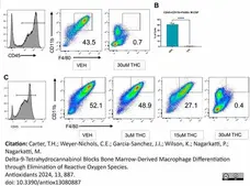
StarBright Blue 615 conjugated Rat anti Mouse F4/80 antibody, clone A3-1 (MCA497SBB615) used to evaluate F4/80 expression on cultured cells by flow cytometry.
Image caption:
In vitro differentiation of BMDCs into macrophages. (A) Representative flow cytometry pseudocolor plots for BMDCs following culture with M-CSF and the addition of LPS at 96 h. CD45+ gated cells were further analyzed for the expression of CD11b+ F4/80+ cells, the percentage of which is shown in each histogram. Unpaired t-test was performed between the two groups. (B) Graphical representation of the percentages of macrophages in vehicle and THC-treated groups at 120h by flow cytometry. (C) Representative flow cytometry pseudocolor plot for CD45+ CD11b+ F4/80+ macrophages cultured with different concentrations of THC.
From: Carter, T.H.; Weyer-Nichols, C.E.; Garcia-Sanchez, J.I.; Wilson, K.; Nagarkatti, P.; Nagarkatti, M.
Delta-9-Tetrahydrocannabinol Blocks Bone Marrow-Derived Macrophage Differentiation through Elimination of Reactive Oxygen Species.
Antioxidants 2024, 13, 887.
doi: 10.3390/antiox13080887
This image is from an open access article distributed under terms of a Creative Commons Attribution License.
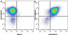
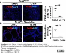
Rat anti Mouse F4/80 antibody, clone A3-1 (MCA497G) used to label macrophages in mouse kidney sections< by immunofluorescence.
Image caption:
Treatment with C-176 decreases the recruitment of macrophages in Pkd1 mutant kidneys. (A) F4/80 staining of kidneys from Pkd1RC/RC mice treated with C-176 (n = 3) or vehicle (n = 3). p < 0.01. Scale bars: 100 μm. (B) F4/80 staining of kidneys from Pkd1fl/fl: Pkhd1-Cre mice treated with C-176 (n = 3) or vehicle (n = 3). p <0.05. Scale bars: 100 μm.
From: Wu, J.; Cheng, S.; Lee, G.; Agborbesong, E.; Li, X.; Zhou, X.; Li, X.
STING Promotes the Progression of ADPKD by Regulating Mitochondrial Function, Inflammation, Fibrosis, and Apoptosis.
Biomolecules 2024, 14, 1215.
doi: 10.3390/biom14101215
This image is from an open access article distributed under terms of a Creative Commons Attribution License.

igure B. StarBright UltraViolet 440 conjugated Rat anti Mouse CD11b (MCA711SBV440) and StarBright Red 815 conjugated Rat anti Mouse F4/80 (MCA497SBR815). All experiments performed on J774.2 cells gated on live single cells, in the presence of 10% mouse serum. Data acquired on the ZE5 Cell Analyzer.
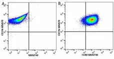
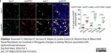
Rat anti Mouse F4/80 antibody (MCA497) used to label macrophages in murine kidneys by immunofluorescence.
Image caption:
Representative images (six to eight visual fields) and quantification of immunolabeling for F4/80, α-SMA and KFP in Trf1+/+ and Trf1flox/flox kidneys.
From: Saraswati S, Martínez P, Serrano R, Mejías D, Graña-Castro O, Álvarez Díaz R, Blasco MA.
Renal fibroblasts are involved in fibrogenic changes in kidney fibrosis associated with dysfunctional telomeres.
Exp Mol Med. 2024 Oct 1.
doi: 10.1038/s12276-024-01318-8.
This image is from an open access article distributed under terms of a Creative Commons Attribution License.
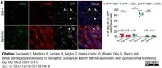
Rat anti Mouse F4/80 antibody (MCA497) used to label macrophages in murine kidneys by immunofluorescence.
Image caption:
Persistent long-term fibroblast-specific deletion of Trf1 leads to acquisition of a mesenchymal phenotype via MMT, partial EMT and EndMT at HEP
a Representative image (six to eight visual fields) and quantifications of immunolabeling for F4/80, α-SMA and KFP in Trf1+/+ and Trf1flox/flox kidneys.
From: Saraswati S, Martínez P, Serrano R, Mejías D, Graña-Castro O, Álvarez Díaz R, Blasco MA.
Renal fibroblasts are involved in fibrogenic changes in kidney fibrosis associated with dysfunctional telomeres.
Exp Mol Med. 2024 Oct 1.
doi: 10.1038/s12276-024-01318-8.
This image is from an open access article distributed under terms of a Creative Commons Attribution License.
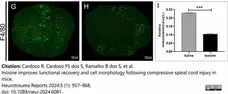
Rat anti Mouse F4/80 antibody, clone A3-1 (MCA497) used to label macrophages in murine spinal cord by immunofluorescence.
Image caption:
Relative immunolabeled area for F4/80. Note that the inosine group showed a reduced immunolabeled area for F4/80 (H) when compared to the saline group (A), as shown in graph (I). *p <0.05; **p <0.01; ***p <0.001; scale bar = 100 μm. GFAP, glial fibrillary acidic protein.
From: Cardoso R, Cardoso FS dos S, Ramalho B dos S, et al.
Inosine improves functional recovery and cell morphology following compressive spinal cord injury in mice,
Neurotrauma Reports 2024:5 (1): 957-68,
doi: 10.1089/neur.2024.0081.
This image is from an open access article distributed under terms of a Creative Commons Attribution License.
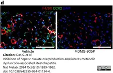
Rat anti Mouse F4/80 antibody, clone A3-1 (MCA497R) used to label macrophages in murine liver sections by immunofluorescence.
Image caption:
(d) Liver samples were collected from mice treated with vehicle (n = 7) or 10 mg kg−1 d−1 (n = 10) of MDMG-935P, stained for F4/80 (red), CCR2 (green) and DAPI (blue).
From: Das S, et al.
Inhibition of hepatic oxalate overproduction ameliorates metabolic dysfunction-associated steatohepatitis.
Nat Metab. 2024 Oct;6(10):1939-1962.
doi: 10.1038/s42255-024-01134-4.
This image is from an open access article distributed under terms of a Creative Commons Attribution License.
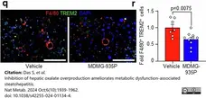
Rat anti Mouse F4/80 antibody, clone A3-1 (MCA497R) used to label macrophages in murine liver sections by immunofluorescence.
Image caption:
(q) Male C57BL/6J mice were fed the MASH diet for 12 weeks, then orally administered vehicle or 10 mg/kg/day of MDMG-935P for an additional 12 weeks on the MASH diet prior to end point analyses. Liver samples were collected from mice treated with vehicle (n=7), or 10 mg/kg/day (n=10) of MDMG-935P, stained for F4/80 (red), TREM2 (green), and DAPI (blue) to visualize nuclei, analysed for (r) F4/80+ and TREM2+ cells and expressed as fold change from vehicle.
From: Das S, et al.
Inhibition of hepatic oxalate overproduction ameliorates metabolic dysfunction-associated steatohepatitis.
Nat Metab. 2024 Oct;6(10):1939-1962.
doi: 10.1038/s42255-024-01134-4.
This image is from an open access article distributed under terms of a Creative Commons Attribution License.
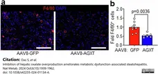
Rat anti Mouse F4/80 antibody, clone A3-1 (MCA497R) used to label macrophages in murine liver sections by immunofluorescence.
Image caption:
Mice were injected with AAV8-GFP or AAV8-AGXT (2x10¹¹ viral genomes per mouse) and placed on the MASH diet for 24 weeks prior to end point analyses. a, Liver samples were collected from mice treated with AAV8-GFP (n=8) or AAV8-AGXT (n=6), stained for F4/80 (red) and DAPI (blue) to visualize nuclei, analysed for (b) F4/80+ cells and expressed as fold change from AAV8-GFP.
From: Das S, et al.
Inhibition of hepatic oxalate overproduction ameliorates metabolic dysfunction-associated steatohepatitis.
Nat Metab. 2024 Oct;6(10):1939-1962.
doi: 10.1038/s42255-024-01134-4.
This image is from an open access article distributed under terms of a Creative Commons Attribution License.

Rat anti Mouse F4/80 antibody, clone A3-1 (MCA497R) used to label macrophages in murine liver sections by immunofluorescence.
Image caption:
Mice were injected with AAV8-GFP or AAV8-AGXT (2 × 10¹¹ viral genomes per mouse) and placed on the MASH diet for 24 weeks before end point analyses. d,e, Liver samples were collected from mice treated with AAV8-GFP (n = 8) or AAV8-AGXT (n = 6), stained for F4/80 (red), CCR2 (green) and DAPI (blue) to visualize nuclei (d), analysed for F4/80+ and CCR2+ cells and expressed as fold change from AAV8-GFP (e), from mice treated with AAV8-GFP (n = 8) or AAV8-AGXT (n = 6). f,g, Liver samples were collected from mice treated with AAV8-GFP (n = 8) or AAV8-AGXT (n = 6), stained for F4/80 (red), TREM2 (green) and DAPI (blue) to visualize nuclei (f), analysed for F4/80+ and TREM2+ cells (g) and expressed as fold change from AAV8-GFP, from mice treated with AAV8-GFP (n = 8) or AAV8-AGXT (n = 6).
From: Das S, et al.
Inhibition of hepatic oxalate overproduction ameliorates metabolic dysfunction-associated steatohepatitis.
Nat Metab. 2024 Oct;6(10):1939-1962.
doi: 10.1038/s42255-024-01134-4.
This image is from an open access article distributed under terms of a Creative Commons Attribution License.
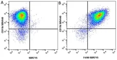
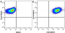
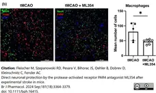
Rat anti mouse F4/80 antibody, clone A3-1 (MCA497G) used to label macrophages in mouse mouse brain by immunofluorescence.
Image caption:
PAR4 blockade reduces macrophage accumulation after experimental stroke. (b) F4/80+ macrophages (red) in ipsilateral cortex sections of mice subjected to tMCAO and 72 h of reperfusion, with and without ML354. Tissue sections were co-stained with neuronal nuclear antigen (NeuN, green) and the nuclear stain DAPI (blue). Data shown are individual values with means &± SD. *P<0.05, significantly different as indicated.
From: Fleischer M, Szepanowski RD, Pesara V, Bihorac JS, Oehler B, Dobrev D, Kleinschnitz C, Fender AC.
Direct neuronal protection by the protease-activated receptor PAR4 antagonist ML354 after experimental stroke in mice.
Br J Pharmacol. 2024 Sep;181(18):3364-3379.
doi: 10.1111/bph.16415.
This image is from an open access article distributed under terms of a Creative Commons Attribution License.

Rat anti Mouse F4/80 antibody, clone A3-1 (MCA497) used to label macrophages in mouse kidneys by immunofluorescence.
Image caption:
MafB and c-Maf were unexpectedly expressed in kidney macrophages after 48 h of IRD. (A): MafB was expressed in WT kidney macrophages after dIRD; immunohistochemistry of frozen sections was conducted, as explained in the Materials and Methods section, and representative slides were chosen; (B): c-Maf was expressed in WT kidney macrophages after dIRD; immunohistochemistry of paraffin sections was conducted, as explained in the Materials and Methods section, and representative slides were chosen; both stainings are shown at 20× magnification; ((A) is zoomed 3 times to illustrate the obvious expression of MafB by the macrophages, and only the merge is zoomed in case of c-Maf). The MafB+F4/80+ and c-Maf+F/40+ bar graphs comparing the MafB and c-Maf expression in wild-type mice kidney macrophages before and after AKI, respectively. The white arrows show the MafB+F4/80+ cells and c-Maf+F4/80+ cells proving the colocalization of both proteins showing the macrophages expressing both transcription factors, respectively, after AKI. NC: negative control: WT mice kidney without antibody staining, *: significant (p-value <0.05).
From: Daassi, D.
MafB Transcription Factor Involved in IRD-Induced AKI (Acute Kidney Injury) Phenotype Attenuation and Inflammation Resolution.
DNA 2025, 5, 5.
https://doi.org/10.3390/dna5010005.
This image is from an open access article distributed under terms of a Creative Commons Attribution License.
Rat anti Mouse F4/80 antibody, clone A3-1 (MCA497G) used to label macrophages in murine femoral artery by immunohistochemistry on formalin fixed, paraffin embedded tissue sections.
Image caption:
Infiltration by F4/80+ macrophages in mouse femoral arteries. The injured femoral arteries were studied two weeks following endothelial cell injury induced by a guidewire. The representative sections (40x magnification) and quantification of the sham or injured arteries. Leukocyte infiltration was mainly neointimal and periadventitial and more marked in the WT NIK+/+ than in the NIKaly/aly mice. *** p <0.0005, vs. NIK+/+ sham or NIK+/+ injured. Mean ± SEM of 4–5 mice per group.
From: Baeza C, Ribagorda M, Maya-Lopez C, Fresno M, Sanchez-Diaz T, Pintor-Chocano A, Sanz AB, Carrasco S, Ortiz A, Sanchez-Niño MD.
NIK Is a Mediator of Inflammation and Intimal Hyperplasia in Endothelial Denudation-Induced Vascular Injury.
Int J Mol Sci. 2024 Oct 25;25(21):11473.
doi: 10.3390/ijms252111473.
This image is from an open access article distributed under terms of a Creative Commons Attribution License.
Rat anti Mouse F4/80 antibody, clone A3-1 (MCA497R) used to label macrophages in mouse pancreata by immunohistochemistry on formalin fixed, paraffin embedded tissue sections.
Image caption:
Representative pictures (from n=5 samples each treatment group) for the quantification shown in Figure 3F. Pancreata were analyzed by IHC for presence of Ly6B.2 positive (neutrophils),) or F4/80 positive (macrophages) cells. The bar indicates 100μm.
From: Bastea LI, Liu X, Fleming AK, Pandey V, Döppler H, Edenfield BH, Krishna M, Zhang L, Thompson EA, Grandgenett PM, Hollingsworth MA, Fairweather D, Clemens D, Storz P.
Coxsackievirus and adenovirus receptor expression facilitates enteroviral infections to drive the development of pancreatic cancer.
Nat Commun. 2024 Dec 4;15(1):10547.
doi: 10.1038/s41467-024-55043-x.
This image is from an open access article distributed under terms of a Creative Commons Attribution License.
Rat anti Mouse F4/80 antibody, clone A3-1 (MCA497GA) used to label macrophages im murine mammary tumors by immunohistochemistry on formalin fixed, paraffin embedded tissue sections.on
Image caption:
A single dose of ErSO-TFPy induces rapid necrosis in tumors, followed by lymphocyte infiltration.
(C) Representative F4/80 images showing macrophage infiltrate over time.
From: Mulligan, M.P. et al.
Single Dose of a Small Molecule Leads to Complete Regressions of Large Breast Tumors in Mice.
ACS Cent Sci. 22 Jan [Epub ahead of print].
doi: 10.1021/acscentsci.4c01628
This image is from an open access article distributed under terms of a Creative Commons Attribution License.
Rat anti Mouse F4/80 antibody, clone A3-1 (MCA497GA) used to macrophages by immunofluorescence.
Image caption:
Representative CLSM image stacks of bone-marrow-derived macrophages cultures on composite scaffolds showing actin distribution (red), macrophage-specific marker F4/80 (green), and cell nuclei (blue) after 7 and 14 d in static and dynamic cultures. Scale bars represent 100 μm.
From: Kontogianni GI, Loukelis K, Bonatti AF, Batoni E, De Maria C, Vozzi G, Naseem R, Dalgarno K, Shin H, Vitale-Brovarone C, Chatzinikolaidou M.
A Mechanically Stimulated Co-culture in 3-Dimensional Composite Scaffolds Promotes Osteogenic and Anti-osteoclastogenic Activity and M2 Macrophage Polarization.
Biomater Res. 2025 Feb 5;29:0135.
doi: 10.34133/bmr.0135.
This image is from an open access article distributed under terms of a Creative Commons Attribution License.
Rat anti Mouse F4/80 antibody, clone A3-1 (MCA497R) used to detect macrophages in mouse skin by immunofluorescence.
Image caption:
Representative images of skin sections from 3-week-old mice of the indicated genotypes, immunostained with antibodies against keratin 10 (K10), keratin 14 (K14) (n = 6), keratin 6 (K6) (n = 6), F4/80 (n = 6) and Gr-1 (n = 6) and counterstained with DAPI (DNA stain). Scale bars: K10, K14, K6 = 20 μm; F4/80, Gr-1 = 30 μm.
From: Schünke H, Göbel U, Dikic I, Pasparakis M.
OTULIN inhibits RIPK1-mediated keratinocyte necroptosis to prevent skin inflammation in mice.
Nat Commun. 2021 Oct 8;12(1):5912.
doi: 10.1038/s41467-021-25945-1.
This image is from an open access article distributed under terms of a Creative Commons Attribution License.
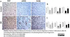
Insert meta-description and applications
Image caption:
Insert image caption
From: Nahmias Blank D, Maimon O, Hermano E, Drai E, Chen O, Popovtzer A, Peretz T, Meirovitz A, Elkin M.
Unraveling the Role of Metabolic Endotoxemia in Accelerating Breast Tumor Progression.
Biomedicines. 2025 Jul 31;13(8):1868.
doi: 10.3390/biomedicines13081868.
This image is from an open access article distributed under terms of a Creative Commons Attribution License.
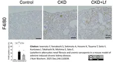
Rat anti Mouse F4/80 antibody, clone A3-1 (MCA497GA) used to identify macrophages in mouse kidney by immunohistochemistry on formalin fixed, paraffin embedded tissue sections.
Image caption:
Lactoferrin administration ameliorated progression of renal impairment.
Representative images of immunohistochemistry staining for F4/80 and quantitative analysis of F4/80 positive area, Tukey-Kramer’s test; *P<.05, ⁎⁎P<.01, ⁎⁎⁎P<.001, Student's t-test; ††P<.01, n=6 per group.
From: Iwamoto Y, Yamakoshi S, Sekimoto A, Hosomi K, Toyama T, Saito Y, Kunisawa J, Takahashi N, Mishima E, Sato E.
Lactoferrin attenuates renal fibrosis and uremic sarcopenia in a mouse model of adenine-induced chronic kidney disease.
J Nutr Biochem. 2025 Dec;146:110039.
doi: 10.1016/j.jnutbio.2025.110039.
This image is from an open access article distributed under terms of a Creative Commons Attribution License.
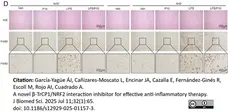
Rat anti Mouse F4/80 antibody, clone A3-1 (MCA497GA) used to identify hepatic macrophages by immunohistochemistry on formalin fixed, paraffin embedded tissue sections.
Image caption:
P10 lessens the inflammatory-liver response in the NRF2-dependent context in mice treated with LPS. C57Bl/6 male mice were treated i.p with vehicle (Tween-80 + PBS, 1:7) or 20 mg/kg P10 for 5 days. Two hours after the last administration, mice received vehicle or 10 mg/kg LPS and were sacrificed after 4 h for liver analysis. D Paraffin-embedded liver section stained with H&E and immunohistochemistry for F4/80.
From: García-Yagüe ÁJ, Cañizares-Moscato L, Encinar JA, Cazalla E, Fernández-Ginés R, Escoll M, Rojo AI, Cuadrado A.
A novel β-TrCP1/NRF2 interaction inhibitor for effective anti-inflammatory therapy.
J Biomed Sci. 2025 Jul 11;32(1):65.
doi: 10.1186/s12929-025-01157-3.
This image is from an open access article distributed under terms of a Creative Commons Attribution License.

Rat anti Mouse F4/80 antibody, clone A3-1 (MCA497GA) used to label macrophages in mouse liver by immunohistochemistry on formalin fixed, paraffin embedded tissue sections.
Image caption:
Global transcription changes in p21Cip1-deficient mice. (E) Representative pictures of F4/80 stained liver sections of WT and p21KO mice under feeding conditions, 24 h and 48 h fasting. Bars correspond to 50 μm.
From: Lopez-Guadamillas E, Fernandez-Marcos PJ, Pantoja C, Muñoz-Martin M, Martínez D, Gómez-López G, Campos-Olivas R, Valverde AM, Serrano M.
p21Cip1 plays a critical role in the physiological adaptation to fasting through activation of PPARα.
Sci Rep. 2016 Oct 10;6:34542.
doi: 10.1038/srep34542. PMID: 27721423; PMCID: PMC5056372..
This image is from an open access article distributed under terms of a Creative Commons Attribution License.
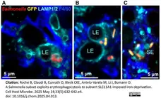
Rat anti Mouse F4/80 antibody, clone A3-1 (MCA497GA) staining of splenic macrophaged by immunofluorescence.
Image caption:
Macrophage erythrophagocytosis provides iron to a subset of Salmonella
(A) Confocal fluorescence micrograph of iron-replete Salmonella (low PryhB-2-gfp activity; constitutive mCherry) in a F4/80-positive red pulp macrophage that contains a large LAMP1/2-positive endosome (LE).
(B) Iron-starved Salmonella (high PryhB-2-gfp activity) in a macrophage that contains LEs.
(C) Iron-starved Salmonella (high PryhB-2-gfp activity) in a macrophage that contains only small endosomes (SEs).
From: Roche B, Claudi B, Cunrath O, Bleck CKE, Antelo-Varela M, Li J, Bumann D.
A Salmonella subset exploits erythrophagocytosis to subvert SLC11A1-imposed iron deprivation.
Cell Host Microbe. 2025 May 14;33(5):632-642.e4.
doi: 10.1016/j.chom.2025.04.013.
This image is from an open access article distributed under terms of a Creative Commons Attribution License.
Filter by Application:
P F C IF WB RE Reset| Rat anti Mouse F4/80 antibody, clone A3-1 recognizes the murine F4/80 antigen, a ~160 kDa cell surface glycoprotein member of the EGF-TM7 family of proteins which shares 68% overall amino acid identity with human EGF module-containing mucin-like hormone receptor 1 (EMR1). Expression of F4/80 is heterogeneous and is modulated during macrophage maturation and activation. The F4/80 antigen is expressed on a wide range of mature tissue macrophages including Kupffer cells, Langerhans cells, microglia, macrophages located in the gut lamina propria, peritoneal cavity, lung, thymus, bone marrow stroma and macrophages in the red pulp of the spleen (Hume, et al. 1984). F4/80 antigen is also expressed on a subpopulation of dendritic cells but is absent from macrophages located in T cell areas of the spleen and lymph node (Gordon, et al. 1994). The ligands and biological functions of the F4/80 antigen have not been fully determined but a role for F4/80 in the generation of efferent CD8+ve regulatory T cells is proposed (Lin, et al. 2005) Rat anti mouse F4/80 antibody, clone Cl:A3-1 modulates cytokine levels released in response to Listeria monocytogenes (Warschkau & Kiderlen, 1999). A Human anti-idiotypic CI:A31 antibody, clone 17867 (HCA154 ) which binds to and blocks activity of Rat anti mouse F4/80 antibody, clone Cl:A3-1 is also available for use as a control in experiments utilizing clone A3-1. |

Our F4/80 (Cl:A3-1) Antibody has been referenced in >719 publications* *Based on June 2020 data from CiteAb's antibody search engine. |
- Target Species
- Mouse
- Product Form
- Purified IgG - liquid
- Preparation
- MCA497RT, MCA497R, MCA497GA: Purified IgG prepared by affinity chromatography on Protein G from tissue culture supernatant.
- MCA497G: Purified IgG prepared by affinity chromatography on Protein G from tissue culture supernatant
- Buffer Solution
- Phosphate buffered saline
- Preservative Stabilisers
0.09% Sodium Azide - Carrier Free
- Yes
- Immunogen
- Thioglycollate stimulated peritoneal macrophages from C57BL/6 mice.
- Approx. Protein Concentrations
- IgG concentration 1.0 mg/ml
- Fusion Partners
- Spleen cells from immunized HOB2 rats were fused with cells of the mouse NS1 myeloma cell line.
- Regulatory
- For research purposes only
- Guarantee
- 12 months from date of despatch
This product is shipped at ambient temperature. It is recommended to aliquot and store at -20°C on receipt. When thawed, aliquot the sample as needed. Keep aliquots at 2-8°C for short term use (up to 4 weeks) and store the remaining aliquots at -20°C.
Avoid repeated freezing and thawing as this may denature the antibody. Storage in frost-free freezers is not recommended.
Avoid repeated freezing and thawing as this may denature the antibody. Storage in frost-free freezers is not recommended.
This product has been reported to work in the following applications. This information is derived from testing within our laboratories, peer-reviewed publications or personal communications from the originators. Please refer to references indicated for further information. For general protocol recommendations, please visit the antibody protocols page.
| Application Name | Verified | Min Dilution | Max Dilution |
|---|---|---|---|
| Flow Cytometry |  |
1/50 | 1/100 |
| Immuno-electron Microscopy | |
||
| Immunofluorescence | |
||
| Immunohistology - Frozen | |
||
| Immunohistology - Paraffin 1 | |
||
| Immunohistology - Resin | |
||
| Immunoprecipitation | |
||
| Radioimmunoassays | |
||
| Western Blotting | |
- 1 Rat anti Mouse F4/80 antibody, clone A3-1 requires pre-treatment of paraffin sections prior to staining. Bio-Rad recommend Proteinase K or Citrate buffer pH 6.0 for this purpose. Please view the protocol at Antigen Retrieval Techniques.
Where this product has not been tested for use in a particular technique this does not necessarily exclude its use in such procedures. Suggested working dilutions are given as a guide only. It is recommended that the user titrates the product for use in their own system using appropriate negative/positive controls.
- Flow Cytometry
- Use 10ul of the suggested working dilution to label 106 cells in 100ul
| Description | Product Code | Applications | Pack Size | List Price | Your Price | Quantity | |
|---|---|---|---|---|---|---|---|
| Goat anti Rat IgG (H/L):HRP | 305005 | C E IB | 1 ml |

|
Log in | ||
| List Price | Your Price | ||||||
|
|
Log in | ||||||
| Description | Goat anti Rat IgG (H/L):HRP | ||||||
| Goat anti Rat IgG2b:HRP | STAR114P | C E P WB | 0.5 mg |
|
Log in | ||
| List Price | Your Price | ||||||
|
|
Log in | ||||||
| Description | Goat anti Rat IgG2b:HRP | ||||||
| Goat F(ab')2 anti Rat IgG:FITC (Mouse Adsorbed) | STAR69 | F | 0.5 ml |
|
Log in | ||
| List Price | Your Price | ||||||
|
|
Log in | ||||||
| Description | Goat F(ab')2 anti Rat IgG:FITC (Mouse Adsorbed) | ||||||
| Goat F(ab')2 anti Rat IgG:RPE (Mouse Adsorbed) | STAR73 | F | 0.5 ml |
|
Log in | ||
| List Price | Your Price | ||||||
|
|
Log in | ||||||
| Description | Goat F(ab')2 anti Rat IgG:RPE (Mouse Adsorbed) | ||||||
| Description | Product Code | Applications | Pack Size | List Price | Your Price | Quantity | |
|---|---|---|---|---|---|---|---|
| Antigen Retrieval Buffer, pH8.0 | BUF025A | P | 500 ml | Log in | |||
| List Price | Your Price | ||||||
| Log in | |||||||
| Description | Antigen Retrieval Buffer, pH8.0 | ||||||
Source Reference
-
Austyn, J.M. & Gordon, S. (1981) F4/80, a monoclonal antibody directed specifically against the mouse macrophage.
Eur J Immunol. 11 (10): 805-15.
Antibody Characterization Reference
-
Hume, D.A. et al. (1984) The mononuclear phagocyte system of the mouse defined by immunohistochemical localisation of antigen F4/80: macrophages associated with epithelia.
Anat Rec. 210 (3): 503-12. -
Lee, S.H. et al. (1985) Quantitative analysis of total macrophage content in adult mouse tissues. Immunochemical studies with monoclonal antibody F4/80.
J Exp Med. 161 (3): 475-89.
References for F4/80 antibody
-
Warschkau, H. & Kiderlen, A.F. (1999) A monoclonal antibody directed against the murine macrophage surface molecule F4/80 modulates natural immune response to Listeria monocytogenes.
J Immunol. 163 (6): 3409-16. -
Moore, K.J. et al. (2000) Divergent response to LPS and bacteria in CD14-deficient murine macrophages.
J Immunol. 165 (8): 4272-80. -
Brown, G.D. et al. (2002) Dectin-1 is a major beta-glucan receptor on macrophages.
J Exp Med. 196: 407-12. -
Biffi, A. et al. (2004) Correction of metachromatic leukodystrophy in the mouse model by transplantation of genetically modified hematopoietic stem cells.
J Clin Invest. 113: 1118-29. -
Dandekar, A.A.et al. (2004) Bystander CD8 T-cell-mediated demyelination is interferon-gamma-dependent in a coronavirus model of multiple sclerosis.
Am J Pathol. 164: 363-9. -
Bevaart, L. et al. (2004) CpG oligodeoxynucleotides enhance FcgammaRI-mediated cross presentation by dendritic cells.
Int Immunol. 16: 1091-8. -
Pizza, F.X. et al. (2005) Neutrophils contribute to muscle injury and impair its resolution after lengthening contractions in mice.
J Physiol. 562 (Pt 3): 899-913. -
Lin, H.H. et al. (2005) The macrophage F4/80 receptor is required for the induction of antigen-specific efferent regulatory T cells in peripheral tolerance.
J Exp Med. 201 (10): 1615-25.
View The Latest Product References
-
Chan, R.J. et al. (2005) Human somatic PTPN11 mutations induce hematopoietic cell hypersensitivity to granulocyte-macrophage colony stimulating factor
Blood. 105: 3737-3742. -
Amano, H. et al. (2005) Absence of recipient CCR5 promotes early and increased allospecific antibody responses to cardiac allografts.
J Immunol. 174: 6499-508. -
Anghelina, M. et al. (2006) Monocytes/macrophages cooperate with progenitor cells during neovascularization and tissue repair: conversion of cell columns into fibrovascular bundles.
Am J Pathol. 168: 529-41. -
Anatelli, F. et al. (2006) Macrophage-targeted photosensitizer conjugate delivered by intratumoral injection.
Mol Pharm. 3: 654-64. -
Dioszeghy, V. et al. (2008) 12/15-Lipoxygenase regulates the inflammatory response to bacterial products in vivo.
J Immunol. 181: 6514-24. -
Buga, G.M. et al. (2008) D-4F reduces EO6 immunoreactivity, SREBP-1c mRNA levels, and renal inflammation in LDL receptor-null mice fed a Western diet.
J Lipid Res. 49: 192-205. -
Mastrangelo, M.A. & Bowers, W.J. (2008) Detailed immunohistochemical characterization of temporal and spatial progression of Alzheimer's disease-related pathologies in male triple-transgenic mice.
BMC Neurosci. 9: 81. -
Davies, M.H. et al. (2008) MCP-1 deficiency delays regression of pathologic retinal neovascularization in a model of ischemic retinopathy.
Invest Ophthalmol Vis Sci. 49: 4195-202. -
Cunningham, O. et al. (2009) Microglia and the urokinase plasminogen activator receptor/uPA system in innate brain inflammation.
Glia. 57: 1802-14. -
Zumsteg, A. et al. (2009) Myeloid cells contribute to tumor lymphangiogenesis.
PLoS One. 4:e7067. -
Hemmi, H. et al. (2009) A new triggering receptor expressed on myeloid cells (Trem) family member, Trem-like 4, binds to dead cells and is a DNAX activation protein 12-linked marker for subsets of mouse macrophages and dendritic cells.
J Immunol. 182:1278-86. -
Devey, L. et al. (2009) Tissue-resident macrophages protect the liver from ischemia reperfusion injury via a heme oxygenase-1-dependent mechanism.
Mol Ther. 17: 65-72. -
González-Périz, A. et al. (2009) Obesity-induced insulin resistance and hepatic steatosis are alleviated by omega-3 fatty acids: a role for resolvins and protectins.
FASEB J. 23: 1946-57. -
Baratta, J.L. et al. (2009) Cellular organization of normal mouse liver: a histological, quantitative immunocytochemical, and fine structural analysis.
Histochem Cell Biol. 131: 713-26. -
deSchoolmeester, M.L. et al. (2009) The mannose receptor binds Trichuris muris excretory/secretory proteins but is not essential for protective immunity.
Immunology. 126: 246-55. -
Choi, K.M. et al. (2010) CD206-positive M2 macrophages that express heme oxygenase-1 protect against diabetic gastroparesis in mice.
Gastroenterology. 138 (7): 2399-409, 2409.e1. -
Seitz, O. et al. (2010) Wound Healing in Mice with High-Fat Diet- or ob Gene-Induced Diabetes-Obesity Syndromes: A Comparative Study
Exp Diabetes Res. 2010: 476969. -
Carlsson, F. et al. (2010) Host-detrimental role of Esx-1-mediated inflammasome activation in mycobacterial infection.
PLoS Pathog. 6(5):e1000895. -
Cougoule, C. et al. (2010) Three-dimensional migration of macrophages requires Hck for podosome organization and extracellular matrix proteolysis.
Blood. 115: 1444-52. -
Zeng, J. & Howard, J.C. (2010) Spontaneous focal activation of invariant natural killer T (iNKT) cells in mouse liver and kidney.
BMC Biol. 8: 142. -
Miao, E.A. et al. (2011) Caspase-1-induced pyroptosis is an innate immune effector mechanism against intracellular bacteria.
Nat Immunol. 11: 1136-42. -
Sun, G. et al. (2011) RIP2-mediated LKB1 deletion causes axon degeneration in the spinal cord and hind-limb paralysis.
Dis Model Mech. 4: 193-202. -
Wang, X. et al. (2011) Activation of the cholinergic antiinflammatory pathway ameliorates obesity-induced inflammation and insulin resistance
Endocrinology. 152: 836-46. -
Chauhan, V.S. et al. (2011) Prophylactic and therapeutic targeting of the neurokinin-1 receptor limits neuroinflammation in a murine model of pneumococcal meningitis.
J Immunol. 186: 7255-63. -
Baudy, A.R. et al. (2011) Non-invasive optical imaging of muscle pathology in mdx mice using cathepsin caged near-infrared imaging.
Mol Imaging Biol. 13: 462-70. -
Kato, J. et al. (2011) ADP-ribosylarginine hydrolase regulates cell proliferation and tumorigenesis.
Cancer Res. 71 (15): 5327-35. -
Ge, Q. et al. (2011) Adipokines identified as new downstream targets for adiponectin: lessons from adiponectin-overexpressing or -deficient mice.
Am J Physiol Endocrinol Metab. 301: E326-35. -
Dioszeghy, V. et al. (2011) Epicutaneous immunotherapy results in rapid allergen uptake by dendritic cells through intact skin and downregulates the allergen-specific response in sensitized mice.
J Immunol. 186: 5629-37. -
Muto, A. et al. (2011) Eph-B4 prevents venous adaptive remodeling in the adult arterial environment.
J Exp Med. 208 (3): 561-75. -
Tarallo, V. et al. (2011) The biflavonoid amentoflavone inhibits neovascularization preventing the activity of proangiogenic vascular endothelial growth factors.
J Biol Chem. 286: 19641-51. -
Kerschbaumer, R.J. et al. (2012) Neutralization of macrophage migration inhibitory factor (MIF) by fully human antibodies correlates with their specificity for the β-sheet structure of MIF.
J Biol Chem. 287 (10): 7446-55. -
Donaldson, D.S. et al. (2012) M cell-depletion blocks oral prion disease pathogenesis.
Mucosal Immunol. 5: 216-25. -
Gornicka, A. et al. (2012) Adipocyte hypertrophy is associated with lysosomal permeability both in vivo and in vitro: role in adipose tissue inflammation.
Am J Physiol Endocrinol Metab. 303: E597-606. -
Rivollier, A. et al. (2012) Inflammation switches the differentiation program of Ly6Chi monocytes from antiinflammatory macrophages to inflammatory dendritic cells in the colon.
J Exp Med. 209: 139-55. -
Hardison, S.E. et al. (2012) Protective immunity against pulmonary cryptococcosis is associated with STAT1-mediated classical macrophage activation.
J Immunol. 189 (8): 4060-8. -
Arima M et al. (2012) Involvement of periostin in regression of hyaloidvascular system during ocular development.
Invest Ophthalmol Vis Sci. 53 (10): 6495-503. -
Henare, K. et al. (2012) Dissection of stromal and cancer cell-derived signals in melanoma xenografts before and after treatment with DMXAA.
Br J Cancer. 106 (6): 1134-47. -
Akbarshahi, H. et al. (2012) Enrichment of Murine CD68(+)CCR2(+) and CD68(+)CD206(+) Lung Macrophages in Acute Pancreatitis-Associated Acute Lung Injury.
PLoS One. 7: e42654. -
Bonde, A.K. et al. (2012) Intratumoral macrophages contribute to epithelial-mesenchymal transition in solid tumors.
BMC Cancer. 12: 35. -
Ubogu, E.E. et al. (2012) Behavioral, electrophysiological, and histopathological characterization of a severe murine chronic demyelinating polyneuritis model.
J Peripher Nerv Syst. 17 (1): 53-61. -
Banda NK et al. (2012) Role of C3a receptors, C5a receptors, and complement protein C6 deficiency in collagen antibody-induced arthritis in mice.
J Immunol. 188 (3): 1469-78. -
Tamaki, S. et al. (2013) Interleukin-16 promotes cardiac fibrosis and myocardial stiffening in heart failure with preserved ejection fraction.
PLoS One. 8: e68893. -
Jain, U. et al. (2013) The C5a receptor antagonist PMX205 ameliorates experimentally induced colitis associated with increased IL-4 and IL-10.
Br J Pharmacol. 168 (2): 488-501. -
Dong, Z. et al. (2013) Specific inhibition of serine/arginine-rich protein kinase attenuates choroidal neovascularization.
Mol Vis. 19: 536-43. -
Uaesoontrachoon, K. et al. (2013) Osteopontin deficiency delays inflammatory infiltration and the onset of muscle regeneration in a mouse model of muscle injury.
Dis Model Mech. 6: 197-205. -
Nygaard, V. et al. (2014) Melanoma brain colonization involves the emergence of a brain-adaptive phenotype.
Oncoscience. 1 (1): 82-94. -
Kihira, Y. et al. (2014) Deletion of hypoxia-inducible factor-1α in adipocytes enhances glucagon-like Peptide-1 secretion and reduces adipose tissue inflammation.
PLoS One. 9(4):e93856. -
Zhang, M.Z. et al. (2015) Inhibition of cyclooxygenase-2 in hematopoietic cells results in salt-sensitive hypertension.
J Clin Invest. 125 (11): 4281-94. -
Chinzei, N. et al. (2015) P21 deficiency delays regeneration of skeletal muscular tissue.
PLoS One. 10 (5): e0125765. -
Sumiyoshi, M. et al. (2015) Antitumor and antimetastatic actions of xanthoangelol and 4-hydroxyderricin isolated from Angelica keiskei roots through the inhibited activation and differentiation of M2 macrophages.
Phytomedicine. 22 (7-8): 759-67. -
Kim, M. et al. (2015) Progression of Alport Kidney Disease in Col4a3 Knock Out Mice Is Independent of Sex or Macrophage Depletion by Clodronate Treatment.
PLoS One. 10 (11): e0141231. -
Lei, B. et al. (2016) Neuroprotective pentapeptide CN-105 improves functional and histological outcomes in a murine model of intracerebral hemorrhage.
Sci Rep. 6: 34834. -
Nagase, M. et al. (2016) Deletion of Rac1GTPase in the Myeloid Lineage Protects against Inflammation-Mediated Kidney Injury in Mice.
PLoS One. 11 (3): e0150886. -
Pepe, G. et al. (2017) Self-renewal and phenotypic conversion are the main physiological responses of macrophages to the endogenous estrogen surge.
Sci Rep. 7: 44270. -
Glastras, S.J. et al. (2017) The renal consequences of maternal obesity in offspring are overwhelmed by postnatal high fat diet.
PLoS One. 12 (2): e0172644. -
Sogawa, Y. et al. (2017) Infiltration of M1, but not M2, macrophages is impaired after unilateral ureter obstruction in Nrf2-deficient mice.
Sci Rep. 7 (1): 8801. -
Crompton, M. et al. (2017) A mutation in Nischarin causes otitis media via LIMK1 and NF-κB pathways.
PLoS Genet. 13 (8): e1006969. -
Suzuki, Y. et al. (2017) Requisite role of vasohibin-2 in spontaneous gastric cancer formation and accumulation of cancer-associated fibroblasts.
Cancer Sci. 108 (12): 2342-51. -
Kawada, S. et al. (2017) Impairment of cold injury-induced muscle regeneration in mice receiving a combination of bone fracture and alendronate treatment.
PLoS One. 12 (7): e0181457. -
Peng, Y. (2018) B cell responses to apoptotic cells in MFG-E8-/- mice.
PLoS One. 13 (10): e0205172. -
Maydan, O. et al. (2018) Uromodulin deficiency alters tubular injury and interstitial inflammation but not fibrosis in experimental obstructive nephropathy.
Physiol Rep. 6 (6): e13654. -
Ubil, E. et al. (2018) Tumor-secreted Pros1 inhibits macrophage M1 polarization to reduce antitumor immune response.
J Clin Invest. 128 (6): 2356-69. -
Morimoto, A. et al. (2019) Hemophagocytosis induced by Leishmania donovani. infection is beneficial to parasite survival within macrophages.
PLoS Negl Trop Dis. 13 (11): e0007816. -
Wang, H. et al. (2019) Embelin can protect mice from thioacetamide-induced acute liver injury.
Biomed Pharmacother. 118: 109360. -
Hanson, K.M. et al. (2019) Apoptosis Resistance in Fibroblasts Precedes Progressive Scarring in Pulmonary Fibrosis and Is Partially Mediated by Toll-Like Receptor 4 Activation.
Toxicol Sci. 170 (2): 489-498. -
Teixeira, D.E. et al. (2019) Lithium ameliorates tubule-interstitial injury through activation of the mTORC2/protein kinase B pathway.
PLoS One. 14 (4): e0215871. -
WasgewatteWijesinghe, D.K. et al. (2019) Normal inflammation and regeneration of muscle following injury require osteopontin from both muscle and non-muscle cells.
Skelet Muscle. 9 (1): 6. -
Saitoh, K. et al. (2019) Effect of dietary fish oil on enhanced inflammation and disturbed lipophagy in white adipose tissue caused by a high fat diet
Fisheries Science. 86 (1): 187-96. -
Ehsanipour, A. et al. (2019) Injectable, Hyaluronic Acid-Based Scaffolds with Macroporous Architecture for Gene Delivery.
Cell Mol Bioeng. 12 (5): 399-413. -
Talamini, L. et al. (2019) Repeated administration of the food additive E171 to mice results in accumulation in intestine and liver and promotes an inflammatory status.
Nanotoxicology. 13 (8): 1087-101. -
Alendar, A. et al. (2020) Gene expression regulation by the Chromodomain helicase DNA-binding protein 9 (CHD9) chromatin remodeler is dispensable for murine development.
PLoS One. 15 (5): e0233394. -
Graff, E.C. et al. (2020) The Absence of Adiponectin Alters Niacin's Effects on Adipose Tissue Inflammation in Mice.
Nutrients. 12 (8): 2427. -
Roche-Molina, M. et al. (2020) The pharmaceutical solvent N-methyl-2-pyrollidone (NMP) attenuates inflammation through Krüppel-like factor 2 activation to reduce atherogenesis.
Sci Rep. 10 (1): 11636. -
Jansen, M.P.B. et al. (2020) Experimental thrombocytopenia does not affect acute kidney injury 24 hours after renal ischemia reperfusion in mice.
Platelets. 31 (3): 383-391. -
Wu, C.Y. et al. (2020) Tris DBA ameliorates IgA nephropathy by blunting the activating signal of NLRP3 inflammasome through SIRT1- and SIRT3-mediated autophagy induction.
J Cell Mol Med.24 (23): 13609-22. -
Liguori, M. et al. (2020) The soluble glycoprotein NMB (GPNMB) produced by macrophages induces cancer stemness and metastasis via CD44 and IL-33.
Cell Mol Immunol. 18 (3): 711-22. -
Robichon, K. et al. (2020) Identification of Interleukin1β as an Amplifier of Interferon alpha-induced Antiviral Responses.
PLoS Pathog. 16 (10): e1008461. -
Zhang, J. et al. (2020) Triptolide attenuates renal damage by limiting inflammatory responses in DOCA-salt hypertension.
Int Immunopharmacol. 89 (Pt A): 107035. -
Hoover, A.A. et al. (2020) Increased canonical NF-kappaB signaling specifically in macrophages is sufficient to limit tumor progression in syngeneic murine models of ovarian cancer.
BMC Cancer. 20 (1): 970. -
Ni, J. et al. (2020) Dual deficiency of angiotensin-converting enzyme-2 and Mas receptor enhances angiotensin II-induced hypertension and hypertensive nephropathy.
J Cell Mol Med.24 (22): 13093-13103. -
Munro, D.A.D. et al. (2020) CNS macrophages differentially rely on an intronic Csf1r enhancer for their development.
Development. 147 (23): dev194449. -
Lai, K. et al. (2020) Triptolide attenuates laser-induced choroidal neovascularization via M2 macrophage in a mouse model.
Biomed Pharmacother. 129: 110312. -
Choi, E.W. et al. (2020) Fas mutation reduces obesity by increasing IL-4 and IL-10 expression and promoting white adipose tissue browning.
Sci Rep. 10 (1): 12001. -
Kerber, E.L. et al. (2020) The Importance of Hypoxia-Inducible Factors (HIF-1 and HIF-2) for the Pathophysiology of Inflammatory Bowel Disease.
Int J Mol Sci. 21 (22): 8551. -
Creed, J. et al. (2020) Argon Inhalation for 24 h After Closed-Head Injury Does not Improve Recovery, Neuroinflammation, or Neurologic Outcome in Mice.
Neurocrit Care. 34 (3): 833-43. -
Jablonski, K. et al. (2020) Physical activity prevents acute inflammation in a gout model by downregulation of TLR2 on circulating neutrophils as well as inhibition of serum CXCL1 and is associated with decreased pain and inflammation in gout patients.
PLoS One. 15 (10): e0237520. -
Bae, C.R. et al. (2020) The endothelial dysfunction blocker CU06-1004 ameliorates choline-deficient L-amino acid diet-induced non-alcoholic steatohepatitis in mice.
PLoS One. 15 (12): e0243497. -
Hitchcock, J.R. et al. (2020) Dynamic architectural interplay between leucocytes and mammary epithelial cells.
FEBS J. 287 (2): 250-66. -
Ellman, D.G. et al. (2020) Conditional Ablation of Myeloid TNF Improves Functional Outcome and Decreases Lesion Size after Spinal Cord Injury in Mice.
Cells. 9 (11): 2407. -
Yamashita, S. et al. (2020) Essential roles of oncostatin M receptor β signaling in renal crystal formation in mice.
Sci Rep. 10 (1): 17150. -
Takahashi, M. et al. (2020) Macrophages fine-tune pupil shape during development.
Dev Biol. 464 (2): 137-44. -
Wong, M.Y. et al. (2020) Semicarbazide-sensitive amine oxidase inhibition ameliorates albuminuria and glomerulosclerosis but does not improve tubulointerstitial fibrosis in diabetic nephropathy.
PLoS One. 15 (6): e0234617. -
Bender, L.H. et al. (2020) Intratumoral Administration of a Novel Cytotoxic Formulation with Strong Tissue Dispersive Properties Regresses Tumor Growth and Elicits Systemic Adaptive Immunity in In Vivo Models.
Int J Mol Sci. 21 (12): 4493. -
Fan, A. et al. (2020) High-salt diet decreases mechanical thresholds in mice that is mediated by a CCR2-dependent mechanism.
J Neuroinflammation. 17 (1): 179. -
Fukushima, H. et al. (2020) Changes in Function and Dynamics in Hepatic and Splenic Macrophages in Non-Alcoholic Fatty Liver Disease.
Clin Exp Gastroenterol. 13: 305-14. -
Otobe, S. et al. (2020) CX3CR1 Deficiency Attenuates DNFB-Induced Contact Hypersensitivity Through Skewed Polarization Towards M2 Phenotype in Macrophages.
Int J Mol Sci. 21 (19): 7401. -
Hayashi, Y. et al. (2020) Cochlear supporting cells function as macrophage-like cells and protect audiosensory receptor hair cells from pathogens.
Sci Rep. 10 (1): 6740. -
Tachibana, M. et al. (2020) Ablation of IL-17A leads to severe colitis in IL-10-deficient mice: implications of myeloid-derived suppressor cells and NO production.
Int Immunol. 32 (3): 187-201. -
Sasaki, Y. et al. (2020) Pemafibrate, a selective PPARα modulator, prevents non-alcoholic steatohepatitis development without reducing the hepatic triglyceride content.
Sci Rep. 10 (1): 7818. -
Zhong, L. et al. (2020) RANKL Is Involved in Runx2-Triggered Hepatic Infiltration of Macrophages in Mice with NAFLD Induced by a High-Fat Diet.
Biomed Res Int. 2020: 6953421. -
Park, J.S. et al. (2020) Clusterin overexpression protects against western diet-induced obesity and NAFLD.
Sci Rep. 10 (1): 17484. -
Tseng, W-C. et al.. (2020) Trichostatin A Alleviates Renal Interstitial Fibrosis Through Modulation of the M2 Macrophage Subpopulation
Int J Mol Sci. 21(17):E5966. -
Guo, X. et al. (2020) Adoptive transfer of Pfkfb3-disrupted hematopoietic cells to wild-type mice exacerbates diet-induced hepatic steatosis and inflammation
Liver Res. 4 (3): 136-44. -
Laurien, L. et al. (2020) Autophosphorylation at serine 166 regulates RIP kinase 1-mediated cell death and inflammation.
Nat Commun. 11 (1): 1747. -
Dong, L. et al. (2021) Deletion of Smad3 protects against diabetic myocardiopathy in db/db mice.
J Cell Mol Med. 25 (10): 4860-9. -
Zhang, J. et al. (2021) A pulsatile release platform based on photo-induced imine-crosslinking hydrogel promotes scarless wound healing.
Nat Commun. 12 (1): 1670. -
Zhu, B. et al. (2021) Adipose tissue inflammation and systemic insulin resistance in mice with diet-induced obesity is possibly associated with disruption of PFKFB3 in hematopoietic cells.
Lab Invest. 101 (3): 328-40. -
Sugita, J. et al. (2021) Cardiac macrophages prevent sudden death during heart stress.
Nat Commun. 12 (1): 1910. -
Hasuzawa, N. et al. (2021) Clodronate, an inhibitor of the vesicular nucleotide transporter, ameliorates steatohepatitis and acute liver injury.
Sci Rep. 11 (1): 5192. -
Makiishi, S. et al. (2021) Carnitine/organic cation transporter 1 precipitates the progression of interstitial fibrosis through oxidative stress in diabetic nephropathy in mice.
Sci Rep. 11 (1): 9093. -
Cheng, P. et al. (2021) Aldose reductase deficiency inhibits LPS-induced M1 response in macrophages by activating autophagy.
Cell Biosci. 11 (1): 61. -
Dalla Pietá, A. et al. (2021) Hyaluronan is a natural and effective immunological adjuvant for protein-based vaccines.
Cell Mol Immunol. 18 (5): 1197-210. -
Kuzumoto, T. et al. (2021) Protective role of resolvin D1, a pro-resolving lipid mediator, in nonsteroidal anti-inflammatory drug-induced small intestinal damage.
PLoS One. 16 (5): e0250862. -
Menz, J. et al. (2021) Increased mortality and altered local immune response in secondary peritonitis after previous visceral operations in mice.
Sci Rep. 11 (1): 16175. -
Xu, H. et al. (2021) Adipocyte inducible 6-phosphofructo-2-kinase suppresses adipose tissue inflammation and promotes macrophage anti-inflammatory activation.
J Nutr Biochem. 95: 108764. -
Fritz, N.M. et al. (2021) Cytomegalovirus chemokine receptor M33 knockout reduces chronic allograft rejection in a murine aortic transplant model.
Transpl Immunol. 64: 101359. -
Kim, J.I. et al. (2021) IDH2 gene deficiency accelerates unilateral ureteral obstructioninduced kidney inflammation through oxidative stress and activation of macrophages.
Korean J Physiol Pharmacol. 25 (2): 139-46. -
Otsuka, H. et al. (2021) Histidine decarboxylase deficiency inhibits NBP-induced extramedullary hematopoiesis by modifying bone marrow and spleen microenvironments.
Int J Hematol. 113 (3): 348-61. -
Wu, W. et al. (2021) Latent TGF-β1 protects against diabetic kidney disease via Arkadia/Smad7 signaling.
Int J Biol Sci. 17 (13): 3583-94. -
de Groot, A.E. et al. (2021) Characterization of tumor-associated macrophages in prostate cancer transgenic mouse models.
Prostate. 81 (10): 629-47. -
Stewart, A.N. et al. (2021) Acute inflammatory profiles differ with sex and age after spinal cord injury.
J Neuroinflammation. 18 (1): 113. -
Iwama, H. et al. (2021) Cathepsin B and D deficiency in the mouse pancreas induces impaired autophagy and chronic pancreatitis.
Sci Rep. 11 (1): 6596. -
Umbarawan, Y. et al. (2021) FABP5 Is a Sensitive Marker for Lipid-Rich Macrophages in the Luminal Side of Atherosclerotic Lesions.
Int Heart J. 62 (3): 666-676. -
Eubler, K. et al. (2021) Exploring the Ion Channel TRPV2 and Testicular Macrophages in Mouse Testis
Int J Mol Sci. 22 (9): 4727. -
Vito, A. et al. (2021) Combined Radionuclide Therapy and Immunotherapy for Treatment of Triple Negative Breast Cancer
Int J Mol Sci. 22 (9): 4843. -
An, C. et al. (2022) Myeloid PTEN deficiency aggravates renal inflammation and fibrosis in angiotensin II-induced hypertension.
J Cell Physiol. 237 (1): 983-91. -
Liu, Y.T. et al. (2021) Seed- and Soil-Dependent Differences in Murine Breast Tumor Microenvironments Dictate Anti-PD-L1 IgG Delivery and Therapeutic Efficacy.
Pharmaceutics. 13(4): 530. -
Cyge, B. et al. (2021) Loss of the ciliary protein Chibby1 in mice leads to exocrine pancreatic degeneration and pancreatitis.
Sci Rep. 11 (1): 17220. -
Schünke, H. et al. (2021) OTULIN inhibits RIPK1-mediated keratinocyte necroptosis to prevent skin inflammation in mice.
Nat Commun. 12 (1): 5912. -
Rabe, D.C. et al. (2021) Tumor Extracellular Vesicles Regulate Macrophage-Driven Metastasis through CCL5.
Cancers (Basel). 13 (14): 3459. -
Nanou, A. et al. (2021) Endothelial Tpl2 regulates vascular barrier function via JNK-mediated degradation of claudin-5 promoting neuroinflammation or tumor metastasis.
Cell Rep. 35 (8): 109168. -
Matsuura, R. et al. (2021) Preexisting heart failure with reduced ejection fraction attenuates renal fibrosis after ischemia reperfusion via sympathetic activation.
Sci Rep. 11 (1): 15091. -
Nosaka, M. et al. (2021) Intrathrombotic appearances of AQP-1 and AQP-3 in relation to thrombus age in murine deep vein thrombosis model.
Int J Legal Med. 135 (2): 547-53. -
Farahat, M. et al. (2021) Effect of Biomechanical Environment on Degeneration of Meckel's Cartilage.
J Dent Res. 100 (2): 171-178. -
Fantin, A. et al. (2021) KIT Is Required for Fetal Liver Hematopoiesis.
Front Cell Dev Biol. 9: 648630. -
Verdoodt, D. et al. (2021) Cochlin Deficiency Protects Aged Mice from Noise-Induced Hearing Loss.
Int J Mol Sci.22 (21): 11549. -
Chang, M.Y. et al. (2022) Metformin induces lactate accumulation and accelerates renal cyst progression in Pkd1-deficient mice.
Hum Mol Genet. 31 (10): 1560-73. -
Wu, W. et al. (2021) Buyang Huanwu Decoction protects against STZ-induced diabetic nephropathy by inhibiting TGF-β/Smad3 signaling-mediated renal fibrosis and inflammation.
Chin Med. 16 (1): 118. -
Teuwen, L.A. et al. (2021) Tumor vessel co-option probed by single-cell analysis.
Cell Rep. 35 (11): 109253. -
Das, J.R. et al. (2021) Circulating fibroblast growth factor-2 precipitates HIV nephropathy in mice.
Dis Model Mech. 14 (7): dmm048980. -
Wada, I. et al. (2021) Retinal VEGF-A Overexpression Is Not Sufficient to Induce Lymphangiogenesis Regardless of VEGF-C Upregulation and Lyve1+ Macrophage Infiltration.
Invest Ophthalmol Vis Sci. 62 (13): 17. -
Schmidtke, L. et al. (2021) Knockout of the KH-Type Splicing Regulatory Protein Drives Glomerulonephritis in MRL-Faslpr Mice.
Cells. 10 (11): 3167. -
Lanigan, L.G. et al. (2021) In Vivo. Tumorigenesis, Osteolytic Sarcomas, and Tumorigenic Cell Lines from Transgenic Mice Expressing the Human T-Lymphotropic Virus Type 1 (HTLV-1) Tax Viral Oncogene.
Am J Pathol. 191 (2): 335-52. -
Fagoonee, S. et al. (2022) Circulating Extracellular Vesicles Contain Liver-Derived RNA Species as Indicators of Severe Cholestasis-Induced Early Liver Fibrosis in Mice.
Antioxid Redox Signal. 36 (7-9): 480-504. -
Kido, Y. et al. (2021) Genetic deletion of Vegfr2 in endothelial cells leads to immediate disruption of tumor vessels and aggravation of hypoxia.
Am J Pathol. 2021: S0002-9440(21)00511-3. -
Aschman, T. et al. (2021) Interferon Lambda Regulates Cellular and Humoral Immunity in Pristane-Induced Lupus.
Int J Mol Sci. 22(21):11747. -
Jala, V.R. et al. (2021) Absence of CCR2 reduces spontaneous intestinal tumorigenesis in the Apc(Min) (/+) mouse model.
Int J Cancer. Jan 26 [Epub ahead of print]. -
Fukase, M. et al. (2022) Intravenous injection of human multilineage-differentiating stress-enduring cells alleviates mouse severe acute pancreatitis without immunosuppressants.
Surg Today. 52 (4): 603-615. -
Chen, C. et al. (2021) Activation of the Unfolded Protein Response (UPR) Is Associated with Cholangiocellular Injury, Fibrosis and Carcinogenesis in an Experimental Model of Fibropolycystic Liver Disease
Cancers. 14 (1): 78. -
Boki, H. et al. (2021) Lymphatic dysfunction exacerbates cutaneous tumorigenesis and psoriasis-like skin inflammation through accumulation of inflammatory cytokines.
J Invest Dermatol. Nov 12; S0022-202X(21)02472-6. -
Ohmura, Y. et al. (2021) Natural Killer T Cells Are Involved in Atherosclerotic Plaque Instability in Apolipoprotein-E Knockout Mice.
Int J Mol Sci. 22(22):12451. -
Hudkins, K.L. et al. (2021) Regression of diabetic nephropathy by treatment with empagliflozin in BTBR ob/ob mice.
Nephrol Dial Transplant. 37 (5): 847-59. -
Suffee, N. et al. (2021) Impacts of a high fat diet on the metabolic profile and the phenotype of atrial myocardium in mice.
Cardiovasc Res.118 (15): 3126-39. -
Yang, S.R. et al. (2021) Cf-02, a novel benzamide-linked small molecule, blunts NF-κB activation and NLRP3 inflammasome assembly and improves acute onset of accelerated and severe lupus nephritis in mice.
FASEB J. 35 (8): e21785. -
Allen, A.C. et al. (2021) Parallel in vivo. experimental evolution reveals that increased stress resistance was key for the emergence of persistent tuberculosis bacilli.
Nat Microbiol. 6 (8): 1082-1093. -
Barboza, P.A. et al. (2021) Acute cylindrospermopsin exposure: Pulmonary and liver harm and mitigation by dexamethasone.
Toxicon. 191: 18-24. -
Sumiyoshi, H. et al. (2021) External administration of moon jellyfish collagen solution accelerates physiological wound healing and improves delayed wound closure in diabetic model mice.
Regen Ther. 18: 223-30. -
Yoon, Y.S. et al. (2021) Activation of the adipocyte CREB/CRTC pathway in obesity.
Commun Biol. 4 (1): 1214. -
Epperly, M.W. et al. (2021) Radiation-Induced Senescence in p16+/LUC Mouse Lung Compared to Bone Marrow Multilineage Hematopoietic Progenitor Cells.
Radiat Res. 196 (3): 235-49. -
Yang, F. et al. (2021) Regulatory role and mechanisms of myeloid TLR4 in anti-GBM glomerulonephritis.
Cell Mol Life Sci. 78 (19-20): 6721-34. -
Suffee, N. et al. (2022) Impacts of a high-fat diet on the metabolic profile and the phenotype of atrial myocardium in mice.
Cardiovasc Res. 118 (15): 3126-39. -
McGonigal, R. et al. (2021) Neuronally expressed a-series gangliosides are sufficient to prevent the lethal age-dependent phenotype in GM3-only expressing mice.
J Neurochem. 158 (2): 217-32. -
Baraibar-Churio, A. et al. (2021) Deficiency of MMP-10 Aggravates the Diseased Phenotype of Aged Dystrophic Mice
Life. 11 (12): 1398. -
Bormann, T. et al. (2021) Role of the COX2-PGE2 axis in S. pneumoniae-induced exacerbation of experimental fibrosis.
Am J Physiol Lung Cell Mol Physiol. 320 (3): L377-L392. -
Wu, L. et al. (2021) The Attenuation of Diabetic Nephropathy by Annexin A1 via Regulation of Lipid Metabolism Through the AMPK/PPARα/CPT1b Pathway.
Diabetes. 70 (10): 2192-203. -
Liu, Y. et al. (2022) Nrf2 deficiency deteriorates diabetic kidney disease in Akita model mice.
Redox Biol. 58: 102525. -
Klein, D. et al. (2022) Early targeting of endoneurial macrophages alleviates the neuropathy and affects abnormal Schwann cell differentiation in a mouse model of Charcot-Marie-Tooth 1A.
Glia. 70 (6): 1100-16. -
Domoto, R. et al. (2022) Role of neuron-derived ATP in paclitaxel-induced HMGB1 release from macrophages and peripheral neuropathy.
J Pharmacol Sci. 148 (1): 156-61. -
Celus, W. et al. (2022) Plexin-A4 Mediates Cytotoxic T-cell Trafficking and Exclusion in Cancer.
Cancer Immunol Res. 10 (1): 126-41. -
Howard, F.H.N. et al. (2022) Nanobugs as Drugs: Bacterial Derived Nanomagnets Enhance Tumor Targeting and Oncolytic Activity of HSV-1 Virus.
Small. : e2104763. -
Alegre, F. et al. (2022) Macrophages Modulate Hepatic Injury Involving NLRP3 Inflammasome: The Example of Efavirenz.
Biomedicines. 10 (1): 109. -
Beuker, C. et al. (2022) Stroke induces disease-specific myeloid cells in the brain parenchyma and pia.
Nat Commun. 13 (1): 945. -
Djudjaj, S. et al. (2022) Activation of Notch3 in Renal Tubular Cells Leads to Progressive Cystic Kidney Disease.
Int J Mol Sci. 23(2):884. -
Bosso, G. et al. (2022) Early differential responses elicited by BRAFV600E in adult mouse models.
Cell Death Dis. 13 (2): 142. -
Leary, N. et al. (2022) Melanoma-derived extracellular vesicles mediate lymphatic remodelling and impair tumour immunity in draining lymph nodes.
J Extracell Vesicles. 11 (2): e12197. -
Xie, H. et al. (2022) An immune-sympathetic neuron communication axis guides adipose tissue browning in cancer-associated cachexia.
Proc Natl Acad Sci U S A. 119 (9): e2112840119. -
Eckes, T. et al. (2022) Sphingosine 1-Phosphate Receptor 5 (S1P 5) Knockout Ameliorates Adenine-Induced Nephropathy
Int J Mol Sci. 23 (7): 3952. -
Schützhold, V. et al. (2022) Knockout of Factor-Inhibiting HIF (Hif1an) in Colon Epithelium Attenuates Chronic Colitis but Does Not Reduce Colorectal Cancer in Mice.
J Immunol. 208 (5): 1280-91. -
Sarkar, T. et al. (2022) Perinatal exposure to synergistic multiple stressors leads to cellular and behavioral deficits mimicking Schizophrenia-like pathology.
Biol Open. 11 (3): bio058870. -
Zheng, Z. et al. (2022) In Vivo. Inhibition of TRPC6 by SH045 Attenuates Renal Fibrosis in a New Zealand Obese (NZO) Mouse Model of Metabolic Syndrome.
Int J Mol Sci. 23 (12): 6870. -
Wu, T-C. et al. (2022) Tolvaptan reduces angiotensin II-induced experimental abdominal aortic aneurysm and dissection.
Vascul Pharmacol. : 106973. -
Okada, K. et al. (2022) Role of Macrophages and Plasminogen Activator Inhibitor-1 in Delayed Bone Repair Induced by Glucocorticoids in Mice
Int J Mol Sci 23 (1): 478 -
Gillard, J. et al. (2022) Enterohepatic Takeda G-Protein Coupled Receptor 5 Agonism in Metabolic Dysfunction-Associated Fatty Liver Disease and Related Glucose Dysmetabolism.
Nutrients. 14 (13): 2707. -
Mendes de Oliveira, E. et al. (2022) Acute Inflammation Is a Predisposing Factor for Weight Gain and Insulin Resistance
Pharmaceutics. 14 (3): 623. -
Mauer, J. et al. (2022) Nintedanib reduces alloimmune-induced chronic airway changes in murine tracheal allografts.
Transpl Immunol. 73: 101608. -
Zhu, B. et al. (2022) Indole Supplementation Ameliorates MCD-induced NASH in mice.
J Nutr Biochem. : 109041. -
Fahlquist-Hagert, C. et al. (2022) Variants of beta-glucan polysaccharides downregulate autoimmune inflammation.
Commun Biol. 5 (1): 449. -
Hussain, K. et al. (2022) HIF activation enhances FcγRIIb expression on mononuclear phagocytes impeding tumor targeting antibody immunotherapy.
J Exp Clin Cancer Res. 41 (1): 131. -
Vlachou, F. et al. (2022) Galectin-3 interferes with tissue repair and promotes cardiac dysfunction and comorbidities in a genetic heart failure model.
Cell Mol Life Sci. 79 (5): 250. -
Zhang, J. et al. (2022) Mesenchymal stem cell-derived extracellular vesicles protect retina in a mouse model of retinitis pigmentosa by anti-inflammation through miR-146a-Nr4a3 axis.
Stem Cell Res Ther. 13 (1): 394. -
Spitzel, M. et al. (2022) Dysregulation of Immune Response Mediators and Pain-Related Ion Channels Is Associated with Pain-like Behavior in the GLA KO Mouse Model of Fabry Disease.
Cells. 11 (11): 1730. -
Chang, M.Y. et al. (2022) Effects of Suramin on Polycystic Kidney Disease in a Mouse Model of Polycystin-1 Deficiency.
Int J Mol Sci. 23 (15) 8499. -
Spix, B. et al. (2022) Whole-body analysis of TRPML3 (MCOLN3) expression using a GFP-reporter mouse model reveals widespread expression in secretory cells and endocrine glands.
PLoS One. 17 (12): e0278848. -
Sitia, G. et al. (2022) Food-Grade Titanium Dioxide Induces Toxicity in the Nematode Caenorhabditis elegans and Acute Hepatic and Pulmonary Responses in Mice.
Nanomaterials (Basel). 12 (10): 1669. -
Sakashita, H. et al. (2022) Spatial and chronological localization of septoclasts in the mouse Meckel's cartilage.
Histochem Cell Biol. 157 (5): 569-80. -
Schepers, M. et al. (2022) Selective PDE4 subtype inhibition provides new opportunities to intervene in neuroinflammatory versus myelin damaging hallmarks of multiple sclerosis.
Brain Behav Immun. 109: 1-22. -
Al Moussawi, K. et al. (2022) Mutant Ras and inflammation-driven skin tumorigenesis is suppressed via a JNK-iASPP-AP1 axis.
Cell Rep. 41 (3): 111503. -
Chaudhary, P. et al. (2022) Optic Nerve Head Myelin-Related Protein, GFAP, and Iba1 Alterations in Non-Human Primates With Early to Moderate Experimental Glaucoma.
Invest Ophthalmol Vis Sci. 63 (11): 9. -
Xie, Q. et al. (2022) Matrix protein Tenascin-C promotes kidney fibrosis via STAT3 activation in response to tubular injury.
Cell Death Dis. 13 (12): 1044. -
Piñeiro-Hermida, S. et al. (2022) Consequences of telomere dysfunction in fibroblasts, club and basal cells for lung fibrosis development.
Nat Commun. 13 (1): 5656. -
Camargo, F.N. et al. (2022) Western Diet-Fed ApoE Knockout Male Mice as an Experimental Model of Non-Alcoholic Steatohepatitis.
Curr Issues Mol Biol. 44 (10): 4692-703. -
Fernández-Ginés, R. et al. (2022) An inhibitor of interaction between the transcription factor NRF2 and the E3 ubiquitin ligase adapter β-TrCP delivers anti-inflammatory responses in mouse liver.
Redox Biol. 55: 102396. -
Kümper, M. et al. (2022) Role of MMP3 and fibroblast-MMP14 in skin homeostasis and repair.
Eur J Cell Biol. 101 (4): 151276. -
van Os, E.A. et al. (2022) Modelling fatty liver disease with mouse liver-derived multicellular spheroids.
Biomaterials. 290: 121817. -
Gawish, R. et al. (2022) A neutrophil-B-cell axis impacts tissue damage control in a mouse model of intraabdominal bacterial infection via Cxcr4.
Elife. 11:e78291. -
Yoon, S.G. et al. (2023) Role of NADPH Oxidases in Renal Aging.
Gerontology. 69 (7): 852-65. -
Wu, Q. et al. (2022) Influenza A virus modulates ACE2 expression and SARS-CoV-2 infectivity in human cardiomyocytes.
iScience. 25 (12): 105701. -
Ryu, K.J. et al. (2023) Effects of time-restricted feeding on letrozole-induced mouse model of polycystic ovary syndrome.
Sci Rep. 13 (1): 1943. -
Liebold, I. et al. (2023) TREM2 Regulates the Removal of Apoptotic Cells and Inflammatory Processes during the Progression of NAFLD
Cells. 12 (3): 341. -
Yamaguchi, M. et al. (2022) Identifying Hyperreflective Foci in Diabetic Retinopathy via VEGF-Induced Local Self-Renewal of CX3CR1+ Vitreous Resident Macrophages.
Diabetes. 71 (12): 2685-2701. -
Lanthier, N. et al. (2022) Liver Fetuin-A at Initiation of Insulin Resistance.
Metabolites. 12 (11): 1023. -
Ahamed, W. et al. (2022) HTRA1 Regulates Subclinical Inflammation and Activates Proangiogenic Response in the Retina and Choroid.
Int J Mol Sci. 23(18): 10206. -
Song, J. et al. (2023) Age-associated adipose tissue inflammation promotes monocyte chemotaxis and enhances atherosclerosis.
Aging Cell. 22 (2): e13783. -
Keul, P. et al. (2022) Sphingosine-1-Phosphate (S1P) Lyase Inhibition Aggravates Atherosclerosis and Induces Plaque Rupture in ApoE(-/-) Mice.
Int J Mol Sci. 23 (17): 9606. -
Zheng, W. et al. (2024) Mycobacterium tuberculosis resides in lysosome-poor monocyte-derived lung cells during chronic infection.
PLoS Pathog. 20 (5): e1012205. -
Riera-Domingo, C. et al. (2023) Breast tumors interfere with endothelial TRAIL at the premetastatic niche to promote cancer cell seeding.
Sci Adv. 9 (12): eadd5028. -
Bani-Hani, M. et al. (2023) Interactions of Carboxylated Nanodiamonds With Mouse Macrophages Cell Line and Primary Cells
Int J Orthopaed Res. 6 (1): 30-43. -
Riera-Domingo, C. et al. (2023) Breast tumors interfere with endothelial TRAIL at the premetastatic niche to promote cancer cell seeding.
Sci Adv. 9 (12): eadd5028. -
Violatto, M.B. et al. (2023) Variations in Biodistribution and Acute Response of Differently Shaped Titania Nanoparticles in Healthy Rodents.
Nanomaterials (Basel). 13 (7): 1174 -
Azulay, M. et al. (2023) Tumor-targeted superantigens produce curative tumor immunity with induction of memory and demonstrated antigen spreading.
J Transl Med. 21 (1): 222. -
Liou, G.Y. et al. (2023) Inflammatory and alternatively activated macrophages independently induce metaplasia but cooperatively drive pancreatic precancerous lesion growth.
iScience. 26 (6): 106820. -
Bobrovskyy, M. et al. (2023) The Type 7b Secretion System of S. aureus and Its Role in Colonization and Systemic Infection.
Infect Immun. 91 (5): e0001523. -
Sakashita, H. et al. (2022) Spatial and chronological localization of septoclasts in the mouse Meckel's cartilage.
Histochem Cell Biol. 157 (5): 569-80. -
Desaki, Y. et al. (2023) Fcα Receptor Type I and Its Association with Atherosclerosis Development
Juntendo Medical Journal. 69 (3): 231-9. -
Weiß, E.M. et al. (2023) Macrophages influence Schwann cell myelin autophagy after nerve injury and in a model of Charcot-Marie-Tooth disease.
J Peripher Nerv Syst. 28 (3): 341-50. -
Otsuka, H. et al. (2023) Histamine deficiency inhibits lymphocyte infiltration in the submandibular gland of aged mice via increased anti-aging factor Klotho.
J Oral Biosci. 65 (3): 243-52. -
Fisch, J. et al. (2024) Effects of different maternal diets on adipose tissue inflammation and liver tissue oxidative stress in dams and their female offspring.
Mol Cell Biochem. 479 (5): 1257-66. -
Upadhyay, K.K. et al. (2023) Hepatocyte-specific loss of LAP2α protects against diet-induced hepatic steatosis, steatohepatitis, and fibrosis in male mice.
Am J Physiol Gastrointest Liver Physiol. 325 (2): G184-G195. -
Araujo, L.C.C. et al. (2023) Estradiol Protects Female ApoE KO Mice against Western-Diet-Induced Non-Alcoholic Steatohepatitis.
Int J Mol Sci. 24 (12): 9845. -
Chien, H.T. et al. (2023) Multi-omics profiling of chemotactic characteristics of brain microglia and astrocytoma.
Life Sci. 330: 121855. -
Healy, S. et al. (2023) Spatial analysis of nanoparticle distribution in human breast xenografts reveals nanoparticles targeted to cancer cells localized with tumor-associated stromal cells.
Nanotheranostics. 7 (4): 393-411. -
Bieber, K. et al. (2023) Forward genetics and functional analysis highlight Itga11 as a modulator of murine psoriasiform dermatitis.
J Pathol. 261 (2): 184-197. -
Zhou, E. et al. (2023) Inhibition of DHCR24 activates LXRα to ameliorate hepatic steatosis and inflammation.
EMBO Mol Med. 15 (8): e16845. -
Kadoya, H. et al. (2023) Activation of the inflammasome drives peritoneal deterioration in a mouse model of peritoneal fibrosis.
FASEB J. 37 (9): e23129. -
Bao, Y. et al. (2023) Sulfated glucuronomannan hexamer inhibits lipid accumulation and ameliorates atherosclerosis in apolipoprotein E-deficient mice
J Funct Foods. 108: 105742. -
Horikami, D. et al. (2023) Roles of lipocalin-type and hematopoietic prostaglandin D synthases in mouse retinal angiogenesis.
J Lipid Res. 64 (10):100439. -
Doha, Z.O. et al. (2023) MYC Deregulation and PTEN Loss Model Tumor and Stromal Heterogeneity of Aggressive Triple-Negative Breast Cancer.
Nat Commun. 14 (1): 5665. -
Itoh, M. et al. (2023) Lysosomal cholesterol overload in macrophages promotes liver fibrosis in a mouse model of NASH.
J Exp Med. 220 (11): e20220681. -
Roger, E. et al. (2023) Connexin-43 hemichannels orchestrate NOD-like receptor protein-3 (NLRP3) inflammasome activation and sterile inflammation in tubular injury.
Cell Commun Signal. 21 (1): 263. -
Saenz-Pipaon, G. et al. (2023) Role of LCN2 in a murine model of hindlimb ischemia and in peripheral artery disease patients, and its potential regulation by miR-138-5P.
Atherosclerosis. 385: 117343. -
Kanhai, A.A. et al. (2023) Short salsalate administration affects cell proliferation, metabolism, and inflammation in polycystic kidney disease
iScience. 26 (11): 108278. -
Zhou, Q. et al. (2023) Thrombospondin1 antagonist peptide treatment attenuates obesity-associated chronic inflammation and metabolic disorders in a diet-induced obese mouse model.
Sci Rep. 13 (1): 20193. -
McFleder, R.L. et al. (2023) Brain-to-gut trafficking of alpha-synuclein by CD11c(+) cells in a mouse model of Parkinson's disease.
Nat Commun. 14 (1): 7529. -
Gogulamudi, V.R. et al. (2023) Heterozygosity for ADP-ribosylation factor 6 suppresses the burden and severity of atherosclerosis.
PLoS One. 18 (5): e0285253. -
Sanchez-Niño, M.D. et al. (2023) Interaction of Fabry Disease and Diabetes Mellitus: Suboptimal Recruitment of Kidney Protective Factors.
Int J Mol Sci. 24 (21): 15853. -
Kadoya, H. et al. (2023) Activation of the inflammasome drives peritoneal deterioration in a mouse model of peritoneal fibrosis.
FASEB J. 37 (9): e23129. -
Kim, M.G. et al. (2023) Altered gut microbiome plays an important role in AKI to CKD transition in aged mice.
Front Med (Lausanne). 10: 1238960. -
Marcadet, L. et al. (2023) RANKL Inhibition Reduces Cardiac Hypertrophy in mdx Mice and Possibly in Children with Duchenne Muscular Dystrophy.
Cells. 12 (11): 1538. -
Sun, Y. et al. (2023) Engineered oncolytic bacteria HCS1 exerts high immune stimulation and safety profiles for cancer therapy.
Theranostics. 13 (15): 5546-60. -
Yiu, W.H. et al. (2023) The long noncoding RNA Meg3 mediates TLR4-induced inflammation in experimental obstructive nephropathy.
Clin Sci (Lond). 137 (5): 317-331. -
Morita, A. et al. (2023) Frequent loss of metastatic ability in subclones of Apc, Kras, Tgfbr2, and Trp53 mutant intestinal tumor organoids.
Cancer Sci. 114 (4): 1437-50. -
Ballesteros-Martínez, C. et al. (2022) Microsomal prostaglandin E synthase-1 is involved in the metabolic and cardiovascular alterations associated with obesity.
Br J Pharmacol. 179 (11): 2733-53. -
Tóvári, J. et al. (2023) Evolving Acquired Vemurafenib Resistance in a BRAF V600E Mutant Melanoma PDTX Model to Reveal New Potential Targets.
Cells. 12 (14): 1919. -
Moschandrea, C. et al. (2024) Mitochondrial dysfunction abrogates dietary lipid processing in enterocytes.
Nature. 625 (7994): 385-92. -
Tarallo, V. et al. (2023) PlGF and VEGF-A/PlGF heterodimer are crucial in the recruitment and activation of immune cells during choroidal neovascularization.
Res Sq. 22 Dec [Epub ahead of print]. -
Turman, J.M. et al. (2021) Accelerated Clearance and Degradation of Cell-Free HIV by Neutralizing Antibodies Occurs via FcγRIIb on Liver Sinusoidal Endothelial Cells by Endocytosis.
J Immunol. 206 (6): 1284-96. -
Kitagawa, K. et al. (2023) Subacute exposure to bisphenol F diglycidyl-ether induces chronic dermatitis characterized by psoriasis-like skin inflammation in mice.
Genes Cells. 28 (1): 42-52. -
Chiozzi, P. et al. (2019) Amyloid β-dependent mitochondrial toxicity in mouse microglia requires P2X7 receptor expression and is prevented by nimodipine.
Sci Rep. 9 (1): 6475. -
Waasdorp, M. et al. (2019) Protease-activated receptor-1 contributes to renal injury and interstitial fibrosis during chronic obstructive nephropathy.
J Cell Mol Med. 23 (2): 1268-1279. -
Hartley, G.P. et al. (2018) Programmed Cell Death Ligand 1 (PD-L1) Signaling Regulates Macrophage Proliferation and Activation.
Cancer Immunol Res. 6 (10): 1260-73. -
Keller, S. et al. (2020) 9-Methyl-β-carboline inhibits monoamine oxidase activity and stimulates the expression of neurotrophic factors by astrocytes.
J Neural Transm (Vienna). 127 (7): 999-1012. -
You, Y.K. et al. (2019) Petchiether A attenuates obstructive nephropathy by suppressing TGF-β/Smad3 and NF-κB signalling.
J Cell Mol Med. 23 (8): 5576-87. -
Doblado, L. et al. (2024) Intestinal Effects of Filtered Alkalinized Water in Lean and Obese Zucker Rats
Microorganisms. 12 (2): 316. -
Li, J. et al. (2024) Mitophagy protects against silver nanoparticle-induced hepatotoxicity by inhibiting mitochondrial ROS and the NLRP3 inflammasome.
Ecotoxicol Environ Saf. 273: 116137. -
Mirhaidari, G.J.M. et al. (2023) Implanted Tissue-Engineered Vascular Graft Cell Isolation with Single-Cell RNA Sequencing Analysis.
Tissue Eng Part C Methods. 29 (2): 72-84. -
Mével, E. et al. (2022) Systemic inhibition or global deletion of CaMKK2 protects against post-traumatic osteoarthritis.
Osteoarthritis Cartilage. 30 (1): 124-36. -
Hilwi, M. et al. (2024) Nuclear localization of heparanase 2 (Hpa2) attenuates breast carcinoma growth and metastasis.
Cell Death Dis. 15 (3): 232. -
Sasaki, I. et al. (2024) A stress sensor, IRE1α, is required for bacterial-exotoxin-induced interleukin-1β production in tissue-resident macrophages.
Cell Rep. 43 (4): 113981. -
Park, A.M. et al. (2020) Galectin-3 as a Therapeutic Target for NSAID-Induced Intestinal Ulcers.
Front Immunol. 11: 550366. -
Martin, K. et al. (2020) Requirement of Mucosa-Associated Lymphoid Tissue Lymphoma Translocation Protein 1 Protease Activity for Fcγ Receptor-Induced Arthritis, but Not Fcγ Receptor-Mediated Platelet Elimination, in Mice.
Arthritis Rheumatol. 72 (6): 919-930. -
Rodrigues-Diez, R. et al. (2024) Resolvin D2 prevents vascular remodeling, hypercontractility and endothelial dysfunction in obese hypertensive mice through modulation of vascular and proinflammatory factors.
Biomed Pharmacother. 174: 116564. -
Bosso, G. et al. (2024) Differential contribution for ERK1 and ERK2 kinases in BRAF(V600E)-triggered phenotypes in adult mouse models.
Cell Death Differ. 31 (6): 804-19. -
Jian, C. et al. (2020) Low-Dose Sorafenib Acts as a Mitochondrial Uncoupler and Ameliorates Nonalcoholic Steatohepatitis.
Cell Metab. 31 (5): 892-908.e11. -
Saravanabavan, S. & Rangan, G.K. (2022) Kidney Cyst Lining Epithelial Cells Are Resistant to Low-Dose Cisplatin-Induced DNA Damage in a Preclinical Model of Autosomal Dominant Polycystic Kidney Disease.
Int J Mol Sci. 23 (20): 12547. -
Rinta-Jaskari, M.M. et al. (2024) Collagen XVIII regulates extracellular matrix integrity in the developing nephrons and impacts nephron progenitor cell behavior.
Matrix Biol. 131: 30-45. -
Hayakawa, K. et al. (2023) Topical application of imatinib mesylate ameliorated psoriasis-like skin lesions in imiquimod-induced murine model via angiogenesis inhibition.
Exp Dermatol. 32 (6): 878-88. -
Dou, Y. et al. (2024) Inhibiting the cGAS‐STING pathway in myeloid cells effectively improves myocardial healing related to TET2 deficiency‐induced DNA damage response
Clinical and Translational Medicine. 22 Jun [Epub ahead of print]. -
Zhao, Y. et al. (2024) Costunolide Inhibits Chronic Kidney Disease Development by Attenuating IKKβ/NF-κB Pathway
Drug Design, Development and Therapy. Volume 18: 2693-712. -
Perretta-Tejedor, N. et al. (2024) Cardiotrophin-1 therapy reduces disease severity in a murine model of glomerular disease.
Physiol Rep. 12 (13): e16129. -
Lopez, D. et al. (2014) Mapping hematopoiesis in a fully regenerative vertebrate: the axolotl.
Blood. 124 (8): 1232-41. -
Yun, M.H. et al. (2015) Recurrent turnover of senescent cells during regeneration of a complex structure.
Elife. 4May 05 [Epub ahead of print]. -
Fujimura, R. et al. (2020) Autophagy protects kidney from phosphate-induced mitochondrial injury.
Biochem Biophys Res Commun. 524 (3): 636-42. -
Xu, M. et al. (2021) DJ-1 Deficiency in Hepatocytes Improves Liver Ischemia-Reperfusion Injury by Enhancing Mitophagy.
Cell Mol Gastroenterol Hepatol. 12 (2): 567-84. -
Mansurov, A. et al. (2022) Masking the immunotoxicity of interleukin-12 by fusing it with a domain of its receptor via a tumour-protease-cleavable linker.
Nat Biomed Eng. 6 (7): 819-829. -
Tóvári, J. et al. (2023) Evolving Acquired Vemurafenib Resistance in a BRAF V600E Mutant Melanoma PDTX Model to Reveal New Potential Targets.
Cells. 12 (14)Jul 24 [Epub ahead of print]. -
Carter, T.H. et al. (2024) Delta-9-Tetrahydrocannabinol Blocks Bone Marrow-Derived Macrophage Differentiation through Elimination of Reactive Oxygen Species
Antioxidants. 13 (8): 887. -
Torrico, S. et al. (2024) PBMC therapy reduces cell death and tissue fibrosis after acute kidney injury by modulating the pattern of monocyte/macrophage survival in tissue.
Biomed Pharmacother. 178: 117186. -
Matthews, D.R. et al. (2021) Methionine- and Choline-Deficient Diet-Induced Nonalcoholic Steatohepatitis Is Associated with Increased Intestinal Inflammation.
Am J Pathol. 191 (10): 1743-53. -
Chan, A.S.L. et al. (2024) Titration of RAS alters senescent state and influences tumour initiation.
Nature. Aug 07 [Epub ahead of print]. -
Jheng, H.F. et al. (2020) Anti-Inflammatory and Antioxidative Properties of Isoflavones Provide Renal Protective Effects Distinct from Those of Dietary Soy Proteins against Diabetic Nephropathy.
Mol Nutr Food Res. 64 (10): e2000015. -
Abed, A. et al. (2021) Endothelial-Specific Deletion of CD146 Protects Against Experimental Glomerulonephritis in Mice.
Hypertension. 77 (4): 1260-72. -
Hermert, D. et al. (2020) The nucleic acid binding protein YB-1-controlled expression of CXCL-1 modulates kidney damage in liver fibrosis.
Kidney Int. 97 (4): 741-52. -
Zaidan, M. et al. (2020) Signaling pathways predisposing to chronic kidney disease progression.
JCI Insight. 5 (9): e126183. -
Chen, C.C. et al. (2024) Transient receptor potential vanilloid 1 inhibition reduces brain damage by suppressing neuronal apoptosis after intracerebral hemorrhage.
Brain Pathol. 34 (5): e13244. -
Kamada, Y. et al. (2024) Core-fucose-specific Pholiota squarrosa lectin decreased hepatic inflammatory macrophage infiltration in steatohepatitis mice.
Glycoconj J. 41 (4-5): 267-78. -
Choi, M. et al. (2024) MicroRNA miR-199a-3p alleviates liver fibrosis by targeting CDK17 in activated hepatic stellate cells
Biochemical and Biophysical Research Communications. : 150727. -
Wu, J. et al. (2024) STING Promotes the Progression of ADPKD by Regulating Mitochondrial Function, Inflammation, Fibrosis, and Apoptosis
Biomolecules. 14 (10): 1215. -
Das, S. et al. (2024) Inhibition of hepatic oxalate overproduction ameliorates metabolic dysfunction-associated steatohepatitis.
Nat Metab. 6 (10): 1939-1962. -
Lepland, A. et al. (2024) Therapeutic Tumor Macrophage Reprogramming in Breast Cancer Through a Peptide-Drug Conjugate
bioRχiv 12 Aug [Epub ahead of print]. -
Saraswati, S. et al. (2024) Renal fibroblasts are involved in fibrogenic changes in kidney fibrosis associated with dysfunctional telomeres.
Exp Mol Med. Oct 01 [Epub ahead of print]. -
Cardoso, R. et al. (2024) Inosine Improves Functional Recovery and Cell Morphology Following Compressive Spinal Cord Injury in Mice
Neurotrauma Reports. 5 (1): 957-68. -
Theil, F. et al. (2024) Repeated CXCR4 Blockade by Plerixafor Attenuates Transplant Vasculopathy in Murine Aortic Allografts.
J Immunol. Oct 09 [Epub ahead of print]. -
Covatti, C. et al. (2024) Photobiomodulation Therapy Effects at Different Stages of the Dystrophic Phenotype: A Histomorphometric Study.
J Manipulative Physiol Ther. Oct 23 [Epub ahead of print]. -
Wang, Q. et al. (2024) RNF13 protects neurons against ischemia-reperfusion injury via stabilizing p62-mediated Nrf2/HO-1 signaling pathway
Cell Communication and Signaling. 22: 535. -
Gottmann, P. et al. (2024) Transcriptomic heterogeneity of non-beta islet cells is associated with type 2 diabetes development in mouse models
Diabetologia. 07 Nov [Epub ahead of print]. -
Tuffs, C. et al. (2024) Genetic Loss of HIF-Prolyl-Hydroxylase (PHD) 1, but not pharmacological Inhibition, mitigates hepatic fibrosis.
Am J Pathol. S0002-9440(24)00411-5. [Epub ahead of print]. -
Lu, J. et al. (2024) Clodronate liposome-mediated macrophage depletion ameliorates iron overload-induced dry eye disease.
Exp Eye Res. : 110204. -
Li, Y. et al. (2024) P46Shc inhibits mitochondrial ACAA2 thiolase, exacerbating mitochondrial injury and inflammation in aging livers
The American Journal of Pathology. 20 Dec [Epub ahead of print]. -
Lin, C.E. et al. (2018) Proteomics analysis of altered proteins in kidney of mice with aristolochic acid nephropathy using the fluorogenic derivatization-liquid chromatography-tandem mass spectrometry method.
Biomed Chromatogr. 32 (3) Dec 05 [Epub ahead of print]. -
Fleischer, M. et al. (2024) Direct neuronal protection by the protease-activated receptor PAR4 antagonist ML354 after experimental stroke in mice.
Br J Pharmacol. 181 (18): 3364-79. -
Daassi, D. (2025) MafB Transcription Factor Involved in IRD-Induced AKI (Acute Kidney Injury) Phenotype Attenuation and Inflammation Resolution
DNA. 5 (1): 5. -
Baeza, C. et al. (2024) NIK Is a Mediator of Inflammation and Intimal Hyperplasia in Endothelial Denudation-Induced Vascular Injury.
Int J Mol Sci. 25 (21): 11473. -
Bastea, L.I. et al. (2024) Coxsackievirus and adenovirus receptor expression facilitates enteroviral infections to drive the development of pancreatic cancer.
Nat Commun. 15 (1): 10547. -
Mulligan, M.P. et al. (2025) Single Dose of a Small Molecule Leads to Complete Regressions of Large Breast Tumors in Mice
ACS Cent Sci. 22 Jan [Epub ahead of print]. -
Kontogianni, G.I. et al. (2025) A Mechanically Stimulated Co-culture in 3-Dimensional Composite Scaffolds Promotes Osteogenic and Anti-osteoclastogenic Activity and M2 Macrophage Polarization.
Biomater Res. 29: 0135. -
Horii, M. et al. (2025) Effects of oral γ-aminobutyric acid intake on muscle regeneration in diabetic mice.
Am J Physiol Cell Physiol. Feb 05 [Epub ahead of print]. -
Van Emmenis, L. et al. (2025) Identification of CCL3 as a Schwann cell chemotactic factor essential for nerve regeneration.
Cell Rep. 44 (2): 115322. -
Choi, M.S. et al. (2025) Anti-liver fibrotic effects of small extracellular vesicle microRNAs from human umbilical cord-derived mesenchymal stem cells and their differentiated hepatocyte-like cells.
Biotechnol Lett. 47 (2): 38. -
Ahmed, S. et al. (2025) Protein-bound uremic toxin clearance as biomarker of kidney tubular function in diabetic kidney disease.
Sci Rep. 15 (1): 23406. -
Nahmias, D.B. et al. (2025) Unraveling the Role of Metabolic Endotoxemia in Accelerating Breast Tumor Progression.
Biomedicines. 13 (8): 1868. -
Iwamoto, Y. et al. (2025) Lactoferrin attenuates renal fibrosis and uremic sarcopenia in a mouse model of adenine-induced chronic kidney disease.
J Nutr Biochem. 146: 110039. -
Al-Huneidi, L. et al. (2025) Quantitative ultrasound analysis using a Nakagami-Gaussian mixture model for automated detection of steatosis and inflammation in mice with chronic liver disease
IEEE Xplore. 1-5. -
Al-Huneidi, L. et al. (2025) Multiparametric imaging of metabolic dysfunction-associated steatotic liver disease using handheld point-of-care ultrasound
IEEE Xplore. 1-4. -
Boziki, M. et al. (2025) Systemic microbial antigen administration ameliorates experimental autoimmune encephalomyelitis via MHC-II downregulation in the CNS and secondary lymphoid organs.
J Neuropathol Exp Neurol. Nov 17 [Epub ahead of print]. -
Cao, H. et al. (2025) Neutrophil elastase-activatable ratiometric photoacoustic nanoprobes for imaging atherosclerotic plaque vulnerability.
Nat Commun. 16 (1): 9635. -
Yuan, X. et al. (2025) Therapeutic sphingosine-1-phosphate receptor modulation by repurposing fingolimod (FTY720) leads to mitigated neuropathy and improved clinical outcome in a mouse model for Charcot-Marie-Tooth 1X disease.
Neuromuscul Disord. 50: 105345. -
Chen, T. et al. (2025) CRISPR/Cas9 gene therapy increases the risk of tumorigenesis in the mouse model of hereditary tyrosinemia type I.
JHEP Rep. 7 (4): 101327. -
Chia, X.X. et al. (2025) Spleen tyrosine kinase signaling in myeloid cells promotes macrophage infiltration, glomerulosclerosis, and interstitial fibrosis in diabetic kidney disease.
Am J Physiol Renal Physiol. 329 (6): F872-F882. -
Choi, M.S. et al. (2025) Anti-liver fibrotic effects of small extracellular vesicle microRNAs from human umbilical cord-derived mesenchymal stem cells and their differentiated hepatocyte-like cells.
Biotechnol Lett. 47 (2): 38. -
Fu, J. et al. (2025) PD-1/PD-L1 inhibitor treatment associated with cardiotoxicity regulated by macrophage polarization and SOCS3/JAK/STAT3 signaling pathway.
Cent Eur J Immunol. 50 (1): 24-37. -
García-Yagüe, Á.J. et al. (2025) A novel β-TrCP1/NRF2 interaction inhibitor for effective anti-inflammatory therapy.
J Biomed Sci. 32 (1): 65. -
Hayashi, K. et al. (2025) Acid-sensing ion channel 3 in macrophages, but not sensory neurons, mediates development of activity-induced muscle pain.
Brain Behav Immun. 130: 106122. -
Jang, H. et al. (2025) Oral PTP1B siRNA Delivery Using Milk-Derived Extracellular Vesicles for Alleviation of Acute Kidney Injury.
ACS Nano. 19 (46): 40085-40099. -
Kretschmer, V. et al. (2023) Deletion of IFT20 exclusively in the RPE ablates primary cilia and leads to retinal degeneration.
PLoS Biol. 21 (12): e3002402. -
Li, Y. et al. (2025) Machine Learning-Integrated Analysis of SULF1, CXCL8, and PBLD Expression as Discriminative Biomarkers for Early Detection and Prognosis in Colorectal Cancer.
Int J Gen Med. 18: 7285-7308. -
Liu, Y. et al. (2025) Disrupting bile acid metabolism by suppressing Fxr causes hepatocellular carcinoma induced by YAP activation.
Nat Commun. 16 (1): 3583. -
Lopez-Guadamillas, E. et al. (2016) p21(Cip1) plays a critical role in the physiological adaptation to fasting through activation of PPARα.
Sci Rep. 6: 34542. -
Marchant, E.D. et al. (2025) Low-frequency ultrasound reverses insulin resistance and diabetes-induced changes in the muscle transcriptome in aged mice.
Am J Physiol Endocrinol Metab. 328 (6): E899-E910. -
Nomura, S. et al. (2025) Imeglimin attenuates liver fibrosis by inhibiting vesicular ATP release from hepatic stellate cells.
FEBS Lett. 599 (23): 3524-3536. -
Park, B. et al. (2025) Repurposing mesalamine for acute kidney injury through supramolecular assembly.
J Control Release. 385: 114041. -
Roche, B. et al. (2025) A Salmonella subset exploits erythrophagocytosis to subvert SLC11A1-imposed iron deprivation.
Cell Host Microbe. 33 (5): 632-642.e4. -
Shin, J.A. et al. (2025) Renoprotective Effects of MIT-001 in Ischemia-Reperfusion Injury: Modulation of Ferroptosis, ROS and Fibrotic Markers.
J Cell Mol Med. 29 (20): e70914. -
Wu, R. et al. (2025) Epigenetic programming of estrogen receptor in adipocytes by high-fat diet regulates obesity-induced inflammation.
JCI Insight. 10 (19): e173423. -
Zhang, Q. et al. (2025) Dual-targeted molecular MRI nanoprobe for precision cardiovascular imaging
Cell Biomaterials. : 100273. -
Wilde, A.C. et al. (2025) DPP4-inhibition reduces pro-inflammatory cytokine production by alpha-beta and gamma-delta T cells in vitro and in the biliary atresia mouse model.
Sci Rep. 15 (1): 30226. -
Soberón, V. et al. (2026) Strong constitutive NF-κB signaling in B cells drives SLL/CLL-like lymphomagenesis and overcomes microenvironmental dependencies.
Leukemia. Jan 16 [Epub ahead of print]. -
Kabashima, M. et al. (2026) Vesicular nucleotide transporter (VNUT)-dependent ATP secretion by hepatic stellate cells promotes liver fibrosis.
Biochim Biophys Acta Mol Basis Dis. 1872 (4): 168180.
Further Reading
-
Gordon, S. et al. (1992) Antigen markers of macrophage differentiation in murine tissues.
Curr Top Microbiol Immunol. 181: 1-37.
- RRID
- AB_872005
- UniProt
- Q61549
- Entrez Gene
- Emr1
- GO Terms
- GO:0005509 calcium ion binding
- GO:0016021 integral to membrane
- GO:0004930 G-protein coupled receptor activity
- GO:0007218 neuropeptide signaling pathway
- GO:0009897 external side of plasma membrane
Request a different product with this specificity
Please Note: All Products are "FOR RESEARCH PURPOSES ONLY"
View all Anti-Mouse ProductsAlways be the first to know.
When we launch new products and resources to help you achieve more in the lab.
Yes, sign me up