CD169 antibody | MOMA-1
Rat anti Mouse CD169
- Product Type
- Monoclonal Antibody
- Clone
- MOMA-1
- Isotype
- IgG2a
- Specificity
- CD169


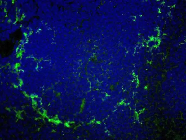
AlexaFluor®647 conjugated Rat anti Mouse CD169 antibody, clone MOMA-1 used to demonstrate marginal zone metallophils by immunofluorescence.
Image caption:
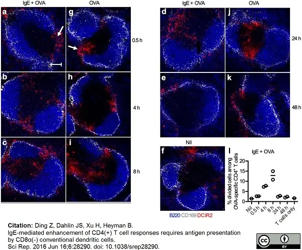
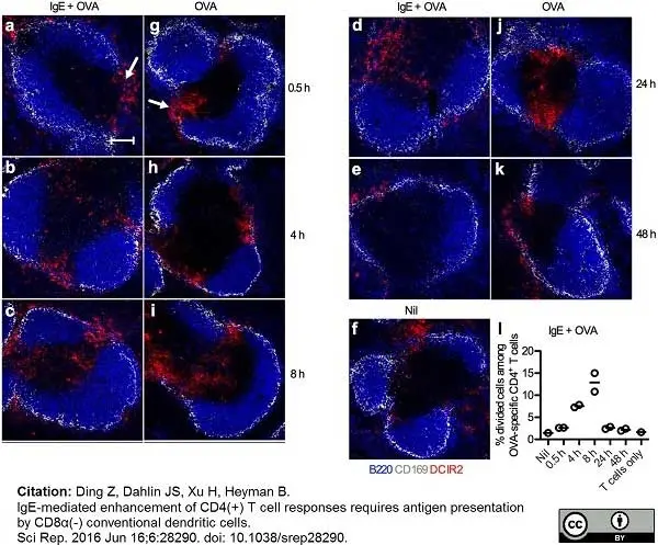
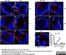
DCIR2+ cDCs migrate from the marginal zone bridging channel to the T cell zone after immunization. Spleens from BALB/c mice (n = 2 per time point) immunized with 250 μg IgE anti-OVA pre-mixed with 100 μg OVA or with 100 μg OVA alone were harvested after 0.5, 4, 8, 24, or 48 h. One unimmunized mouse (Nil) was used as control. (a–k) Half of each spleen was snap-frozen and non-consecutive spleen sections were stained and analyzed by confocal microscopy. Localization of DCIR2+ cDCs in spleens harvested at indicated time points after immunization was followed. B220+ B cells, blue; CD169+ metallophilic macrophages, grey; DCIR2+ cDCs, red. Marginal zone bridging channels are indicated with arrows in (a,g). Images show representative areas (640 μm × 640 μm) of 3–4 T cell zones from 2 non-consecutive sections of each sample in every group. Scale bar represents 100 μm. Data represent one experiment where mice were immunized with IgE-OVA or OVA alone and one where they were immunized with IgE-OVA. (l) The other halves of the spleens from mice immunized with IgE-OVA complexes in (a–e) were prepared into single cell suspensions and 6 × 10⁵ cells were used as APCs in co-cultures with 105 CFSE-labeled CD4+ T cells isolated from DO11.10 splenocytes. Percentages of divided cells among OVA-specific CD4+ T cells after incubation for 3 days with APCs taken from an unimmunized mouse (Nil) or from mice immunized with IgE-OVA complexes are quantified by flow cytometry as shown in Fig. 3. CD4+ T cells cultured alone were used as negative control. Each circle represents one mouse and the lines represent the mean values.
From: Ding Z, Dahlin JS, Xu H, Heyman B.
IgE-mediated enhancement of CD4(+) T cell responses requires antigen presentation by CD8α(-) conventional dendritic cells.
Sci Rep. 2016 Jun 16;6:28290.
doi: 10.1038/srep28290.
This image is from an open access article distributed under terms of a Creative Commons Attribution License.
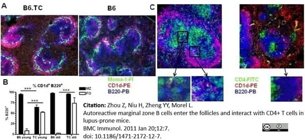
FITC conjugated Rat anti Mouse CD169 antibody, clone MOMA-1 (MCA947F) used for the detection of CD169 expressing cells in murine spleen by immunofluorescence.
Image caption:
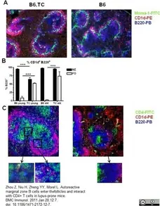
Intrafollicular location of MZB cells in B6.TC spleens. A. Representative spleen sections from 3 mo old B6.TC (left) and B6 (right) mice stained with Moma-1-FITC, CD1d-PE and B220-PB. The B220+ CD1d+ MZB cells show as bright pink while the other B cells show as blue. The ring of Moma-1+ metallophillic macrophages delineates the MZ inner edge. B. Percentage of CD1d+ B220+ B cells relative to total B220+ B cells outside (MZ) and inside (FO) the Moma-1+ ring in 3 mo and 10 mo old B6 and B6.TC mice. The data show means and standard errors of the mean (SEM) calculated of 4 MZ and FO areas for each mouse. ***: p < 0.001 for t tests. C. Representative spleen sections from 10 mo old B6.TC (left) and B6 (right) mice stained with CD4-FITC, CD1d-PE and B220-PB. Boxed areas show multiple contacts between green B6.TC CD4 T cells and pink MZB cells, but not in the B6 spleen. Original magnification: 200X.
From: Zhou et al..
Autoreactive marginal zone B cells enter the follicles and interact with CD4+ T cells in lupus-prone mice
BMC Immunology 2011 12:7.
doi: 10.1186/1471-2172-12-7.
This is from an open access article distributed under the terms of the Creative Commons Attribution License.
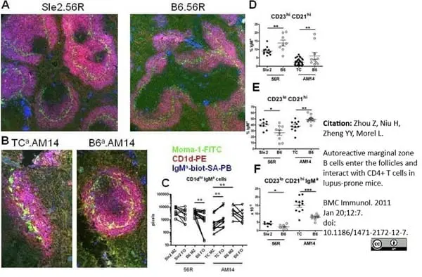
FITC conjugated Rat anti Mouse CD169 antibody, clone MOMA-1 (MCA947F) used for the detection of CD169 expressing cells in murine spleen by immunofluorescence.
Image caption:
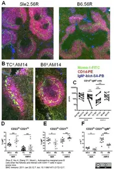
Intrafollicular location of 56R and AM14 HC Tg MZB cells. Representative spleen sections of B6.Sle2.56R and B6.56R (A, 100X) and B6.TC.AM14.IgHa/b and B6.AM14.IgHa/b (B, 200X) mice stained with Moma-1-FITC, CD1d-PE, and IgMa-biotin-SA-PB. C. Quantitation in pixels corresponding to the stain combination specific for each cell type of CD1d+ IgMa cells in the MZ (pink) and corresponding FO (blue) B cell areas in B6.Sle2.56R, B6.56R, B6.TC.AM14.IgHa/b and B6.AM14.IgHa/b mice. Paired MZ and FO values within a strain were compared with the Wilcoxon signed ranked test, and the B6.TC.AM14.IgHa/b and B6.AM14.IgHa/b MZ values were compared with the Mann-Whitney test. CD21hi CD23hi representing the T2 and MZB precursor cells expressed as the percentage of transgenic IgMa cells (D), and percentage of CD21hi CD23lo MZ B cells expressed as the percentage of transgenic IgMa cells (E) and their absolute numbers (F) in B6.Sle2.56R and B6.56R and B6.TC.AM14.IgHa/b and B6.AM14.IgHa/b mice. Graphs show means and SEMs, and the statistical significance of Student t tests. *: p <0.05, **: p <0.01, ***: p <0.001.
From: Zhou et al..
Autoreactive marginal zone B cells enter the follicles and interact with CD4+ T cells in lupus-prone mice
BMC Immunology 2011 12:7.
doi: 10.1186/1471-2172-12-7.
This is from an open access article distributed under the terms of the Creative Commons Attribution License.
FITC conjugated Rat anti Mouse CD169 antibody, clone MOMA-1 (MCA947F) used for the detection of CD169 expressing cells in murine spleen by immunofluorescence.
Image caption:
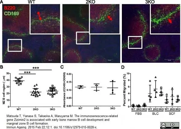
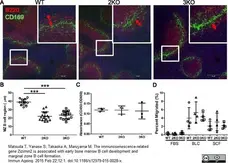
Marginal zone B cell regions were narrowed in Ziz2 KO mice. (A) Spleen sections were stained with anti-B220 (Red) and anti-CD169 (Green) antibodies for the MZ B cell region (B220-positive region outside CD169-positive cells). All mice were 10 weeks old. Scale bars: 100 μm. (B) MZ B cell regions were narrower in Ziz2 and Ziz3 KO mice than in wild type mice. (C) The proliferative activity of MZ B cells in response to LPS was not altered in both KO mice. Three mice per group were used. Data from three independent experiments (one mouse per group per experiment was used) were summarized. (D) The migratory activity of MZ B cells was analyzed using a transwell and flow cytometry. Activity against BLC or SDF1 (SDF) was not altered in both KO mice. Four mice per group were used. Each plot indicates data from one mouse. Data from four independent experiments (one mouse per group per experiment was used) were summarized. 2KO: Ziz2 KO. 3KO: Ziz3 KO. ***: P < 0.0001.
From: Matsuda T, Yanase S, Takaoka A, Maruyama M.
The immunosenescence-related gene Zizimin2 is associated with early bone marrow B cell development and marginal zone B cell formation.
Immun Ageing. 2015 Feb 22;12:1.
doi: 10.1186/s12979-015-0028-x.
This image is from an open access article distributed under terms of a Creative Commons Attribution License.
FITC conjugated Rat anti Mouse CD169 antibody, clone MOMA-1 (MCA947F) used for the detection of CD169 expressing cells in murine spleen by immunofluorescence.
Image caption:
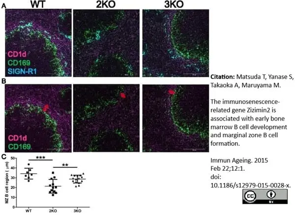
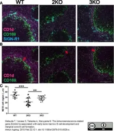
Marginal zone B cell regions were narrowed in Ziz2 KO mice. (A-B) Splenic sections were stained with anti-CD1d (Magenta), anti-CD169 (Green) (B), and anti-SIGN-R1(Blue) antibodies (A) Three mice per group were used. Between one and eight follicles per mouse (per section) were captured. All mice were 10 weeks old. Scale bars: 100 μm. (C) The CD1d-positive region outside CD169-positive cells was significantly narrower in Ziz2 KO mice than in the other groups. 2KO: Ziz2 KO. 3KO: Ziz3 KO. **: P < 0.01 ***: P < 0.0001 N = 7–15.
From: Matsuda T, Yanase S, Takaoka A, Maruyama M.
The immunosenescence-related gene Zizimin2 is associated with early bone marrow B cell development and marginal zone B cell formation.
Immun Ageing. 2015 Feb 22;12:1.
doi: 10.1186/s12979-015-0028-x.
This image is from an open access article distributed under terms of a Creative Commons Attribution License.
FITC conjugated Rat anti Mouse CD169 antibody, clone MOMA-1 (MCA947F) used for the detection of CD169 expressing cells in murine spleen sections by immunofluorescence.
Image caption:
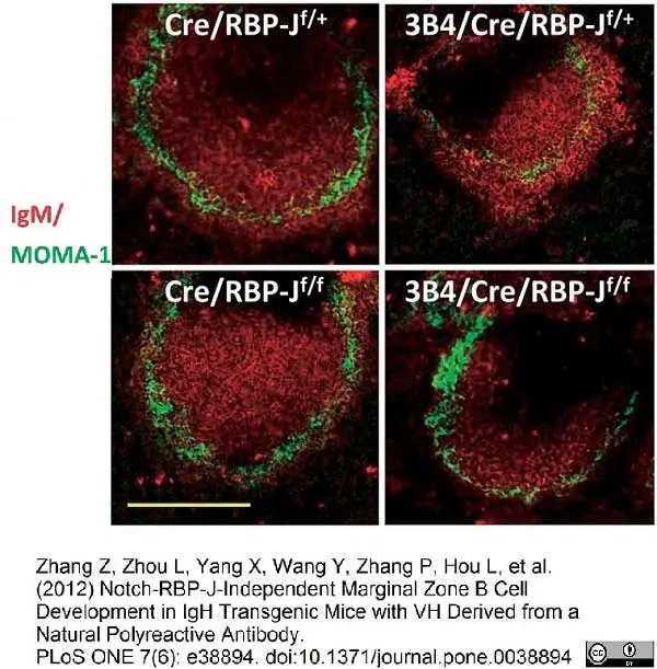
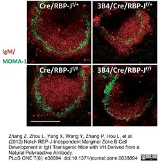
Immunofluorescent staining of splenic cryosections with Cy3-anti-IgM and FITC-anti–MOMA-1. MOMA-1+ metalophilic macrophages define the border between the MZ B and FO B cells. Data are representative of five independent experiments. Scale bar, 100 μm.
From: Zhang Z, Zhou L, Yang X, Wang Y, Zhang P, et al. (2012) Notch-RBP-J-Independent Marginal Zone B Cell Development in IgH Transgenic Mice with VH Derived from a Natural Polyreactive Antibody.
PLoS ONE 7(6): e38894.
doi: 10.1371/journal.pone.0038894
This is from an open access article distributed under the terms of the Creative Commons Attribution License.
FITC conjugated Rat anti Mouse CD169 antibody, clone MOMA-1 (MCA947F) used for the detection of sialoadhesin positive cells in the spleen of mice using immunofluorescence.
Image caption:
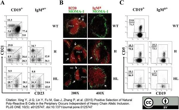
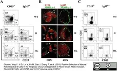
Analysis of B cells development in spleen and PEC of transgenic mice. (A) The phenotype of B cells in the spleen of TgVH3B4I and TgVH/L3B4 mice. CD1+ or IgMa+ splenic B cells from indicated mice were analyzed for the expression of CD21 and CD23 by FACS. Numbers next to each gate indicate the percentage of cells in that gate in total CD19+ or IgM+ cells. At least 7 mice from each genotype were analyzed. (B) Spleen sections from the indicated mice were stained with anti-MOMA1-FITC and anti-B220-Biotin (left panels) or anti-IgMa-biotin (right panel) followed by streptavidin-Cy3, and acquired using fluorescence microscopy. Follicular (Fo) areas around PALS are shown and MZ is indicated with arrow. The original magnitude was ×200 or ×400 as indicated. (C) CD19+ or IgMa+ B cells in PEC of indicated mice were evaluated for the expression of CD5 by FACS. At least 5 mice from each genotype were analyzed. Numbers next to each gate indicate the percentage of cells in that gate in total CD19+ or IgMa+ cells.
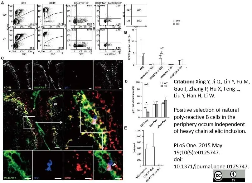
From: Xing Y, Ji Q, Lin Y, Fu M, Gao J, Zhang P, et al. (2015)
Positive Selection of Natural Poly-Reactive B Cells in the Periphery Occurs Independent of Heavy Chain Allelic Inclusion.
PLoS ONE 10(5): e0125747.
doi: 10.1371/journal.pone.0125747.
This is from an open access article distributed under the terms of the Creative Commons Attribution License.
AlexaFluor 647® Rat anti Mouse CD169 antibody, clone MOMA-1 (MCA947A647) used for the detection of marginal zone metallophils in mouse spleen by immunofluorescence.
Image caption:
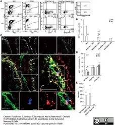
A fraction of BEC is CDH17+. (A) Spleens were treated with dispase and collagenase IV to obtain single stromal cells, and cell surface markers were analyzed by flow cytometry. Stromal cells were separated into four subpopulations: fibroblastic reticular cells (FRCs, gp38+CD31-), lymphatic endothelial cells (LEC, gp38+CD31+), BEC (gp38-CD31+), and double-negative (DN) cells (far right panel). Each fraction was then tested for CDH17 expression. The numbers adjacent to the gates indicate the percentage (%) of the indicated cells within their respective parental gates (shown on top of each panel). (B) The percentages of CDH17+ stromal cells within the respective parental gates are plotted on a bar graph. The percentages were calculated by subtracting the values of KO mice from those of WT mice (n = 7 (MAdCAM-1+ BEC); n = 8 (others)). *P≤0.05 (Student's t-test). (C) WT mouse spleen was stained with anti-CD169 (yellow), anti-MAdCAM-1 (green), anti-mouse IgG1 (blue), anti-CDH17 (red, BD1B), and analyzed by confocal microscopy. The middle-right micrograph is an enlarged view of the boxed area shown in the middle-left micrograph. The bottom row of images shows each separate color channel and an expanded view of the merged image shown within the box in the middle-right panel. The white arrowhead shown in the bottom right image indicates an IgG1+ cell that is adjacent to a CDH17+ cell in the MZ sinus. Data are representative of three independent experiments. Scale bars, 50 μm (top and middle row of images) or 10 μm (bottom row). (D) The localization of IgG1+ cells in the MZ, red pulp, and white pulp in WT and KO mice are plotted on a bar graph. Values are expressed as the percentage of IgG1+ cells localized within each sub-region of the spleen (n = 6; *P≤0.05 (Student's t-test)). (E) The number of MZ sinus (MAdCAM-1+) cells, CDH17+ cells, and CDH17+ MZ sinus cells (Cdh17+MAdCAM-1+) in the spleen of WT mice are plotted on a bar graph. Cell numbers were counted in six histological sections of WT spleen. The mean and standard deviation are plotted.
From: Funakoshi S, Shimizu T, Numata O, Ato M, Melchers F, Ohnishi K (2015)
BILL-Cadherin/Cadherin-17 Contributes to the Survival of Memory B Cells.
PLoS ONE 10(1): e0117566.
This is from an open access article distributed under the terms of the Creative Commons Attribution License.

FITC conjugated Rat anti Mouse CD169 antibody, clone MOMA-1 (MCA947F) used for the detection of marginal zone macrophages in mouse spleen by immunofluorescence of formalin fixed tissue cryosections
Image caption:
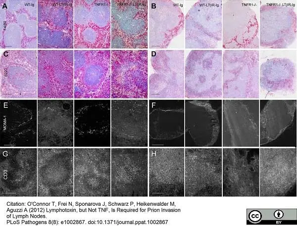
Common hematopoietic markers in lymphoid organs do not correlate with prion deposition. Frozen sections from spleens (A, C, E & G) and mesenteric lymph nodes (B, D, F & H) from C57BL/6 (WT) Ig-treated, C57BL/6 (WT) LTβR-Ig-treated, TNFR1−/− Ig-treated, or TNFR1−/− LTβR-Ig-treated mice were analyzed by immunohistochemistry and developed with alkaline phosphatase (A–D) or immunofluorescence (E–H) for macrophages (F4/80; A & B), B-cells (B-cells; C & D), metallophilic macrophages (MOMA-1; E & F), and T-cells (CD3; G & H). Size bars in A & C = 100 μm; B & D = 200 μm; E–H = 100 μm.
From: O'Connor T, Frei N, Sponarova J, Schwarz P, Heikenwalder M, Aguzzi A (2012)
Lymphotoxin, but Not TNF, Is Required for Prion Invasion of Lymph Nodes.
PLoS Pathogens 8(8): e1002867.
This is from an open access article distributed under the terms of the Creative Commons Attribution License.
FITC-conjugated Rat anti Mouse CD169 antibody, clone MOMA-1 (MCA947F) used for the identification of marginal zone metallophilic macrophages in murine spleen by immunofluorescence on formalin fixed cryosections.
Image caption:
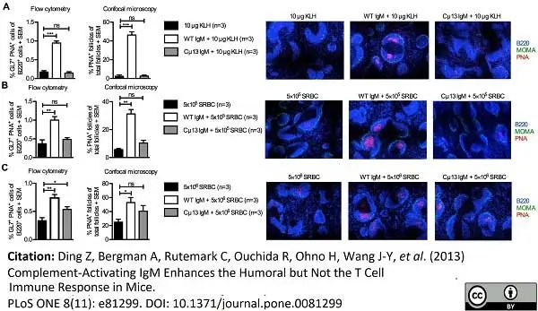
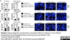
IgM from BALB/c mice enhances germinal center responses. On day 0, BALB/c mice were immunized i.v. with WT or Cμ13 IgM specific for KLH (50 μg/mouse) or SRBC (0.2 ml of a solution with HA titer 1:32) 30 min before 10 μg KLH (A), 5×10⁵ (B) or 5×10⁶ SRBC (C) were administered via the same route; controls received antigens or specific IgM alone. Spleens were harvested on day 10. Splenocytes from half of each spleen were analyzed by flow cytometry; germinal center B cells were gated as GL7+PNA+ amongst B220+ cells (Figure S1) and the percentages of germinal center B cells were quantified (A-C, upper left panels). The other halves of the spleens were sectioned, stained with anti-B220 (blue), anti-MOMA (green) and PNA (red), and analyzed for number of PNA+ germinal centers in B cell follicles by confocal microscopy (A-C, upper right panels); each image is a representative area (1725 μm × 1295 μm) for 2-3 whole sections with original magnification ×10 (A-C, lower panels). Germinal center responses of mice immunized with specific IgM alone were always lower than the responses of mice immunized with antigens alone (not shown). Data are representative of two experiments with each antigen dose. ns = not significant; * = p < 0.05; ** = p < 0.01.
From: Ding Z, Bergman A, Rutemark C, Ouchida R, Ohno H, Wang J-Y, et al. (2013)
Complement-Activating IgM Enhances the Humoral but Not the T Cell Immune Response in Mice.
PLoS ONE 8(11): e81299.
doi: 10.1371/journal.pone.0081299.
This is from an open access article distributed under the terms of the Creative Commons Attribution License.
FITC-conjugated Rat anti Mouse antibody, clone MOMA-1 (MCA947F) used to demonstrate marginal zone metalophilic macrophages in murine spleen by immunofluorescence of acetone fixed cryosections.
Image caption:
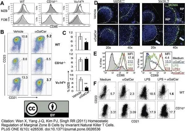
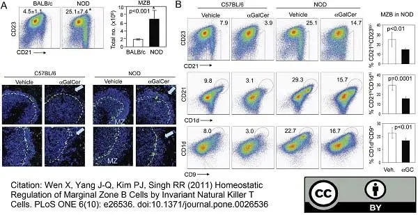
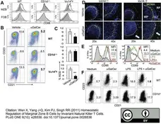
Effect of αGalCer treatment on activation and numbers of MZBs in vivo and in vitro. A–D. Four-mo-old female CD1d−/−, WT and Vα14Tg BALB/c mice were injected i.p. with 4 μg of αGalCer or vehicle. Their spleens were harvested 3–4 d later and analyzed by flow cytometry and immunohistochemistry for MZBs. Results using three mice per group from one representative of at least three independent experiments are shown. A. CD86 expression on gated CD19+CD21hiCD23−/low (MZBs) and CD19+CD21+CD23+ (FoBs) in αGalCer (thick line) or vehicle (shaded area) injected mice. B. MZBs (CD21hiCD23−/low) were analyzed on gated CD19+ lymphocytes. MZB frequency is expressed as % of CD19+ lymphocytes in representative dotplots (B) and as the mean±SE from three each of vehicle or αGalCer-injected WT, CD1d−/− and Vα14Tg mice (C). A significant reduction of MZBs was found in αGalCer-treated WT (*p<0.05) and Vα14Tg mice (**p<0.01), but not in CD1d−/− mice. D. Frozen spleen sections from αGalCer or vehicle treated mice were stained for APC-IgM (blue) and FITC-MOMA1 (green). Confocal images show IgM+ cells (blue) in the marginal zone between red pulp and MOMA-1 (green) in CD1d−/− and vehicle-injected Vα14Tg mice. IgM+ cells in the marginal zone are reduced in αGalCer-treated Vα14Tg mice (as indicated by a blue arrow). MZ, marginal zone; RP, red pulp; WP, white pulp. 20× and 40× magnification. E, F, Spleen cells (2×10⁶ cells per ml) from 3-mo-old BALB/c mice were cultured with or without LPS in the absence or presence of αGalCer (100 ng/ml). Results represent five independent experiments, each time using cells from one mouse per group. E. Expression of CD69 and CD86 are shown on gated MZBs (CD19+CD21hiCD23−/low) in spleen cells cultured with medium alone (shaded area), αGalCer (blue line), LPS (green line) or LPS+αGalCer (red line) for 24 h. The MFI of CD69 and CD86 are shown. F. Spleen cells from WT and CD1d−/− BALB/c mice were cultured without or with LPS and/or αGalCer for 72 h. MZB cells are expressed as % of mature B cells (AA4.1− IgM+).
From: Wen X, Yang J-Q, Kim PJ, Singh RR (2011)
Homeostatic Regulation of Marginal Zone B Cells by Invariant Natural Killer T Cells.
PLoS ONE 6(10): e26536.
doi: 10.1371/journal.pone.0026536.
This is from an open access article distributed under the terms of the Creative Commons Attribution License.
FITC-conjugated Rat anti Mouse antibody, clone MOMA-1 (MCA947) used to demonstrate marginal zone metalophilic macrophages in murine spleen by immunofluorescence of acetone fixed cryosections.
Image caption:
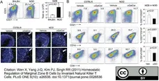
αGalCer treatment limits MZB expansion in NOD mice. A. Spleen cells from NOD or normal BALB/c mice were analyzed for MZBs. Comparison of MZB frequency between NOD and BALB/c mice (n = 6 NOD and 3 BALB/c mice, 8–10-week-old females). Numbers on dot plots indicate MZBs (CD21hiCD23−/low) as the mean ± S.E. % of CD19+ cells (*p<0.001). Bar diagram shows the mean ± S.E. absolute numbers of MZBs per spleen (*p±0.001). (B, C) Spleens were harvested from 2-mo-old NOD or C57BL/6 mice at 3 d after a single i.p. injection with αGalCer or vehicle (n = 6 mice per group). B, Cells were analyzed for MZBs as CD21hi CD23−/low CD1dhi CD9+ cells on gated CD19+ B cells. Numbers on dotplots indicate MZBs as % of CD19+ B cells. Bar diagrams show the mean ± S.E. % MZBs in NOD mice. Compared to vehicle-injected mice, αGalCer-treated NOD mice had a significant reduction in MZBs, defined as CD19+CD21hiCD23−/low (p = 0.007), CD19+CD21hiCD1dhi (p = 0.0001), CD19+CD1dhiCD9+ (p = 0.009). C, Frozen spleen sections were stained for APC-IgM (blue) and FITC-MOMA1 (green). Confocal images at 20× and 40× magnification show the reduced thickness of marginal zone (MZ) IgM+ cells (blue) in αGalCer-treated mice (as indicated by blue arrows).Data represent three independent experiments.
From: Wen X, Yang J-Q, Kim PJ, Singh RR (2011)
Homeostatic Regulation of Marginal Zone B Cells by Invariant Natural Killer T Cells.
PLoS ONE 6(10): e26536.
doi: 10.1371/journal.pone.0026536
This is from an open access article distributed under the terms of the Creative Commons Attribution License.
Rat anti Mouse CD169 antibody clone MOMA-1 (MCA947) used for the identification of marginal zone metallophilic macrophages in murine spleen by immunofluorescence on formalin fixed cryosections.
Image caption:
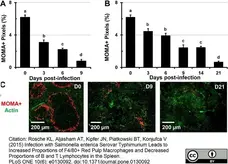
Decreased proportions of MOMA+ MZ macrophages during infection with Salmonella. C57BL/6 mice were infected i.v. with (A) 5x10⁵ CFUs (high dose) or (B) 1x10⁵ CFUs (low dose) of Salmonella and spleen sections were stained with MOMA antibodies. Images were analyzed using Volocity software and data are expressed as the percentage of pixels positive for the MOMA cell marker per total image area. Data is expressed as the mean ± standard deviation. Means within a group that do not share superscripts are significantly different from each other (p< 0.05). (C) Representative images showing the changes in the distribution of MOMA+ macrophages (red) at day 0 (control), 6, and 9 post-infection in spleens of mice infected with 5x10⁵ CFUs of Salmonella. Actin staining with phalloidin-Alexa350 (green) highlights the tissue architecture of the spleen.
From: Rosche KL, Aljasham AT, Kipfer JN, Piatkowski BT, Konjufca V (2015)
Infection with Salmonella enterica Serovar typhimurium Leads to Increased Proportions of F4/80+ Red Pulp Macrophages and Decreased Proportions of B and T Lymphocytes in the Spleen.
PLoS ONE 10(6): e0130092.
doi: 10.1371/journal.pone.0130092.
This is from an open access article distributed under the terms of the Creative Commons Attribution License.
AlexaFluor® 647 conjugated Rat anti Mouse CD169 antibody, clone MOMA-1 (MCA947A647) used to demonstrate marginal zone metallophils by immunofluorescence.
Image caption:
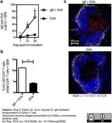
a) BALB/c mice were immunized with 50 μg IgE anti-OVA pre-mixed with 20 μg OVA (n = 7), or 20 μg OVA alone (n = 7). Sera from d 7, 21, and 35 after immunization were analysed for IgG anti-OVA by ELISA. (b) BALB/c mice were adoptively transferred with splenocytes from DO11.10 mice one day before administration of 50 μg IgE anti-OVA pre-mixed with 20 μg OVA (n = 3) or 20 μg OVA alone (n = 3). Spleens were harvested 3 days after immunization and half of each spleen was analysed for proliferation of OVA-specific CD4+ T cells by flow cytometry. The gating strategy is shown in Supplementary Fig. S2. Percentages of KJ1-26+CD4+ T cells among total CD4+ T cells of each group were then quantified. (c) The other half of each spleen as in (b) was frozen and spleen sections were stained and analysed by confocal microscopy. B220, blue; CD169, grey; DO11.10 TCR, red. Images show T cell areas (640 μm × 640 μm) representative of 6 T cell zones from 2 non-consecutive sections per sample in each group. Scale bar represents 100 μm. (a,b) Data are representative of three independent experiments and are shown as mean + SEM. Significance was determined between the group immunized with IgE-OVA complexes and the group immunized with OVA alone by Student's t-test. *p < 0.05; **p < 0.01; ***p < 0.001.
From: Ding Z, Dahlin JS, Xu H, Heyman B.
IgE-mediated enhancement of CD4(+) T cell responses requires antigen presentation by CD8α(-) conventional dendritic cells.
Sci Rep. 2016 Jun 16;6:28290.
doi: 10.1038/srep28290.
This is from an open access article distributed under the terms of the Creative Commons Attribution License.
AlexaFluor® 647 conjugated Rat anti Mouse CD169 antibody, clone MOMA-1 (MCA947A647) used to demonstrate marginal zone metallophils by immunofluorescence.
Image caption:
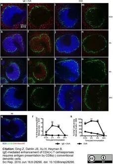
BALB/c mice were immunized with 50 μg IgE anti-OVA pre-mixed with 150 μg OVA-Alexa 647 (n = 2 per time point) (a–h), 150 μg OVA-Alexa 647 alone (n = 2 per time point) (i–p), or left unimmunized (q). Spleens were harvested 0.5, 2, 4, or 24 h after immunization. Non-consecutive sections of spleens were stained and analysed by confocal microscopy. B220+ B cells, blue; CD169+ metallophilic macrophages, green; OVA-Alexa 647, red. (a–d,i–l,q) All colors are shown. (e–h,m–p) All colors expect blue are shown. (a–q) Images show follicular areas (640 μm × 640 μm) representative of 3–4 follicular areas from 2 non-consecutive sections per sample in each group. Scale bar represents 100 μm. Data represent one experiment at 0.5 h and 2 h and two experiments at 4 h and 24 h. (r) Quantification of the Ag+ area within the B220+ follicular area. (s) Percentages of Ag+ cells among follicular B cells was analysed by flow cytometry on splenocytes from the other half of each spleen in (a–p). Follicular B cells are gated as B220+CD21+CD23high cells (Supplementary Fig. S3). (r,s) Data are shown as mean + SEM. Significance was determined between the group immunized with IgE-OVA complexes and the group immunized with OVA alone by Student's t-test. *p < 0.05; ***p < 0.001.
From: Ding Z, Dahlin JS, Xu H, Heyman B.
IgE-mediated enhancement of CD4(+) T cell responses requires antigen presentation by CD8α(-) conventional dendritic cells.
Sci Rep. 2016 Jun 16;6:28290.
doi: 10.1038/srep28290.
This is from an open access article distributed under the terms of the Creative Commons Attribution License.

AlexaFluor® 647 conjugated Rat anti Mouse CD169 antibody, clone MOMA-1 (MCA947A647) used to demonstrate marginal zone metallophils by immunofluorescence.
Image caption:
DCIR2+ cDCs migrate from the marginal zone bridging channel to the T cell zone after immunization. Spleens from BALB/c mice (n = 2 per time point) immunized with 250 μg IgE anti-OVA pre-mixed with 100 μg OVA or with 100 μg OVA alone were harvested after 0.5, 4, 8, 24, or 48 h. One unimmunized mouse (Nil) was used as control. (a–k) Half of each spleen was snap-frozen and non-consecutive spleen sections were stained and analyzed by confocal microscopy. Localization of DCIR2+ cDCs in spleens harvested at indicated time points after immunization was followed. B220+ B cells, blue; CD169+ metallophilic macrophages, grey; DCIR2+ cDCs, red. Marginal zone bridging channels are indicated with arrows in (a,g). Images show representative areas (640 μm × 640 μm) of 3–4 T cell zones from 2 non-consecutive sections of each sample in every group. Scale bar represents 100 μm. Data represent one experiment where mice were immunized with IgE-OVA or OVA alone and one where they were immunized with IgE-OVA. (l) The other halves of the spleens from mice immunized with IgE-OVA complexes in (a–e) were prepared into single cell suspensions and 6 × 10⁵ cells were used as APCs in co-cultures with 105 CFSE-labeled CD4+ T cells isolated from DO11.10 splenocytes. Percentages of divided cells among OVA-specific CD4+ T cells after incubation for 3 days with APCs taken from an unimmunized mouse (Nil) or from mice immunized with IgE-OVA complexes are quantified by flow cytometry as shown in Fig. 3. CD4+ T cells cultured alone were used as negative control. Each circle represents one mouse and the lines represent the mean values.
From: Ding Z, Dahlin JS, Xu H, Heyman B.
IgE-mediated enhancement of CD4(+) T cell responses requires antigen presentation by CD8α(-) conventional dendritic cells.
Sci Rep. 2016 Jun 16;6:28290.
doi: 10.1038/srep28290.
This is from an open access article distributed under the terms of the Creative Commons Attribution License.

Rat anti Mouse CD169 antibody, clone MOMA-1 (MCA947G) used to identify CD169 expressing microglia in murine optic nerve tissue by immunofluorescence.
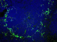
Image caption:
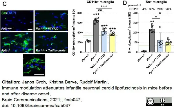
Therapeutic fingolimod and teriflunomide treatment attenuates neuroinflammation in CLN1 mice.
(C) Representative immune fluorescence microscopy and quantification of CD11b+ and (D) Sn+ microglia in longitudinal optic nerve sections (n = 5 mice per group, One-way ANOVA and Tukey's post hoc test). Blue and yellow lines indicate mean values from previously published preventive (from 1 to 6 months of age) fingolimod and teriflunomide treatment approaches, respectively. Scale bars: 20µm. *P <0.05, **P <0.01, ***P <0.001.
From: Groh J, Berve K, Martini R,
Immune modulation attenuates infantile neuronal ceroid lipofuscinosis in mice before and after disease onset,
Brain Communications, 2021;, fcab047,
doi: 10.1093/braincomms/fcab047
This image is from an open access article distributed under terms of a Creative Commons Attribution License.
FITC conjugated Rat anti Mouse CD169 antibody, clone MOMA-1 (MCA947F) used for the identification of marginal zone metalophilic macrophages in mouse spleen by immunofluorescence on acetone fixed cryosections.
Image caption:
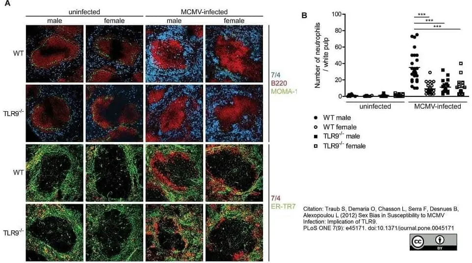
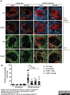
Increased number of neutrophils in the splenic white pulp of MCMV-infected WT male mice. WT or TLR9−/− male and female mice were left untreated of infected with 1×10⁵ PFU of MCMV and 4 days later spleens were collected and processed for immunofluorescent analysis. (A) Murine spleen sections were incubated with antibodies specific to neutrophils (7/4, blue or red), B cells (B220, red), marginal metalophilic macrophages (MOMA-1, green) and reticular fibroblasts (ER-TR7, green). MCMV infection led to (A) a dramatic increase in the number of neutrophils that were present in the splenic red pulp and disappearance of the MZ metallophilic macrophages and (B) an increase in the number of neutrophils in the splenic white pulp areas compared to uninfected mice. These phenomena were more prominent in WT male than in WT female or TLR9−/− male and female mice. (B) Number of neutrophils per white pulp area were counted on slides stained in A. *** p<0.001. Data are representative of 9 mice per group.
From: Traub S, Demaria O, Chasson L, Serra F, Desnues B, Alexopoulou L (2012)
Sex Bias in Susceptibility to MCMV Infection: Implication of TLR9.
PLoS ONE 7(9): e45171.
doi: 10.1371/journal.pone.0045171
This is from an open access article distributed under the terms of the Creative Commons Attribution License.
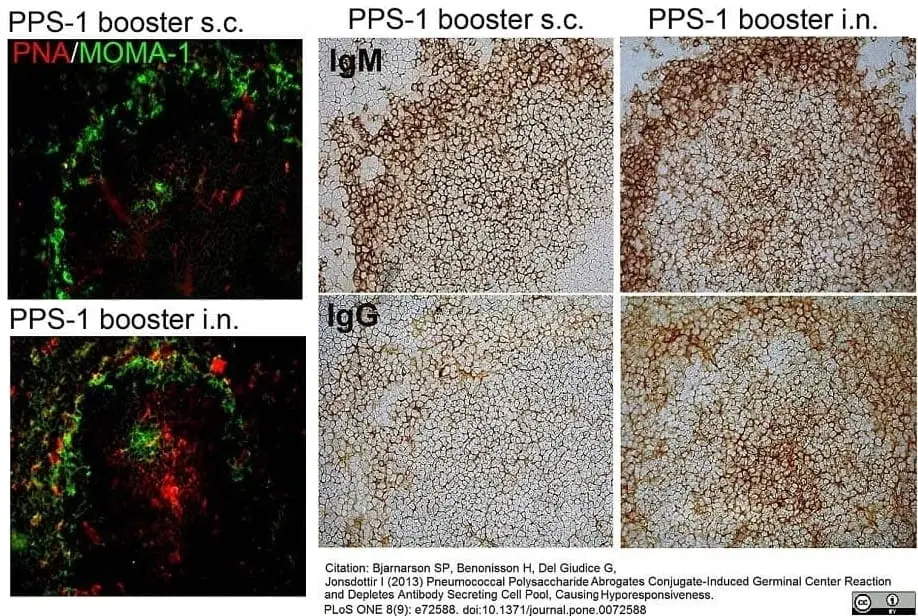
FITC conjugated Rat anti Mouse CD169 (MCA947F) antibody used to demonstrate marginal zone metalophilic macrophages in murine spleen by immunofluorescence on acetone fixed cryosections.
Image caption:
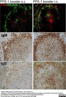
Plain PPS-1 booster s.c. abrogates the Pnc1-TT-induced GC reaction in mice primed as neonates. Active germinal centers in spleen sections were enumerated with PNA staining (upper panels). Double fluorescent staining was performed with PNA and MOMA-1 (metallophilic marginal macrophages) to show the follicular structure (top panel). IgM+ and IgG+ follicles were identified with anti-IgM (middle panel) and anti-IgG (lower panel) staining, 7 days after booster with 5.0 μg PPS-1 and 5.0 μg LT-K63 s.c. (left) or i.n. (right). Spleen sections, 7 μm, were prepared from four different levels in the spleen, starting 700 μm into the tissue and each level separated by 210 μm. One representative section per group is shown. Results are from one representative of two independent experiments (8 mice per group) showing comparable results.
From: Bjarnarson SP, Benonisson H, Del Giudice G, Jonsdottir I (2013)
Pneumococcal Polysaccharide Abrogates Conjugate-Induced Germinal Center Reaction and Depletes Antibody Secreting Cell Pool, Causing Hyporesponsiveness.
PLoS ONE 8(9): e72588.
doi: 10.1371/journal.pone.0072588.
This is from an open access article distributed under the terms of the Creative Commons Attribution License.
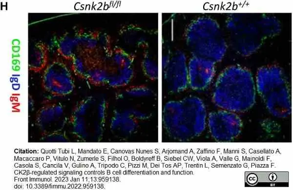
FITC conjugated Rat anti Mouse CD169, clone MOMA-1 (MCA947F) used to label CD169 expressing cells in mouse spleen by immunofluorescence.
Image caption:
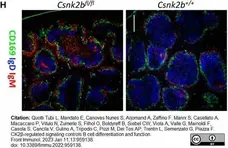
Phenotypic characterization of splenic B cell subsets in CK2βCTRL and CK2βκOsup> mice.
(H) CD169 (green), IgD (blue) and IgM (red) expression in spleen sections from CK2βCTRL and CK2βκOsup> mice was analyzed by IF. One representative mouse out of 3 per genotype is shown; 3 independent experiments. Bar, 50μm. Images were acquired with Zeiss LSM 700 confocal microscope and ZEN software. Pictures were acquired using 10x/0.3 dry and 20x/0.8 dry objectives at room temperature and merged in three-color images with ImageJ software.
From: Quotti Tubi L, Mandato E, Canovas Nunes S, Arjomand A, Zaffino F, Manni S, Casellato A, Macaccaro P, Vitulo N, Zumerle S, Filhol O, Boldyreff B, Siebel CW, Viola A, Valle G, Mainoldi F, Casola S, Cancila V, Gulino A, Tripodo C, Pizzi M, Dei Tos AP, Trentin L, Semenzato G, Piazza F.
CK2β-regulated signaling controls B cell differentiation and function.
Front Immunol. 2023 Jan 11;13:959138.
doi: 10.3389/fimmu.2022.959138.
This image is from an open access article distributed under terms of a Creative Commons Attribution License.
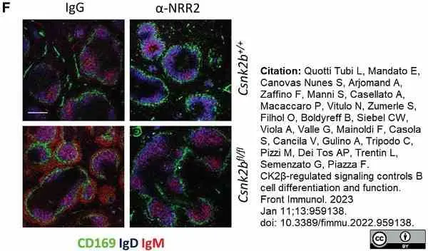
FITC conjugated Rat anti Mouse CD169, clone MOMA-1 (MCA947F) used to label CD169 expressing cells in mouse spleen by immunofluorescence.
Image caption:
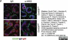
(F) IF images of spleen sections showing CD169 (green), IgD (blue) and IgM (red) expression in IgG and α-NRR2 treated mice. Bar, 50μm. One representative mouse out of two per group is shown. Images were acquired with Zeiss LSM 700 confocal microscope and analyzed with ZEN software. Pictures were acquired using objectives 10x/0.3 dry and 20x/0.8 dry at room temperature and merged in three-color images with ImageJ software.
From: Quotti Tubi L, Mandato E, Canovas Nunes S, Arjomand A, Zaffino F, Manni S, Casellato A, Macaccaro P, Vitulo N, Zumerle S, Filhol O, Boldyreff B, Siebel CW, Viola A, Valle G, Mainoldi F, Casola S, Cancila V, Gulino A, Tripodo C, Pizzi M, Dei Tos AP, Trentin L, Semenzato G, Piazza F.
CK2β-regulated signaling controls B cell differentiation and function.
Front Immunol. 2023 Jan 11;13:959138.
doi: 10.3389/fimmu.2022.959138.
This image is from an open access article distributed under terms of a Creative Commons Attribution License.
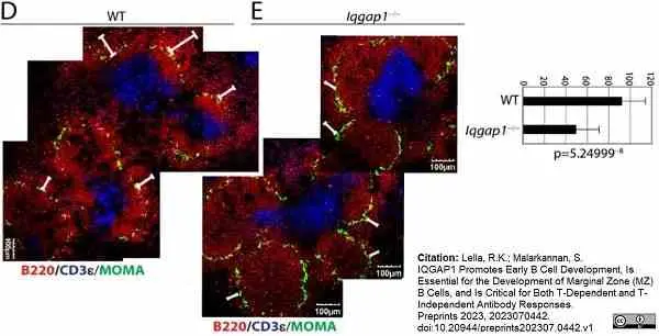
Rat anti Mouse CD169 antibody, clone MOMA-1 (MCA947G) used to stain metallophilic macrophages in mouse spleen sections by immunofluorescence.
Image caption:
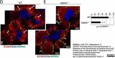
Lack of IQGAP1 leads to increased NF-B, FO-B but decreased MZ-B cells in the spleen and the dysregulated early B cell development in the spleen of Iqgap1−/− mice is B cell-intrinsic. D, E) Immunohistochemical analyses of splenic sections from WT and Iqgap1−/− mice. Spleens were isolated, embedded in paraffin, 7-micron thick sections were cut, and stained for total B cells (anti-B220), T cells (anti-CD3ε), and metallophilic macrophages (anti-MOMA antibody). Panels show the locations of T and B cells as part of the B cell follicles and metalophillic antigen-1+ macrophages surrounding the follicles.
From: Lella, R.K. and Malarkannan, S.
IQGAP1 Promotes Early B Cell Development, Is Essential for the Development of Marginal Zone (MZ) B Cells, and Is Critical for Both T-Dependent and T-Independent Antibody Responses.
Cell Mol Life Sci . 2024 Nov 25;81(1):462.
doi: 10.1007/s00018-024-05509-4.
This image is from an open access article distributed under terms of a Creative Commons Attribution License.
Filter by Application:
IF F Reset| Rat anti Mouse CD169, clone MOMA-1 recognizes murine CD169, also known as sialoadhesin or Siglec-1. CD169 is a lectin-like receptor expressed by certain populations of macrophages including marginal zone metallophils of the spleen, subcapsular macrophages of lymph nodes and stromal macrophages in bone marrow (Morris et al. 1991). CD169 is a ~185 kDa sialic acid binding receptor containing 17 immunoglobulin-like domains (Crocker et al. 1992). Expression of CD169 can be induced on macrophages in culture by a serum factor and further modulated by cytokine exposure (McWilliam et al. 1992). Rat anti mouse CD169, clone MOMA-1 has been used for the in vivo depletion of specific macrophage populations (Kraal et al. 1988). |
- Target Species
- Mouse
- Species Cross-Reactivity
-
Target Species Cross Reactivity Human Rat - N.B. Antibody reactivity and working conditions may vary between species.
- Product Form
- Purified IgG - liquid
- Preparation
- MCA947G: Purified IgG prepared by affinity chromatography on Protein A from tissue culture supernatant
- MCA947GA: Purified IgG prepared by affinity chromatography on Protein G from tissue culture supernatant
- Buffer Solution
- Phosphate buffered saline
- Preservative Stabilisers
- <0.1% Sodium Azide (NaN3)
- Immunogen
- Stromal (reticular) elements from mouse lymph nodes.
- Approx. Protein Concentrations
- IgG concentration 1.0 mg/ml
- Fusion Partners
- Spleen cells from hyperimmunized mice were fused with cells from the murine SP2/0 myeloma.
- Regulatory
- For research purposes only
- Guarantee
- 12 months from date of despatch
This product is shipped at ambient temperature. It is recommended to aliquot and store at -20°C on receipt. When thawed, aliquot the sample as needed. Keep aliquots at 2-8°C for short term use (up to 4 weeks) and store the remaining aliquots at -20°C.
Avoid repeated freezing and thawing as this may denature the antibody. Storage in frost-free freezers is not recommended.
Avoid repeated freezing and thawing as this may denature the antibody. Storage in frost-free freezers is not recommended.
This product has been reported to work in the following applications. This information is derived from testing within our laboratories, peer-reviewed publications or personal communications from the originators. Please refer to references indicated for further information. For general protocol recommendations, please visit the antibody protocols page.
| Application Name | Verified | Min Dilution | Max Dilution |
|---|---|---|---|
| Immunofluorescence |  |
||
| Immunohistology - Frozen | |
Where this product has not been tested for use in a particular technique this does not necessarily exclude its use in such procedures. Suggested working dilutions are given as a guide only. It is recommended that the user titrates the product for use in their own system using appropriate negative/positive controls.
- Histology Positive Control Tissue
- Lymphoid tissue
Source Reference
-
Kraal, G. and Janse, M. (1986) Marginal metallophilic cells of the mouse spleen identified by a monoclonal antibody.
Immunology. 58: 665-9.
References for CD169 antibody
-
Kaisho, T. et al. (2001) IkappaB kinase alpha is essential for mature B cell development and function.
J Exp Med. 193: 417-26. -
Alcamo, E. et al. (2002) Requirement for the NF-κB family member RelA in the development of secondary lymphoid organs.
J Exp Med. 195: 233-44. -
Miosge, L.A. et al. (2002) Analysis of an ethylnitrosourea-generated mouse mutation defines a cell intrinsic role of nuclear factor kappaB2 in regulating circulating B cell numbers.
J Exp Med.196: 1113-9. -
Karlsson, M.C. et al. (2003) Macrophages control the retention and trafficking of B lymphocytes in the splenic marginal zone.
J Exp Med. 198: 333-40. -
Whipple, E.C. et al. (2004) Analyses of the in vivo trafficking of stoichiometric doses of an anti-complement receptor 1/2 monoclonal antibody infused intravenously in mice.
J Immunol. 173 (4): 2297-306. -
Benlagha, K. et al. (2004) Mechanisms governing B cell developmental defects in invariant chain-deficient mice.
J Immunol. 172: 2076-83. -
Girkontaite, I. et al. (2004) The sphingosine-1-phosphate (S1P) lysophospholipid receptor S1P3 regulates MAdCAM-1+ endothelial cells in splenic marginal sinus organization.
J Exp Med. 200 (11): 1491-501. -
Ferguson, A.R. et al. (2004) Marginal zone B cells transport and deposit IgM-containing immune complexes onto follicular dendritic cells.
Int Immunol. 16 (10): 1411-22.
View The Latest Product References
-
Höpken, U.E. et al. (2004) Distinct and overlapping roles of CXCR5 and CCR7 in B-1 cell homing and early immunity against bacterial pathogens.
J Leukoc Biol. 76 (3): 709-18. -
Cariappa, A. et al. (2005) The CD9 tetraspanin is not required for the development of peripheral B cells or for humoral immunity.
J Immunol. 175: 2925-30. -
Acevedo-Suárez, C.A. et al. (2005) Uncoupling of anergy from developmental arrest in anti-insulin B cells supports the development of autoimmune diabetes.
J Immunol. 174 (2): 827-33. -
Rolf, J. et al. (2005) The enlarged population of marginal zone/CD1d(high) B lymphocytes in nonobese diabetic mice maps to diabetes susceptibility region Idd11.
J Immunol. 174: 4821-7. -
Kanayama, N. et al. (2005) Analysis of marginal zone B cell development in the mouse with limited B cell diversity: role of the antigen receptor signals in the recruitment of B cells to the marginal zone.
J Immunol. 174 (3): 1438-45. -
Oetke, C. et al. (2006) The antigen recognized by MOMA-I is sialoadhesin.
Immunol Lett. 106: 96-98. -
Caton, M.L. et al. (2007) Notch-RBP-J signaling controls the homeostasis of CD8- dendritic cells in the spleen.
J Exp Med. 204 (7): 1653-64. -
Cadman, E.T. et al. (2008) Alterations of splenic architecture in malaria are induced independently of Toll-like receptors 2, 4, and 9 or MyD88 and may affect antibody affinity.
Infect Immun. 76: 3924-31. -
Gangadharan, B. et al. (2008) Murine gamma herpesvirus-induced fibrosis is associated with the development of alternatively activated macrophages.
J Leukoc Biol. 84: 50-8. -
Awasthi, A. et al. (2010) Rap1b facilitates NK cell functions via IQGAP1-mediated signalosomes.
J Exp Med. 207: 1923-38. -
Tumanov, A.V. et al. (2010) Cellular source and molecular form of TNF specify its distinct functions in organization of secondary lymphoid organs.
Blood. 116 (18): 3456-64. -
Mattsson, J. et al. (2011) Complement activation and complement receptors on follicular dendritic cells are critical for the function of a targeted adjuvant.
J Immunol. 187: 3641-52. -
Carnrot, C. et al. (2011) Marginal zone B cells are naturally reactive to collagen type II and are involved in the initiation of the immune response in collagen-induced arthritis.
Cell Mol Immunol. 8 (4): 296-304. -
Rehm, A. et al. (2011) Cooperative function of CCR7 and lymphotoxin in the formation of a lymphoma-permissive niche within murine secondary lymphoid organs.
Blood. 118 (4): 1020-33. -
Birjandi, S.Z. et al. (2011) Alterations in marginal zone macrophages and marginal zone B cells in old mice.
J Immunol. 186: 3441-51. -
Bhattacharyya, S. et al. (2011) NFATc1 affects mouse splenic B cell function by controlling the calcineurin-NFAT signaling network.
J Exp Med. 208 (4): 823-39. -
Muppidi, J.R. et al. (2011) Cannabinoid receptor 2 positions and retains marginal zone B cells within the splenic marginal zone.
J Exp Med. 208 (10): 1941-8. -
Zhou, Z. et al. (2011) Autoreactive marginal zone B cells enter the follicles and interact with CD4+ T cells in lupus-prone mice.
BMC Immunol. 12:7. -
Jang, I.K. et al. (2011) Growth-factor receptor-bound protein-2 (Grb2) signaling in B cells controls lymphoid follicle organization and germinal center reaction.
Proc Natl Acad Sci U S A. 108: 7926-31. -
Zhang, Z. et al. (2012) Notch-RBP-J-Independent Marginal Zone B Cell Development in IgH Transgenic Mice with V(H) Derived from a Natural Polyreactive Antibody.
PLoS One. 7: e38894. -
Flores, M. et al. (2015) FcγRIIB prevents inflammatory type I IFN production from plasmacytoid dendritic cells during a viral memory response.
J Immunol. 194 (9): 4240-50. -
Matsuda T et al. (2015) The immunosenescence-related gene Zizimin2 is associated with early bone marrow B cell development and marginal zone B cell formation.
Immun Ageing. 12: 1. -
Funakoshi, S. et al. (2015) BILL-cadherin/cadherin-17 contributes to the survival of memory B cells.
PLoS One. 10 (1): e0117566. -
Xing Y et al. (2015) Positive Selection of Natural Poly-Reactive B Cells in the Periphery Occurs Independent of Heavy Chain Allelic Inclusion.
PLoS One. 10 (5): e0125747. -
Bradford, B.M. et al. (2016) Prion pathogenesis is unaltered following down-regulation of SIGN-R1.
Virology. 497: 337-345. -
Ding, Z. et al. (2016) IgE-mediated enhancement of CD4(+) T cell responses requires antigen presentation by CD8α(-) conventional dendritic cells.
Sci Rep. 6: 28290. -
Oh, D.S. et al. (2017) Transient Depletion of CD169+ Cells Contributes to Impaired Early Protection and Effector CD8+ T Cell Recruitment against Mucosal Respiratory Syncytial Virus Infection.
Front Immunol. 8: 819. -
Bogie, J.F. et al. (2018) CD169 is a marker for highly pathogenic phagocytes in multiple sclerosis.
Mult Scler. 24 (3): 290-300. -
Tsai, C.Y. et al. (2018) Bystander inhibition of humoral immune responses by Epstein-Barr virus LMP1.
Int Immunol. 30 (12): 579-90. -
Dekker, J.D. et al. (2019) Loss of the FOXP1 Transcription Factor Leads to Deregulation of B Lymphocyte Development and Function at Multiple Stages.
Immunohorizons. 3 (10): 447-62. -
Vanderkerken, M. et al. (2020) TAO-kinase 3 governs the terminal differentiation of NOTCH2-dependent splenic conventional dendritic cells.
Proc Natl Acad Sci U S A. 117 (49): 31331-31342. -
Groh, J. et al. (2021) Immune modulation attenuates infantile neuronal ceroid lipofuscinosis in mice before and after disease onset.
Brain Commun. 3 (2): fcab047. -
Quotti Tubi, L. et al. (2022) CK2β-regulated signaling controls B cell differentiation and function.
Front Immunol. 13: 959138. -
Ghilas, S. et al. (2021) Natural killer cells and dendritic epidermal γδ T cells orchestrate type 1 conventional DC spatiotemporal repositioning toward CD8+ T cells
iScience. 24 (9): 103059. -
Lella, R.K. & Malarkannan, S. (2023) IQGAP1 Promotes Early B Cell Development, Is Essential for the Development of Marginal Zone (MZ) B Cells, and Is Critical for Both T-Dependent and T-Independent Antibody Responses.
Preprints 2023, 2023070442. -
Glaubitz, J. et al. (2020) Experimental pancreatitis is characterized by rapid T cell activation, Th2 differentiation that parallels disease severity, and improvement after CD4(+) T cell depletion.
Pancreatology. 20 (8): 1637-47. -
Van, K.J.D. et al. (2018) Dendritic Cell Targeting mRNA Lipopolyplexes Combine Strong Antitumor T-Cell Immunity with Improved Inflammatory Safety.
ACS Nano. 12 (10): 9815-29. -
Ozawa, M. et al. (2022) Micro- and Macro-Anatomical Frameworks of Lymph Nodes Indispensable for the Lymphatic System Filtering Function.
Front Cell Dev Biol. 10: 902601.
- Synonyms
- Sialoadhesin
- UniProt
- Q62230
- Entrez Gene
- Siglec1
- GO Terms
- GO:0005886 plasma membrane
- GO:0005515 protein binding
- GO:0007155 cell adhesion
- GO:0016021 integral to membrane
- GO:0005576 extracellular region
- GO:0005529 sugar binding
- GO:0006897 endocytosis
View more products with CD169 specificity
Please Note: All Products are "FOR RESEARCH PURPOSES ONLY"
View all Anti-Mouse ProductsAlways be the first to know.
When we launch new products and resources to help you achieve more in the lab.
Yes, sign me up