Protocol: Measuring Apoptosis Using Annexin V
FC22
This method provides a general procedure for staining cells using Annexin V Kits to measure apoptosis. Early in apoptosis, phosphatidylserine (PS) is exposed at the cell surface, and annexin V binds to this membrane phospholipid in the presence of Ca2+. The addition of a viability dye allows cells in early and late-stage apoptosis to be identified. Annexin V is available in different excitation and emission wavelengths, and the most suitable for your experimental setup and cytometer configuration should be selected.
This method provides a general staining procedure for use with Bio-Rad’s Annexin V reagents. A certain level of technical skill is required for the successful design and implementation of these techniques; these are guidelines only and may need to be adjusted for particular cell types or applications.
Reagents
- Phosphate Buffered Saline, 10x (PBS) (BUF036A) containing 1% bovine serum albumin (PBS/BSA)
-
Annexin V Assay Kit (ANNEX200APC, ANNEX50APC, ANNEXINF, ANNEXINPE, ANNEXINSBB675, ANNEXINSBB765, ANNEXINSBUV400, ANNEXINSBUV795, ANNEXINSBV440, ANNEXINSBV515, ANNEXINSBV790, ANNEXINSBY800) containing:
- Annexin V conjugated to a fluorescent fluorophore
- 10x Binding Buffer
- dH2O
- DNA-binding viability dye, e.g., ReadiDrop Propidium Iodide (1351101)
-
Required when staining cell surface antigens:
- Staining Buffer (BUF073)
Method
- Dilute the 10x Binding Buffer to 1x in dH2O (1 mL 10x Binding Buffer + 9 mL dH2O).
- Prepare cells appropriately. Refer to protocol FC1 for information on preparation of cells from tissue culture cell lines, FC2 for isolation of peripheral blood mononuclear cells from whole blood, and FC3 for preparation of cells from tissues.
- Resuspend cells in cold (4°C) PBS/BSA and adjust to 1 x 10⁶ cells/mL.
- Aliquot 100 μL of the cell suspension into as many wells of a 96-well plate as required.
- Centrifuge at 300–400 x g for 5 min and discard supernatant.
- Optional: Perform staining of cell surface antigens and associated wash steps. Refer to protocols FC4 and FC21 for more details.
- Resuspend cells in 95 μL of 1x Binding Buffer (10x Binding Buffer diluted to 1x with dH2O) and 5 μL Annexin V. Mix well and incubate at room temperature for 10 min in the dark.
- Centrifuge at 300–400 x g for 5 min and discard supernatant.
- Resuspend cells in 200 μL 1x Binding Buffer, centrifuge at 300–400 x g for 5 min, and discard supernatant.
- Resuspend cells in 200 μL 1x Binding Buffer and store in the dark at 4°C for up to 4 hr before acquisition.
- Stain with a DNA-binding viability dye 5 min prior to acquiring samples on a flow cytometer.
Notes
- Annexin V Kit is available in several formats, including StarBright™ Dyes and traditional fluorophores. The most appropriate format for your assay should be used, taking into account the cytometer configuration and any other fluorophores in the experiment
- The viability dye used should have minimal spillover and spread with the annexin V format. Common viability dyes used are ReadiDrop Propidium Iodide (1351101), ReadiDrop 7-AAD (1351102), and PureBlu DAPI (1351303)
- Staining can also be performed in 5 mL tubes
- Staining for annexin V and cell surface antigens using antibodies can be performed at the same time. Staining should be performed in Binding Buffer, with an incubation time not exceeding 15 min
- All antibodies should be titrated prior to use to ensure the optimal concentration is used
- Appropriate controls should be included. A positive control can be generated by treating cells with 1 μM staurosporine for 4–6 hr to induce apoptosis
Flow Cytometry Resources
- Annexin V Kits
- StarBright Dyes
- Flow Cytometry Protocols
- Fluorescence Spectraviewer
- Flow supporting reagents
Support
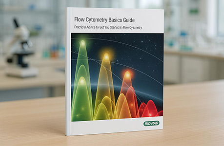
Flow Cytometry Basics Guide
Practical advice to get you started in flow cytometry
Keep up to date with recent advances by downloading our popular Flow Cytometry Basics Guide, which not only provides practical advice and best practice examples when planning multicolor experiments, but also a basic overview of all the important flow cytometry principles.
This comprehensive guide is split into nine easy-to-read chapters covering 54 pages, making this ideal for beginners wanting to gain confidence when planning experiments, and can also be used as a teaching aid.