Live/Dead Exclusion in Flow Cytometry
Author: Mike Blundell | Reviewer: Chloe Fenton
Why Dead Cell Exclusion Is Important
The presence of dead cells in your sample can greatly affect your staining and therefore the quality of your data. This is because dead cells have greater autofluorescence and increased nonspecific antibody binding, which can lead to false positives and reduce the dynamic range. This may make identification of weakly positive samples and rare populations difficult.
While using gates based on the forward and side scatter can help to remove debris and dead cells, it will not exclude them all. Because of this, dyes have been developed to distinguish live from dead cells.
Methods for Live/Dead Discrimination
Nucleic Acid Binding Dyes
One group of viability dyes are the nucleic acid binding dyes. Examples of these include propidium iodide (PI) and 7-AAD, which are excitable by both the 488 nm and 561 nm lasers. When they bind to double-stranded nucleic acid, they fluoresce. They are excluded by live cells, as these dyes are not membrane-permeable.
They can be added directly to samples after being stained with antibodies and after a brief incubation acquired as normal. The dead cells can then be identified and removed from the final analysis by gating on the unstained population (live cells). As these dyes rely on membrane integrity, it is not possible to fix the samples.
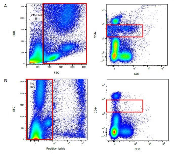
Fig. 24. Using a live/dead stain can improve your staining. A. Use of forward and side scatter gating (red rectangle) may not remove all dead cells and some nonspecific binding may still be present. B. Exclusion of dead cells using propidium iodide staining (red rectangle) means less nonspecific binding and easier identification of positively-stained populations. Images shown here are human peripheral blood stained with CD14 and CD3.
Protein Binding Dyes
There are a second group of viability dyes available to discriminate dead cells from your samples. These are protein binding dyes rather than DNA binding dyes. These dyes will bind to both live and dead cells (Figure 25). However, when a cell has a compromised membrane, as seen in dead and dying cells, there is access to a greater amount of protein, therefore they have higher fluorescence.
Similar to the DNA binding dyes, the dead cells can be excluded by gating on the less-stained population (live cells).

Fig. 25. Protein binding viability dyes. A. Live cell with dye bound to surface primary amines. B. Dead cell with dye bound to surface and intracellular primary amines.
The benefit of these dyes is that, once the cells are stained with the viability dyes, they can be fixed (they can also be used unfixed) without any reduction in the resolution between live and dead cells. In addition, they are available in a broader range of excitation and emission spectra than DNA binding dyes for convenient addition to multicolor flow cytometry panels.
Comparison of Live/Dead Viability Dyes
The key differences between nucleic acid binding dyes and protein binding dyes are summarized below.
| Feature | Nucleic Acid Binding Dyes (e.g., PI, 7-AAD) | Protein Binding Dyes |
|---|---|---|
| Mechanism | Bind to nucleic acids in cells with compromised membranes, excluded by live cells | Bind to cellular proteins, with higher staining in dead cells |
| Fixation compatibility | Not compatible with fixation | Compatible with fixation |
| Panel flexibility | Limited spectral range | Broad range of excitation and emission spectra |
| Typical use case | Live cell exclusion in unfixed samples | Multicolor panels and workflows requiring fixation |
Frequently Asked Questions
Why is dead cell exclusion important in flow cytometry?
Dead cells can exhibit increased autofluorescence and nonspecific antibody binding, which may lead to false positive signals and reduced resolution between cell populations. Excluding dead cells improves data quality and reliability.
What dyes are used for live/dead discrimination?
Common dyes include nucleic acid binding dyes such as propidium iodide (PI) and 7-AAD, and protein binding dyes that distinguish cells based on membrane integrity and protein accessibility.
Can propidium iodide be used on fixed cells?
No, propidium iodide relies on membrane integrity and is therefore not suitable for use with fixed samples.
What is the difference between fixable and nonfixable viability dyes?
Nonfixable dyes such as nucleic acid stains rely on membrane exclusion and cannot be used after fixation. Fixable dyes, such as protein binding dyes, form stable interactions that allow cells to be fixed without losing the ability to distinguish live and dead populations.
Is forward and side scatter gating sufficient to remove dead cells?
Forward and side scatter gating can help remove debris and some dead cells, but it will not exclude all nonviable cells. Viability dyes are recommended to improve accuracy.
Flow Cytometry Basics Guide Download
Get your own copy to peruse at your leisure
Download our updated Flow Cytometry Basics Guide to have practical advice, best practice examples, and a basic overview of all the important flow cytometry principles in one handy location.

Resources
Bio-Rad is committed to helping you succeed in Flow Cytometry by sharing knowledge and best practices from our experts. Below are some extremely useful resources to enhance your journey of discovery and support your success.