Flow Cytometry Guide: Autofluorescence
Author: Mike Blundell | Reviewer: Chloe Fenton
What Is Autofluorescence in Flow Cytometry?
Autofluorescence is the natural fluorescence emitted by cells due to endogenous cellular compounds. It creates background signal that can interfere with the detection of fluorescently labeled targets.
What Causes Autofluorescence?
Cellular Compounds
Cells have a natural level of fluorescence, called autofluorescence, which can be a problem in flow cytometry data analysis. Cellular autofluorescence can be due to the presence of collagen and elastin, cyclic ring compounds such as NADPH and riboflavin, aromatic amino acids, and cellular organelles such as mitochondria and lysosomes.
Cell Type and Structure
The level in cells can vary due to differences in the amount of these cellular compounds and organelles that give rise to fluorescence. In general, larger cells and more granular cells have increased autofluorescence due to an increase in the number of fluorescent compounds.
Autofluorescence Wavelengths
Most autofluorescence is detected at shorter light wavelengths with most absorbing at 350–500 nm and emitting at 350–550 nm. This is especially true for mammalian cells, which contain many compounds that are excited by the 488 nm laser and emit in the FITC range. Autofluorescence can therefore be a problem in these light ranges as the signal-to-noise ratio is decreased, resulting in reduced sensitivity and false positives. In addition, autofluorescence spillover into another channel can mask low expressers.
The level of autofluorescence can be determined using unstained controls. As there is less autofluorescence at longer light wavelengths, fluorophores that emit above 600 nm will have less autofluorescence interference. The use of a very bright fluorophore will also reduce the impact of autofluorescence.
Why Autofluorescence Is a Problem in Flow Cytometry
- Reduces signal-to-noise ratio
- Decreases sensitivity for low-expression markers
- Causes false positives
- Can result in spillover into neighboring channels
How to Measure Autofluorescence
The level of autofluorescence can be determined using unstained controls.
How to Reduce Autofluorescence in Flow Cytometry
- Use fluorophores that emit above 600 nm
- Use brighter fluorophores to increase signal over background
- Include proper unstained controls for reference
- Optimize experimental design to reduce background signal
Autofluorescence Across Different Cell Types
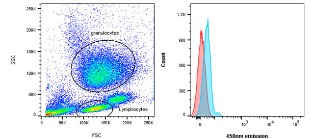
Fig. 23. Autofluorescence. Natural levels of autofluorescence can vary between different cells types. Lymphocytes (red) have less fluorescence than granulocytes (blue).
Flow Cytometry Autofluorescence FAQs
What causes autofluorescence in flow cytometry?
Autofluorescence is caused by endogenous cellular compounds such as NADPH, collagen, elastin, and aromatic amino acids.
Why is autofluorescence a problem?
Autofluorescence increases background signal, reduces sensitivity, and can mask low-expressing cell populations.
How can autofluorescence be reduced?
It can be reduced by choosing fluorophores with emission wavelengths above 600 nm and using bright fluorophores to improve signal-to-noise ratio.
How is autofluorescence measured?
Autofluorescence levels are determined using unstained controls.
Flow Cytometry Basics Guide Download
Get your own copy to peruse at your leisure
Download our updated Flow Cytometry Basics Guide to have practical advice, best practice examples, and a basic overview of all the important flow cytometry principles in one handy location.

Resources
Bio-Rad is committed to helping you succeed in Flow Cytometry by sharing knowledge and best practices from our experts. Below are some extremely useful resources to enhance your journey of discovery and support your success.